

Plasmacytoid melanoma of the urinary bladder and lymph nodes with immunohistochemical expression of plasma cell markers revealing primary esophageal melanoma
- PMID: 23133774
- PMCID: PMC3485897
- DOI: 10.1155/2012/916256
Plasmacytoid melanoma of the urinary bladder and lymph nodes with immunohistochemical expression of plasma cell markers revealing primary esophageal melanoma
Abstract
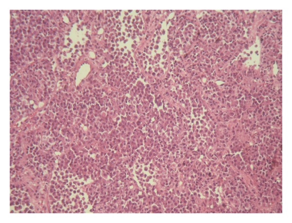
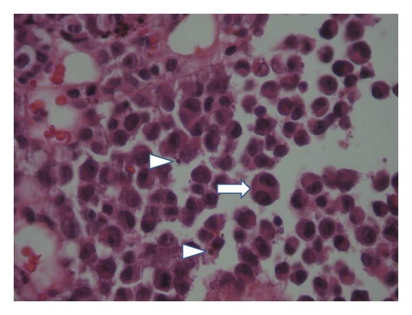
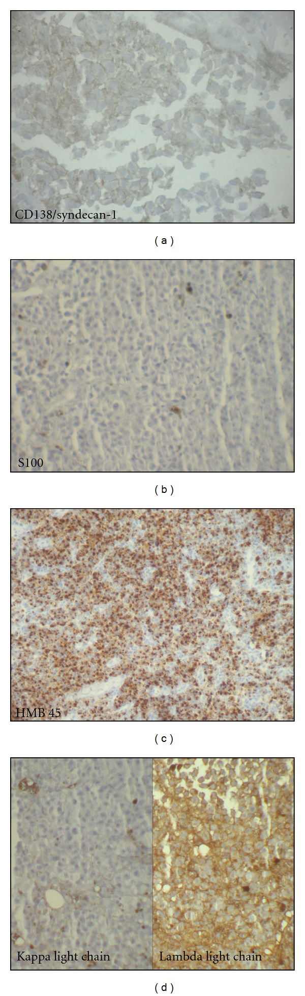
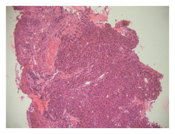
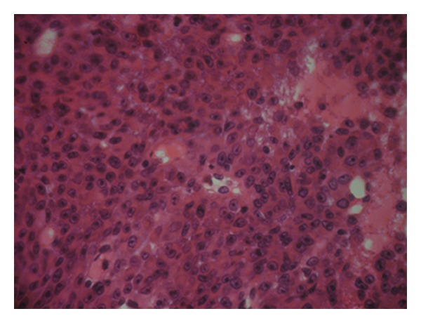
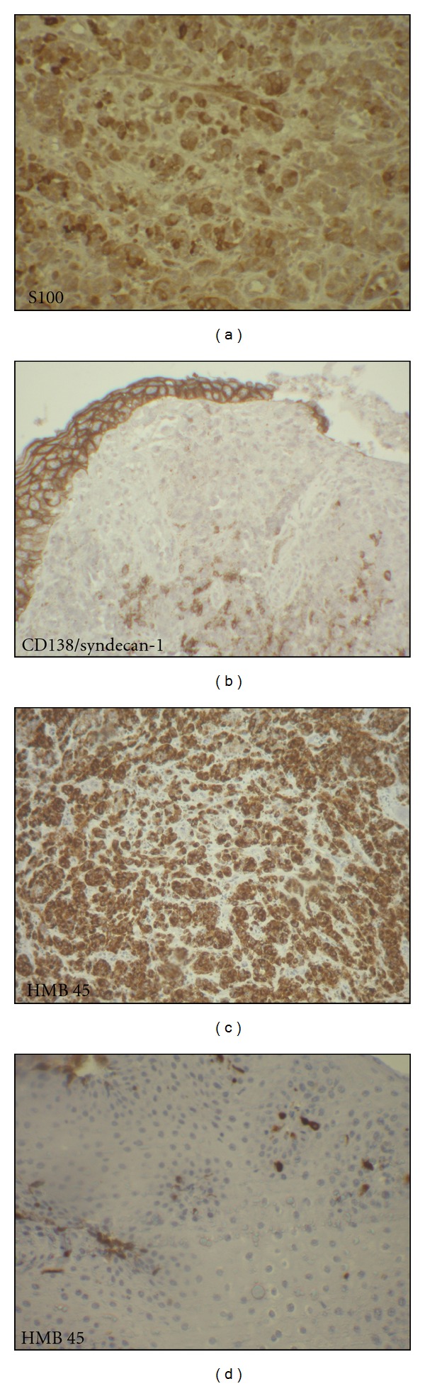
Plasmacytoid variant of melanoma is reported in only rare cases. We present the case of a 54-years-old man admitted for enlarged lymph nodes in the lumbar region. Initial diagnosis of plasmablastic lymphoma/plasma cell myeloma was considered. At our institute, a bladder polyp was removed. Microscopic exam demonstrated dense plasmacytoid cells infiltration with pigment deposits. Immunohistochemical study showed strong expression of HMB45, Melan A, and vimentin. There was focal positivity with S100 protein and CD138/syndecan-1. The diagnosis of metastatic plasmacytoid melanoma was finally established. Clinical exam revealed an esophageal melanoma with melanosis supporting its primary location. Although rarely, melanoma especially plasmacytoid variant may express plasma cell markers which may lead to erroneous diagnosis of plasma cell proliferation. Careful morphological examination for melanin pigment and the use of panel of melanocytic markers are helpful for diagnosis.
Figures
Similar articles
-
Melanoma of the urinary bladder: a review of the literature.Surg Res Pract. 2014;2014:605802. doi: 10.1155/2014/605802. Epub 2014 Jan 12. Surg Res Pract. 2014. PMID: 25374957 Free PMC article. Review.
-
A Case of Amelanotic Plasmacytoid Melanoma with Strong CD138 Expression, Mimicking Multiple Myeloma - A Case Report.Iran J Pathol. 2025 Spring;20(2):225-230. doi: 10.30699/ijp.2025.2040460.3354. Epub 2025 Mar 10. Iran J Pathol. 2025. PMID: 40487251 Free PMC article.
-
Can SOX-10 or KBA.62 Replace S100 Protein in Immunohistochemical Evaluation of Sentinel Lymph Nodes for Metastatic Melanoma?Appl Immunohistochem Mol Morphol. 2016 Jan;24(1):26-9. doi: 10.1097/PAI.0000000000000146. Appl Immunohistochem Mol Morphol. 2016. PMID: 25611246
-
[Plasmacytoid urothelial carcinoma of the urinary bladder: a clinicopathologic study of 16 cases].Zhonghua Bing Li Xue Za Zhi. 2013 Jul;42(7):433-7. doi: 10.3760/cma.j.issn.0529-5807.2013.07.001. Zhonghua Bing Li Xue Za Zhi. 2013. PMID: 24246859 Chinese.
-
Metastatic malignant melanoma of the urinary bladder: A case report and review of the literature.Indian J Pathol Microbiol. 2016 Oct-Dec;59(4):532-534. doi: 10.4103/0377-4929.191820. Indian J Pathol Microbiol. 2016. PMID: 27721291 Review.
Cited by
-
Melanoma of the urinary bladder: a review of the literature.Surg Res Pract. 2014;2014:605802. doi: 10.1155/2014/605802. Epub 2014 Jan 12. Surg Res Pract. 2014. PMID: 25374957 Free PMC article. Review.
-
Complete remission in metastatic primary malignant melanoma of the esophagus with nivolumab: a case report.J Med Case Rep. 2021 Jul 14;15(1):345. doi: 10.1186/s13256-021-02928-w. J Med Case Rep. 2021. PMID: 34256852 Free PMC article.
-
A Case of Amelanotic Plasmacytoid Melanoma with Strong CD138 Expression, Mimicking Multiple Myeloma - A Case Report.Iran J Pathol. 2025 Spring;20(2):225-230. doi: 10.30699/ijp.2025.2040460.3354. Epub 2025 Mar 10. Iran J Pathol. 2025. PMID: 40487251 Free PMC article.
-
Dermal melanoma with plasmacytoid differentiation in a dog.J Vet Diagn Invest. 2024 Nov;36(6):941-944. doi: 10.1177/10406387241273907. Epub 2024 Aug 20. J Vet Diagn Invest. 2024. PMID: 39165111 Free PMC article.
-
Metastasis of Malignant Melanoma to Urinary Bladder: A Case Report and Review of the Literature.Case Rep Pathol. 2015;2015:173870. doi: 10.1155/2015/173870. Epub 2015 May 28. Case Rep Pathol. 2015. PMID: 26106499 Free PMC article.
References
-
- Banerjee SS, Harris M. Morphological and immunophenotypic variations in malignant melanoma. Histopathology. 2000;36(5):387–402. - PubMed
-
- Siddaraju N, Yaranal PJ, Mishra MM, Soundararaghavan J. Fine needle aspiration cytology in recurrent amelanotic melanoma: a case report. Acta Cytologica. 2007;51(5):829–832. - PubMed
-
- Parwani AV, Chan TY, Mathew S, Ali SZ. Metastatic malignant melanoma in liver aspirate: cytomorphologic distinction from hepatocellular carcinoma. Diagnostic Cytopathology. 2004;30(4):247–250. - PubMed
-
- Ortega KL, de Araújo NS, de Souza FB, Magalhães MHCG. Primary malignant melanoma of the oral cavity: a case report. International Journal of Dermatology. 2004;43(10):750–752. - PubMed
-
- Riddle ND, Bui MM. When melanoma is negative for S100: diagnostic pitfalls. Archives of Pathology & Laboratory Medicine. 2012;136:237–239. - PubMed
LinkOut - more resources
Full Text Sources
