

Medulloblastoma with excessive nodularity: radiographic features and pathologic correlate
- PMID: 23133782
- PMCID: PMC3485496
- DOI: 10.1155/2012/310359
Medulloblastoma with excessive nodularity: radiographic features and pathologic correlate
Abstract
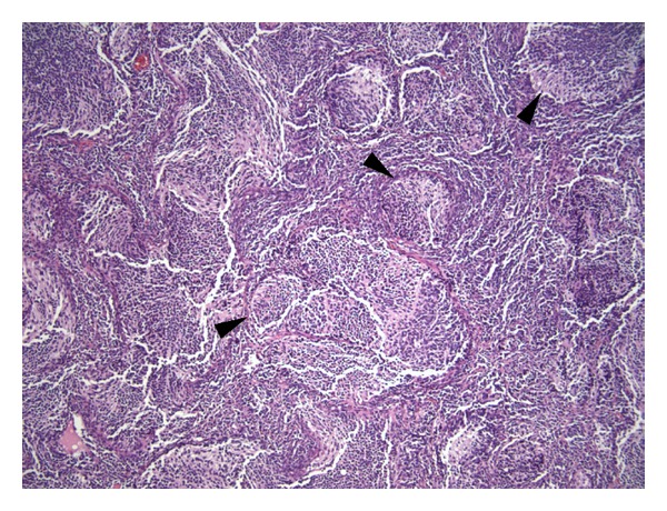
Medulloblastoma with extensive nodularity is a rare subtype of the most common malignant childhood brain tumor and has been associated with more favorable prognosis. The authors report the case of a 10-month-old girl with a posterior fossa tumor of excessive nodularity with decreased diffusivity on diffusion-weighted magnetic resonance imaging sequences and robust grape-like postgadolinium contrast enhancing features. The unique neuroradiographic features were confirmed by histopathology and a diagnosis of medulloblastoma with extensive nodularity was made. This case highlights the importance of recognizing this unique medulloblastoma subtype preoperatively, as the more favorable outcome may preclude less aggressive medical management.
Figures

Similar articles
-
Cerebellopontine angle medulloblastoma with extensive nodularity in a child: case report and review of the literature.Childs Nerv Syst. 2017 May;33(5):839-842. doi: 10.1007/s00381-016-3325-6. Epub 2016 Dec 24. Childs Nerv Syst. 2017. PMID: 28013334
-
Medulloblastoma with extensive nodularity: single photon emission CT study with iodine-123 metaiodobenzylguanidine.AJNR Am J Neuroradiol. 2002 Oct;23(9):1564-7. AJNR Am J Neuroradiol. 2002. PMID: 12372749 Free PMC article.
-
Gyriform differentiation in medulloblastoma - a radiological predictor of histology.Pediatr Neurosurg. 2007;43(2):142-5. doi: 10.1159/000098390. Pediatr Neurosurg. 2007. PMID: 17337929
-
Medulloblastomas with favorable versus unfavorable histology: how many small blue cell tumor types are there in the brain?Adv Anat Pathol. 2002 Nov;9(6):345-50. doi: 10.1097/00125480-200211000-00003. Adv Anat Pathol. 2002. PMID: 12409643 Review.
-
Medulloblastoma with extensive nodularity undergoing post-therapeutic maturation to a gangliocytoma: a case report and literature review.Pediatr Neurosurg. 2010;46(5):381-4. doi: 10.1159/000322896. Epub 2011 Mar 9. Pediatr Neurosurg. 2010. PMID: 21389751 Review.
Cited by
-
Hedgehog Pathway Inhibitors as Targeted Cancer Therapy and Strategies to Overcome Drug Resistance.Int J Mol Sci. 2022 Feb 3;23(3):1733. doi: 10.3390/ijms23031733. Int J Mol Sci. 2022. PMID: 35163655 Free PMC article. Review.
-
Genome sequencing of SHH medulloblastoma predicts genotype-related response to smoothened inhibition.Cancer Cell. 2014 Mar 17;25(3):393-405. doi: 10.1016/j.ccr.2014.02.004. Cancer Cell. 2014. PMID: 24651015 Free PMC article.
-
Compartments in medulloblastoma with extensive nodularity are connected through differentiation along the granular precursor lineage.Nat Commun. 2024 Jan 8;15(1):269. doi: 10.1038/s41467-023-44117-x. Nat Commun. 2024. PMID: 38191550 Free PMC article.
References
-
- CBTRUS. CBTRUS Statistical Report. Hindsdale, Ill, USA: Central Brain Tumor Registry of the United States; 2012. Primary brain and central nervous system tumors diagnosed in the United States in 2004–2008.
LinkOut - more resources
Full Text Sources
