. 2012;2(2):221-31.
Epub 2012 Mar 28.

Pictorial review of SPECT/CT imaging applications in clinical nuclear medicine

Affiliations

Pictorial review of SPECT/CT imaging applications in clinical nuclear medicine

Peeyush Bhargava et al. Am J Nucl Med Mol Imaging. 2012.

Abstract

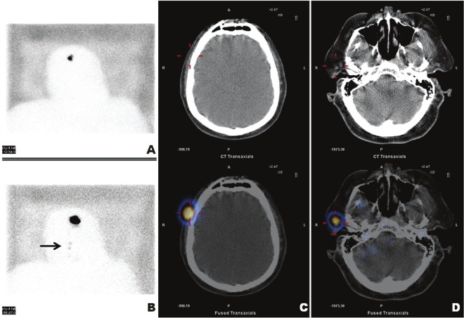
Integrated SPECT/CT scanners are gaining popularity as hybrid molecular imaging devices which can acquire SPECT and CT in a single exam. CT can be a low dose non-contrast enhanced scan for attenuation correction and anatomical localization, or a contrast enhanced diagnostic quality scan for additional anatomical characterization. We present a pictorial review highlighting the usefulness of this emerging technology. We present SPECT/CT images of 13 patients where additional information was provided by the co-registered low dose non-contrast enhanced CT scan. They belong to 12 male and 1 female patients with age ranging from 28 to 76 yrs, who were referred to the Nuclear Medicine Department for various indications. We describe these cases under in the following categories: bone scintigraphy (2), leukocyte scintigraphy (2), nuclear oncology (5), nuclear cardiology (1), and general nuclear medicine (3). Additional information provided by the co-registered low dose CT improves the diagnostic confidence in image interpretation of SPECT imaging.

Keywords: CT; Hybrid SPECT/CT; SPECT.


Figures

Figure 1
Figure 1
Planar whole body bone scan images (A & B) show foci of uptake in the thoracic and lumbar spine. SPECT/CT localizes foci of increased uptake to degenerative osteophytes (C & D) and facet joint arthritis (E), ruling out osteoblastic bone metastases.
Figure 2
Figure 2
Planar whole body bone scan images (A & B) show bone metastasis in the sternum, L2, L3, left ilium. SPECT/CT localizes bone metastasis (C & D) and finds unsuspected liver metastasis (E) from a previously unknown synchronous lung cancer.
Figure 3
Figure 3
Planar images from a three phase bone scan (A & B) and In-111 leukocyte scan (C) with uptake in the left heel ulcer/calcaneus. 111In-leukocyte SPECT/CT images confirm osteomyelitis of the left calcaneus (D) and soft tissue infection in the right leg (E).
Figure 4
Figure 4
Planar whole body 111In-leukocyte images (A & B) show abnormal focus abutting the spleen. Intra-abdominal abscess is localized on SPECT/CT (C).
Figure 5
Figure 5
Planar early and delayed 99mTc-sestamibi images (A & B) show persistent focal uptake in the mediastinum. SPECT/CT localizes ectopic parathyroid adenoma to para-aortic/prevascular region (C).
Figure 6
Figure 6
Planar whole body 111In-pentetreotideimages (A & B) show abnormalfocus in the inferior aspect ofliver. Physiologic gall bladder activity wasconfirmed on 111In-pentetreotide SPECT/CT imaging (C).
Figure 7
Figure 7
Planar whole body 123I-MIBG images (A & B) show focal uptake in the right midabdomen. SPECT/CT localizes recurrent paraganglioma (C) and shows physiologic uptake in the left adrenal (D).
Figure 8
Figure 8
Planar whole body 131I-NaI images (A & B) show three foci in the neck. Residual thyroid tissue (C) and lingual thyroid tissue (D) localized on SPECT/CT.
Figure 9
Figure 9
Planar whole body 111In-capromab pendetide (Prostascint) images (A & B) show abnormal focal uptake in the mid abdomen. Metastatic mesenteric lymph nodes confirmed on SPECT/CT (C).
Figure 10
Figure 10
Top two rows (A) of vertical long axis images (non attenuation corrected) from myocardial perfusion imaging show partially reversible inferior wall defect. Bottom two rows (B) of (attenuation corrected) images show the defect to be predominantly reversible.
Figure 11
Figure 11
Planar (right lateral) images (A & B) from 99mTc-sulfur colloid lymphoscintigraphy for a right temple melanoma. Arrow shows focal sentinel node activity. SPECT/CT localizes the site of injection (C), and a pre-auricular lymph node (D).
Figure 12
Figure 12
Planar anterior and posterior images (A & B) from 99mTc-sulfur colloid liver and spleen scan show multiple extrahepatic foci (splenosis). SPECT/CT localizes these foci to soft tissue nodules (C & D).
Figure 13
Figure 13
Planar images (A & B) from a cisternogram in anterior and lateral projections show CSF leak at the base of skull. SPECT/CT localizes CSF leak to the soft tissues between C1 and C2 vertebrae (C).

References

    1. Mariani G, Bruselli L, Kuwert T, Kim EE, Flotats A, Israel O, Dondi M, Watanabe N. A review on the clinical uses of SPECT/CT. Eur J Nucl Med Mol Imaging. 2010;37:1959–1985. - PubMed
    1. Buck AK, Nekolla S, Ziegler S, Beer A, Krause BJ, Herrmann K, Scheidhauer K, Wester HJ, Rummeny EJ, Schwaiger M, Drzezga A. SPECT/CT. J Nucl Med. 2008;49:1305–1319. - PubMed
    1. Bybel B, Brunken RC, DiFilippo FP, Neumann DR, Wu G, Cerqueira MD. SPECT/CT imaging: clinical utility of an emerging technology. Radiographics. 2008;28:1097–1113. - PubMed
    1. Patel CN, Chowdhury FU, Scarsbrook AF. Hybrid SPECT/CT: the end of "unclear" medicine. Post-grad Med J. 2009;85:606–613. - PubMed
    1. Delbeke D, Schöer H, Martin WH, Wahl RL. Hybrid imaging (SPECT/CT and PET/CT): improving therapeutic decisions. Semin Nucl Med. 2009;39:308–340. - PubMed

LinkOut - more resources