Ionizing radiation causes active degradation and reduces matrix synthesis in articular cartilage
- PMID: 23134087
- PMCID: PMC3788671
- DOI: 10.3109/09553002.2013.747015
Ionizing radiation causes active degradation and reduces matrix synthesis in articular cartilage
Abstract
Purpose: Little is known regarding radiation effects on adult articular (joint) cartilage, though joint damage has been reported following cancer treatment or occupational exposures. The aim of this study was to determine if radiation can reduce cartilage matrix production, induce cartilage degradation, or interfere with the anabolic effects of IGF-1.
Materials and methods: Isolated chondrocytes cultured in monolayers and whole explants harvested from ankles of human donors and knees of pigs were irradiated with 2 or 10 Gy γ-rays, with or without IGF-1 stimulation. Proteoglycan synthesis and IGF-1 signaling were examined at Day 1; cartilage degradation throughout the first 96 hours.
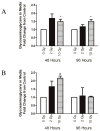
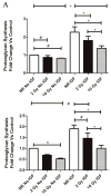
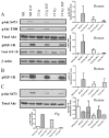
Results: Human and pig cartilage responded similarly to radiation. Cell viability was unchanged. Basal and IGF-1 stimulated proteoglycan synthesis was reduced following exposure, particularly following 10 Gy. Both doses decreased IGF-induced Akt activation and IGF-1 receptor phosphorylation. Matrix metalloproteinases (ADAMTS5, MMP-1, and MMP-13) and proteoglycans were released into media after 2 and 10 Gy.
Conclusions: Radiation induced an active degradation of cartilage, reduced proteoglycan synthesis, and impaired IGF-1 signaling in human and pig chondrocytes. Lowered Akt activation could account for decreased matrix synthesis. Radiation may cause a functional decline of cartilage health in joints after exposure, contributing to arthropathy.
Conflict of interest statement
The authors report no declarations of interest.
Figures


Similar articles
-
Effects of physical stimulation with electromagnetic field and insulin growth factor-I treatment on proteoglycan synthesis of bovine articular cartilage.Osteoarthritis Cartilage. 2004 Oct;12(10):793-800. doi: 10.1016/j.joca.2004.06.012. Osteoarthritis Cartilage. 2004. PMID: 15450529
-
The combination of insulin-like growth factor 1 and osteogenic protein 1 promotes increased survival of and matrix synthesis by normal and osteoarthritic human articular chondrocytes.Arthritis Rheum. 2003 Aug;48(8):2188-96. doi: 10.1002/art.11209. Arthritis Rheum. 2003. PMID: 12905472
-
Extracellular nicotinamide phosphoribosyltransferase (NAMPT/visfatin) inhibits insulin-like growth factor-1 signaling and proteoglycan synthesis in human articular chondrocytes.Arthritis Res Ther. 2012 Jan 30;14(1):R23. doi: 10.1186/ar3705. Arthritis Res Ther. 2012. PMID: 22289259 Free PMC article.
-
Effects of growth factors and cytokines on proteoglycan turnover in articular cartilage.Br J Rheumatol. 1992;31 Suppl 1:1-6. Br J Rheumatol. 1992. PMID: 1532522 Review.
-
[Regulation of cartilage matrix synthesis by chondrocytes].Rev Rhum Ed Fr. 1994 Nov 15;61(9 Pt 2):93S-98S. Rev Rhum Ed Fr. 1994. PMID: 7858613 Review. French.
Cited by
-
Reducing hand radiation during renal access for percutaneous nephrolithotomy: a comparison of radiation reduction techniques.Urolithiasis. 2024 Jan 13;52(1):27. doi: 10.1007/s00240-023-01510-x. Urolithiasis. 2024. PMID: 38217570 Free PMC article.
-
Pilot screening of potential matrikines resulting from collagen breakages through ionizing radiation.Radiat Environ Biophys. 2024 Aug;63(3):337-350. doi: 10.1007/s00411-024-01086-z. Epub 2024 Aug 8. Radiat Environ Biophys. 2024. PMID: 39115696 Free PMC article.
-
A reproducible radiation delivery method for unanesthetized rodents during periods of hind limb unloading.Life Sci Space Res (Amst). 2015 Jul;6:10-4. doi: 10.1016/j.lssr.2015.05.002. Life Sci Space Res (Amst). 2015. PMID: 26097807 Free PMC article.
-
Knee and Hip Joint Cartilage Damage from Combined Spaceflight Hazards of Low-Dose Radiation Less than 1 Gy and Prolonged Hindlimb Unloading.Radiat Res. 2019 Jun;191(6):497-506. doi: 10.1667/RR15216.1. Epub 2019 Mar 29. Radiat Res. 2019. PMID: 30925135 Free PMC article.
-
MakoTM robotic-arm-assisted total hip and total knee arthroplasty outcomes in an orthopedic oncology setting: A case series.J Orthop. 2023 Oct 26;46:70-77. doi: 10.1016/j.jor.2023.10.021. eCollection 2023 Dec. J Orthop. 2023. PMID: 37942217 Free PMC article.
References
-
- Baxter NN, Habermann EB, Tepper JE, Durham SB, Virnig BA. Risk of pelvic fractures in older women following pelvic irradiation. Journal of the American Medical Association. 2005;294(20):2587–93. - PubMed
-
- Cornelissen M, de Ridder L. Dose- and time-dependent increase of lysosomal enzymes in embryonic cartilage in vitro after ionizing radiation. Scanning Microscopy. 1990;4(3):769–73. discussion 773–4. - PubMed
Publication types
MeSH terms
Substances
Grants and funding
LinkOut - more resources
Full Text Sources
Miscellaneous
