Impaired neurovascular coupling to ictal epileptic activity and spreading depolarization in a patient with subarachnoid hemorrhage: possible link to blood-brain barrier dysfunction
- PMID: 23134492
- PMCID: PMC3625730
- DOI: 10.1111/j.1528-1167.2012.03699.x
Impaired neurovascular coupling to ictal epileptic activity and spreading depolarization in a patient with subarachnoid hemorrhage: possible link to blood-brain barrier dysfunction
Abstract
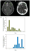
Spreading depolarization describes a sustained neuronal and astroglial depolarization with abrupt ion translocation between intraneuronal and extracellular space leading to a cytotoxic edema and silencing of spontaneous activity. Spreading depolarizations occur abundantly in acutely injured human brain and are assumed to facilitate neuronal death through toxic effects, increased metabolic demand, and inverse neurovascular coupling. Inverse coupling describes severe hypoperfusion in response to spreading depolarization. Ictal epileptic events are less frequent than spreading depolarizations in acutely injured human brain but may also contribute to lesion progression through increased metabolic demand. Whether abnormal neurovascular coupling can occur with ictal epileptic events is unknown. Herein we describe a patient with aneurysmal subarachnoid hemorrhage in whom spreading depolarizations and ictal epileptic events were measured using subdural opto-electrodes for direct current electrocorticography and regional cerebral blood flow recordings with laser-Doppler flowmetry. Simultaneously, changes in tissue partial pressure of oxygen were recorded with an intraparenchymal oxygen sensor. Isolated spreading depolarizations and clusters of recurrent spreading depolarizations with persistent depression of spontaneous activity were recorded over several days followed by a status epilepticus. Both spreading depolarizations and ictal epileptic events where accompanied by hyperemic blood flow responses at one optode but mildly hypoemic blood flow responses at another. Of note, quantitative analysis of Gadolinium-diethylene-triamine-pentaacetic acid (DTPA)-enhanced magnetic resonance imaging detected impaired blood-brain barrier integrity in the region where the optode had recorded the mildly hypoemic flow responses. The data suggest that abnormal flow responses to spreading depolarizations and ictal epileptic events, respectively, may be associated with blood-brain barrier dysfunction.
Wiley Periodicals, Inc. © 2012 International League Against Epilepsy.
Conflict of interest statement
The authors have no financial interest in this manuscript and no affiliations (relationships) to disclose. The authors confirm that they have read the Journal’s position on issues involved in ethical publication and affirm that this report is consistent with those guidelines.
Figures


Similar articles
-
Cortical spreading ischaemia is a novel process involved in ischaemic damage in patients with aneurysmal subarachnoid haemorrhage.Brain. 2009 Jul;132(Pt 7):1866-81. doi: 10.1093/brain/awp102. Epub 2009 May 6. Brain. 2009. PMID: 19420089 Free PMC article.
-
Inverse neurovascular coupling to cortical spreading depolarizations in severe brain trauma.Brain. 2014 Nov;137(Pt 11):2960-72. doi: 10.1093/brain/awu241. Epub 2014 Aug 24. Brain. 2014. PMID: 25154387
-
Correlates of spreading depolarization in human scalp electroencephalography.Brain. 2012 Mar;135(Pt 3):853-68. doi: 10.1093/brain/aws010. Brain. 2012. PMID: 22366798 Free PMC article.
-
Recording, analysis, and interpretation of spreading depolarizations in neurointensive care: Review and recommendations of the COSBID research group.J Cereb Blood Flow Metab. 2017 May;37(5):1595-1625. doi: 10.1177/0271678X16654496. Epub 2016 Jan 1. J Cereb Blood Flow Metab. 2017. PMID: 27317657 Free PMC article. Review.
-
The continuum of spreading depolarizations in acute cortical lesion development: Examining Leão's legacy.J Cereb Blood Flow Metab. 2017 May;37(5):1571-1594. doi: 10.1177/0271678X16654495. Epub 2016 Jan 1. J Cereb Blood Flow Metab. 2017. PMID: 27328690 Free PMC article. Review.
Cited by
-
Vasoconstriction and Impairment of Neurovascular Coupling after Subarachnoid Hemorrhage: a Descriptive Analysis of Retinal Changes.Transl Stroke Res. 2018 Jun;9(3):284-293. doi: 10.1007/s12975-017-0585-8. Epub 2017 Nov 8. Transl Stroke Res. 2018. PMID: 29119370
-
The Relationship Between Seizures and Spreading Depolarizations in Patients with Severe Traumatic Brain Injury.Neurocrit Care. 2022 Jun;37(Suppl 1):31-48. doi: 10.1007/s12028-022-01441-2. Epub 2022 Feb 16. Neurocrit Care. 2022. PMID: 35174446
-
Characterization of the relationship between intracranial pressure and electroencephalographic monitoring in burst-suppressed patients.Neurocrit Care. 2015 Apr;22(2):212-20. doi: 10.1007/s12028-014-0059-8. Neurocrit Care. 2015. PMID: 25142827 Free PMC article.
-
Dexamethasone-Enhanced Continuous Online Microdialysis for Neuromonitoring of O2 after Brain Injury.ACS Chem Neurosci. 2023 Jul 19;14(14):2476-2486. doi: 10.1021/acschemneuro.2c00703. Epub 2023 Jun 27. ACS Chem Neurosci. 2023. PMID: 37369003 Free PMC article.
-
Anatomy and physiology of the blood-brain barrier.Semin Cell Dev Biol. 2015 Feb;38:2-6. doi: 10.1016/j.semcdb.2015.01.002. Epub 2015 Feb 11. Semin Cell Dev Biol. 2015. PMID: 25681530 Free PMC article. Review.
References
-
- Abbott NJ, Ronnback L, Hansson E. Astrocyte-endothelial interactions at the blood–brain barrier. Nat Rev Neurosci. 2006;7:41–53. - PubMed
-
- Attwell D, Iadecola C. The neural basis of functional brain imaging signals. Trends Neurosci. 2002;25:621–625. - PubMed
-
- Butzkueven H, Evans AH, Pitman A, Leopold C, Jolley DJ, Kaye AH, Kilpatrick CJ, Davis SM. Onset seizures independently predict poor outcome after subarachnoid hemorrhage. Neurology. 2000;55:1315–1320. - PubMed
Publication types
MeSH terms
Grants and funding
LinkOut - more resources
Full Text Sources

