. 2012 Dec 28;287(53):44164-72.
doi: 10.1074/jbc.M112.401521. Epub 2012 Nov 7.

PAX2 protein induces expression of cyclin D1 through activating AP-1 protein and promotes proliferation of colon cancer cells

Affiliations

PAX2 protein induces expression of cyclin D1 through activating AP-1 protein and promotes proliferation of colon cancer cells

Hai-Sheng Zhang et al. J Biol Chem. .

Abstract

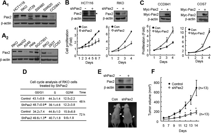
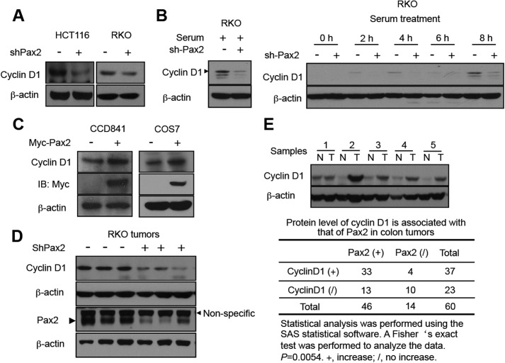
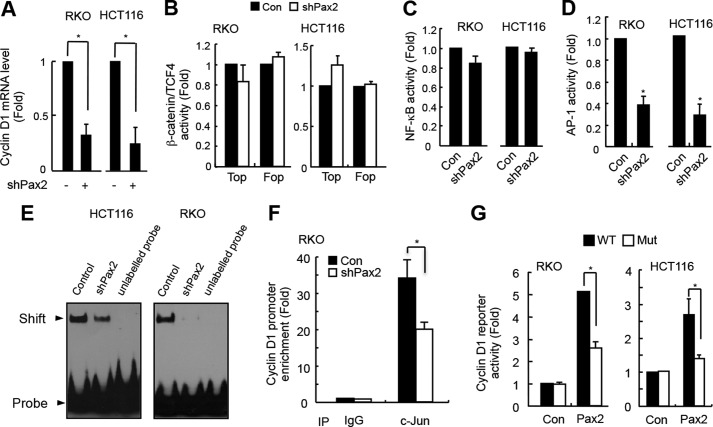
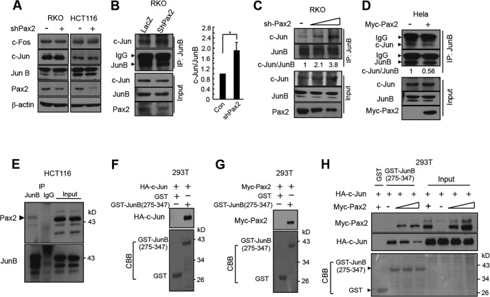
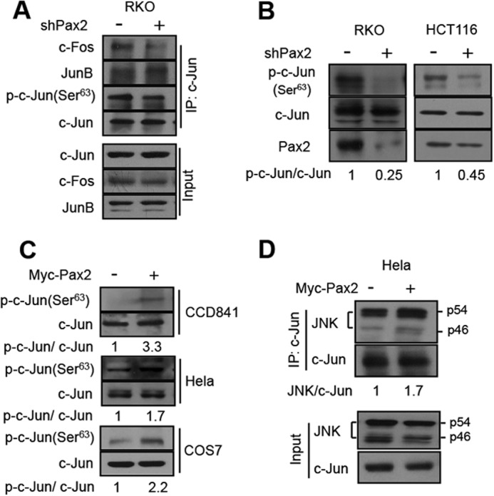
Paired box (PAX) 2, a transcription factor, plays a critical role in embryogenesis. When aberrantly expressed in adult tissues, it generally exhibits oncogenic properties. However, the underlying mechanisms remain unclear. We reported previously that the expression of PAX2 was up-regulated in human colon cancers. However, the role of PAX2 in colon cancer cells has yet to be determined. The aim of this study is to determine the function of PAX2 in colon cancer cells and to investigate the possible mechanisms underlain. We find that knockdown of PAX2 inhibits proliferation and xenograft growth of colon cancer cells. Inhibition of PAX2 results in a decreased expression of cyclin D1. Expression of cyclin D1 is found increased in human primary colon malignant tumors, and its expression is associated with that of PAX2. These data indicate that PAX2 is a positive regulator of expression of cyclin D1. We find that knockdown of PAX2 inhibits the activity of AP-1, a transcription factor that induces cyclin D1 expression, implying that PAX2 induces cyclin D1 through AP-1. PAX2 has little effect on expression of AP-1 members including c-Jun, c-Fos, and JunB. Our data show that PAX2 prevents JunB from binding c-Jun and enhances phosphorylation of c-Jun, which may elevate the activity of AP-1. Taken together, these results suggest that PAX2 promotes proliferation of colon cancer cells through AP-1.


Figures

FIGURE 1.
FIGURE 1.
PAX2 promotes proliferation of colon cancer cells. A, expression of PAX2 in colon cancer cells and normal colon epithelial cells (A1) and in other types of cells (A2). Expression of PAX2 was determined by Western blot as described under “Materials and Methods.” B, knockdown of PAX2 inhibited proliferation of colon cancer cells. HCT116 and RKO cells were infected with control (Con) or shPAX2 adenovirus. Cell proliferation was determined as described under “Materials and Methods.” The data are the mean ± S.E. (n = 3). *, p < 0.05 versus control. C, overexpression PAX2 increased cell proliferation. To determine the effect of PAX2 on proliferation of COS7 cell, a pool of COS7 cells that stably expressed PAX2 was prepared as described under “Materials and Methods.” CCD841 cells were transfected with the vector expressing Myc-tagged PAX2. *, p < 0.05 versus control (n = 3). D, knockdown of PAX2 led to cell G1 arrest. RKO cells were infected with control or shPAX2 adenovirus. The infected cells were subjected for cell cycle analysis as described under “Materials and Methods.” *, p < 0.05 versus control (n = 3). E, knockdown of PAX2 inhibited tumor growth of RKO cells. RKO cells were infected with control or shPAX2 adenovirus. 3 × 10⁶ of the infected cells were injected subcutaneously into the flank of nude mice. The upper panel shows the knockdown efficiency of PAX2. The lower panel shows representative mice. F, the volume of the tumors. The tumor volumes were measured 5 days after injection. The data are mean ± S.E. (n = 13). *, p < 0.05 versus control.
FIGURE 2.
FIGURE 2.
PAX2 induces expression of cyclin D1. A, knockdown of PAX2 decreased expression of cyclin D1. HCT116 and RKO cells infected with control or shPAX2 adenovirus were harvested for immunoblotting. B, knockdown of PAX2 decreased expression of cyclin D1. RKO cells were infected as described above. In 48 h, the cells were switched to serum-free medium and incubated for 24 h. Serum was added, and the cells were harvested at different times as indicated. The cells incubated in serum-containing medium were used as a control. C, overexpression of PAX2 increased expression of cyclin D1. The CCD841 and COS7 cells described in the legend for Fig. 1 (panel C) were used for determination of cyclin D1 by immunoblotting. D, expression of PAX2 and cyclin D1 in xenografts of RKO cells. E, expression of cyclin D1 is associated with that of PAX2 in human colon tumors. Sixty pairs of human primary colon tumors were analyzed. The immunoblotting of the tissue extracts was described previously (13). The upper panel indicates a representative Western blot. N, normal; T, tumor. A Fisher's exact test was performed to analyze the data.
FIGURE 3.
FIGURE 3.
PAX2 induces cyclin D1 through AP-1. A, knockdown of PAX2 decreased cyclin D1 mRNA level. HCT116 and RKO cells were infected with control or shPAX2 adenovirus. In 48 h, the cells were harvested for real-time PCR analysis. B, PAX2 did not affect the transcriptional activity of β-catenin/TCF4. HCT116 and RKO cells were transfected with TOP/FOP reporter and β-galactosidase plasmids. The next day, the cells were infected with control (Con) or shPAX2 adenovirus. In 48 h, the luciferase activity was determined. C, PAX2 did not affect NF-κB activity. HCT116 and RKO cells were transfected with NF-κB reporter and β-galactosidase plasmids. The cells were incubated overnight followed by infection with control or shPAX2 adenovirus. In 48 h, the relative NF-κB activity was determined by measuring luciferase activity. D, knockdown of PAX2 repressed AP-1 activity. HCT116 and RKO cells were transfected with AP-1 reporter and β-galactosidase plasmids. The transfected cells were incubated overnight and then infected with control or shPAX2 adenovirus. In 48 h, the cells were harvested for luciferase assay. E, knockdown of PAX2 inhibited AP-1 DNA binding activity. HCT116 and RKO cells were infected with control or shPAX2 adenovirus. In 72 h, the infected cells were harvested for EMSA assay. F, ChIP assay was performed as described under “Materials and Methods.” RKO cells were used in the experiment. G, PAX2 regulated expression of cyclin D1 via AP-1. The cyclin D1 promoter reporter and mutated cyclin D1 promoter reporter plasmids were prepared as described under “Materials and Methods.” RKO and HCT116 cells were transfected with cyclin D1 promoter reporter, PAX2, and β-galactosidase plasmids. In 24 h, the cells were harvested for luciferase activity assay. *, p < 0.05 versus control (n = 3).
FIGURE 4.
FIGURE 4.
PAX2 inhibits the interaction between JunB and c-Jun. A, PAX2 had little effect on expression of c-Jun, c-Fos, and JunB. HCT116 and RKO cells were infected with control or shPAX2 adenovirus. Forty-eight hours after infection, the cells were harvested for immunoblotting. B, knockdown of PAX2 enhanced the c-Jun-JunB interaction. RKO cells were infected as above. Forty-eight hours after infection, the cells were harvested for immunoprecipitation (IP). The c-Jun/JunB ratio (c-Jun/JunB) was determined by measuring the density of co-immunoprecipitated c-Jun protein band and normalized to that of immunoprecipitated JunB. The c-Jun/JunB ratio in control (Con) cells was designated as 1. The data are mean ± S.E. (n = 3). *, p < 0.05 versus control. C, knockdown of PAX2 enhanced the c-Jun-JunB interaction in a dose-dependent manner. RKO cells were infected with different amounts of shPAX2 virus. Forty-eight hours after infection, the cells were harvested for immunoprecipitation. D, PAX2 inhibited the interaction between c-Jun and JunB. HeLa cells were transfected as indicated. In 24 h, the cells were harvested for immunoprecipitation. The c-Jun/JunB ratio was determined as above. E, the endogenous PAX2 and JunB were co-immunoprecipitated. HCT116 cell lysates were used for immunoprecipitation. F, c-Jun bound to JunB(275–347). 293T cells were transfected with HA-c-Jun. Equal amounts of cell lysates containing HA-c-Jun were incubated with the glutathione-Sepharose beads that already captured GST or GST-JunB(275–347). The beads were washed, and HA-c-Jun retained on beads was determined. G, PAX2 bound to GST-JunB(275–347). 293T cells were transfected with Myc-PAX2. Equal amounts of cell lysates containing Myc-PAX2 were incubated with the glutathione-Sepharose beads that captured GST or GST-JunB(275–347). The beads were washed, and Myc-PAX2 retained on beads was determined. H, PAX2 competed with c-Jun for binding JunB(275–347). 293T cells were transfected with HA-c-Jun or different amounts of Myc-PAX2 plasmids. Equal amounts of lysates containing HA-c-Jun were incubated with glutathione-Sepharose beads containing GST-JunB(275–347) at 4 °C for 3 h. The beads were washed, and equal amounts of lysates containing different amounts of Myc-PAX2 were added. The beads were incubated at 4 °C for 3 h. The beads were washed and incubated in SDS-PAGE loading buffer, and the resolved proteins were analyzed by immunoblotting.
FIGURE 5.
FIGURE 5.
PAX2 enhances phosphorylation of c-Jun. A, knockdown of PAX2 decreased the c-Jun-c-Fos interaction and c-Jun phosphorylation. RKO cells were infected with control or shPAX2 virus. In 48 h, the cells were harvested, and cell lysates were prepared for immunoprecipitation (IP) and immunoblotting. B, knockdown of PAX2 suppressed the phosphorylation of c-Jun. HCT116 and RKO cells were infected with control or shPAX2 adenovirus. In 48 h, the cells were harvested for immunoblotting. The antibody against phospho-c-Jun at Ser-63 was used. The p-c-Jun(Ser-63) to c-Jun ratio (p-c-Jun/c-Jun) was determined by measuring the density of the p-c-Jun(Ser-63) band and normalized to that of c-Jun. C, overexpression of PAX2 increased phosphorylation of c-Jun. CCD841, HeLa, and COS7 cells were transfected with Myc-PAX2 plasmid. The lysates of these cells were used to determine the level of p-c-Jun(Ser-63) by Western blot. p-c-Jun/c-Jun ratio was determined as described above. D, PAX2 enhanced the interaction of JNK and c-Jun. HeLa cells were transfected with control or Myc-PAX2 plasmid. In 24 h, the cells were harvested for determination of the interaction of JNK and c-Jun by means of immunoprecipitation.

References

    1. Robson E. J., He S. J., Eccles M. R. (2006) A PANorama of PAX genes in cancer and development. Nat. Rev. Cancer 6, 52–62 - PubMed
    1. Lang D., Powell S. K., Plummer R. S., Young K. P., Ruggeri B. A. (2007) PAX genes: roles in development, pathophysiology, and cancer. Biochem. Pharmacol. 73, 1–14 - PubMed
    1. Muratovska A., Zhou C., He S., Goodyer P., Eccles M. R. (2003) Paired-box genes are frequently expressed in cancer and often required for cancer cell survival. Oncogene 22, 7989–7997 - PubMed
    1. Luu V. D., Boysen G., Struckmann K., Casagrande S., von Teichman A., Wild P. J., Sulser T., Schraml P., Moch H. (2009) Loss of VHL and hypoxia provokes PAX2 up-regulation in clear cell renal cell carcinoma. Clin. Cancer Res. 15, 3297–3304 - PubMed
    1. Torban E., Eccles M. R., Favor J., Goodyer P. R. (2000) PAX2 suppresses apoptosis in renal collecting duct cells. Am. J. Pathol. 157, 833–842 - PMC - PubMed

Publication types

MeSH terms