. 2012 Dec 6;492(7427):104-7.
doi: 10.1038/nature11589. Epub 2012 Nov 7.

Identification of a rudimentary neural crest in a non-vertebrate chordate

Affiliations

Identification of a rudimentary neural crest in a non-vertebrate chordate

Philip Barron Abitua et al. Nature. .

Abstract

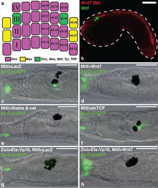
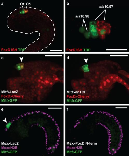
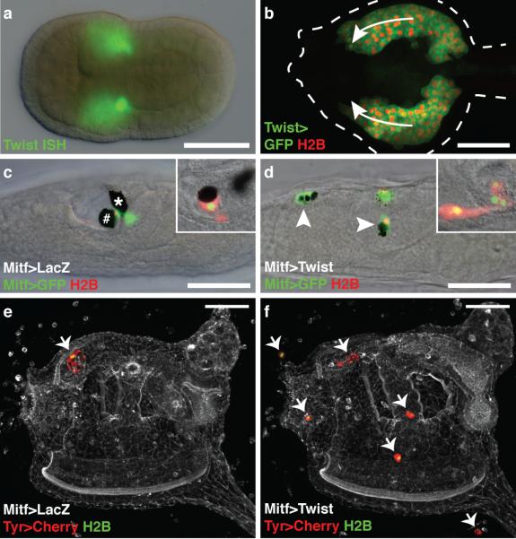
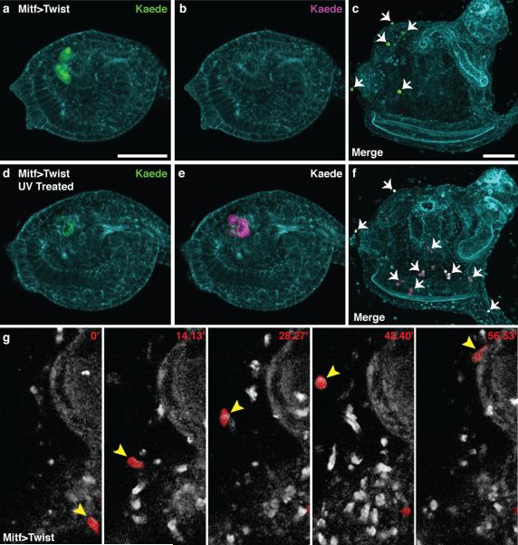
Neural crest arises at the neural plate border, expresses a core set of regulatory genes and produces a diverse array of cell types, including ectomesenchyme derivatives that elaborate the vertebrate head. The evolution of neural crest has been proposed to be a key event leading to the appearance of new cell types that fostered the transition from filter feeding to active predation in ancestral vertebrates. However, the origin of neural crest remains controversial, as homologous cell types have not been unambiguously identified in non-vertebrate chordates. Here we show that the tunicate Ciona intestinalis possesses a cephalic melanocyte lineage (a9.49) similar to neural crest that can be reprogrammed into migrating 'ectomesenchyme' by the targeted misexpression of Twist (also known as twist-like 2). Our results suggest that the neural crest melanocyte regulatory network pre-dated the divergence of tunicates and vertebrates. We propose that the co-option of mesenchyme determinants, such as Twist, into the neural plate ectoderm was crucial to the emergence of the vertebrate 'new head'.


Figures

Figure 1
Figure 1. Wnt signaling promotes ocellus formation
a, Gastrula stage schematic indicates the lineage specific expression of enhancers in this study. b, Tailbud electroporated with Mitf>LacZ detected with an antibody (green), and hybridized with a Wnt7 probe (red). c-f, Larvae electroporated with βγ-crystallin>GFP marks the otolith and anterior palps. c, Co-electroporated with Mitf>LacZ (166/196 had an otolith and ocellus). d, Co-electroporated with Mitf>Wnt7 (172/205 had two ocelli). e, Co-electroporated with Mitf>stable β-catenin (189/205 had two ocelli). f, Coelectroporated with Mitf>dnTCF (100/205 had two otoliths). g,h, Larvae electroporated with Zicl>Ets:VP16 and βγ-crystallin>GFP. g, Co-electroporated with Mitf>LacZ (75/100 had extra otoliths). h, Co-electroporated with Mitf>Wnt7 (only 11/100 had extra otoliths). Scale bars, 50 μm.
Figure 2
Figure 2. FoxD represses Mitf in the ocellus
a, Tailbud electroporated with TRP>LacZ detected with an antibody (green) marking the precursors of the otolith and ocellus, and hybridized with a FoxD probe (red). b, FoxD is expressed in the posterior a10.97 cell. c-d, Tailbuds electroporated with FoxD>mCherry and Mitf>GFP. Arrowheads mark the presumptive ocellus. c, Co-electroporated with Mitf>LacZ (126/180 expressed mCherry). d, Co-electroporated with Mitf>dnTCF (only 30/180 expressed mCherry). e-f, Tailbuds electroporated with Msx>H2B mCherry, and Mitf>GFP. Arrowhead shows GFP expression in a9.49 derivatives. e, Co-electroporated with Msx>LacZ. f, Co-electroporated with Msx>FoxD N-term. Scale bars 50 μm (a, c-f); 25 μm (b).
Figure 3
Figure 3. Twist reprograms the a9.49 lineage
a, Neurula hybridized with a Twist probe. b, Tailbud during mesenchyme migration (arrows) co-electroporated with Twist>GFP and Twist>H2B mCherry. c,d, Larvae electroporated with Mitf>GFP, and Mitf>H2B mCherry. Insets show lineage marked with Tyr>mCherry, and Tyr>H2B YFP. c, Co-electroporated with Mitf>LacZ. # and * mark the otolith and ocellus, respectively. d, Co-electroporated with Mitf>Twist. Arrowheads indicate ectopic position of a9.49 derivatives. e,f, Juveniles electroporated Tyr>mCherry, and Tyr>H2B YFP. Arrows indentify the position of a9.49 derivatives. e, Co-electroporated with Mitf>LacZ. f, Coelectroporated with Mitf>Twist. Scale bars, 50 μm.
Figure 4
Figure 4. Lineage tracing of reprogrammed a9.49 cells
a-f, Ciona electroporated with Mitf>Twist and Tyr>Kaede. a,b, Non-UV treated tailbud shows only green fluorescence c, Embryo never exposed to UV results in a juvenile with only green ectopic cells (arrows). d,e, UV treated tailbud shows green and red fluorescence respectively. f, UV treated embryo results in a juvenile with green and red ectopic cells (arrows). g, Time-lapse frames of Supplemental Movie 2 (minutes indicated) shows the migration of reprogrammed a9.49 cell labeled with Tyr>mCherry (arrowhead). Scale bars, 50 μm.

References

    1. Bronner ME, Le Douarin NM. Evolution and development of the neural crest: An overview. Dev. Biol. 2012;366:2–9. - PMC - PubMed
    1. Le Douarin NM, et al. Neural crest cell plasticity and its limits. Development. 2004;131:4637–4650. - PubMed
    1. Gans C, Northcutt RG. Neural crest and the origin of vertebrates: a new head. Science. 1983;220:268–273. - PubMed
    1. Yu JK, Meulemans D, McKeown SJ, Bronner-Fraser M. Insights from the amphioxus genome on the origin of vertebrate neural crest. Genome Res. 2008;18:1127–1132. - PMC - PubMed
    1. Delsuc F, Brinkmann H, Chourrout D, Philippe H. Tunicates and not cephalochordates are the closest living relatives of vertebrates. Nature. 2006;439:965–968. - PubMed

Publication types

MeSH terms

Substances