. 2012 Nov 15;491(7424):413-7.
doi: 10.1038/nature11602. Epub 2012 Nov 7.

The μ transpososome structure sheds light on DDE recombinase evolution

Affiliations

The μ transpososome structure sheds light on DDE recombinase evolution

Sherwin P Montaño et al. Nature. .

Abstract

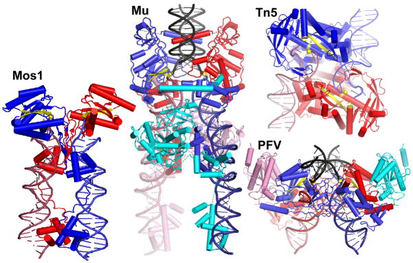
Studies of bacteriophage Mu transposition paved the way for understanding retroviral integration and V(D)J recombination as well as many other DNA transposition reactions. Here we report the structure of the Mu transpososome--Mu transposase (MuA) in complex with bacteriophage DNA ends and target DNA--determined from data that extend anisotropically to 5.2 Å, 5.2 Å and 3.7 Å resolution, in conjunction with previously determined structures of individual domains. The highly intertwined structure illustrates why chemical activity depends on formation of the synaptic complex, and reveals that individual domains have different roles when bound to different sites. The structure also provides explanations for the increased stability of the final product complex and for its preferential recognition by the ATP-dependent unfoldase ClpX. Although MuA and many other recombinases share a structurally conserved 'DDE' catalytic domain, comparisons among the limited set of available complex structures indicate that some conserved features, such as catalysis in trans and target DNA bending, arose through convergent evolution because they are important for function.


Conflict of interest statement

Author Information:

Coordinates and structure factors were deposited at the PDB, with ID 4fcy. Reprints and permissions information is available at www.nature.com/reprints. The authors have no competing financial interests.

Figures

Figure 1
Figure 1
Transposition pathway and structure determination. a) Cartoon of transposition. The transposase (MuA) pairs the phage genome ends (blue and red). At each end, the same active site catalyzes the attack of H2O at the phage-host junction and then the direct attack of the phage 3′-OH on target DNA (“strand transfer”). Target binding is nonspecific, and there is a 5 bp stagger between the sites of attack. Host and target DNAs may be entire circular replicons. After the ATP-dependent unfoldase ClpX disassembles the final strand transfer complex, the 3′ hydroxyls are used as replication primers, resulting in duplication of the phage genome. Our crystals contain the strand transfer product (3rd panel). b) Domain structure of MuA. c) Experimental electron density map after phase improvement with Parrot superimposed on the model (contours are 1.2 and 2σ). Note: part a was originally drawn in Illustrator, and an editable pdf can be supplied if needed. Part b was drawn in Word.
Figure 2
Figure 2
Transpososome structure. The complex sits on a crystallographic twofold (vertical) that relates the blue and red halves. The pale- and dark-colored subunits adopt different conformations within the homotetramer. DNA colors match Figure 1. a) Cartoon. Catalytic sites are marked as yellow and tan stars (facing the viewer or the background, respectively) and domains of the blue subunits are labeled. b) Ribbon drawing, with the scissile phosphate groups shown as yellow spheres. c) Same drawing as in B, rotated ~90° about a vertical axis.
Figure 3
Figure 3
This is a stereo pair. Stereo close-up of interactions near the Mu DNA-target junction. Colors are the same as in Figure 2. A segment of DNA from a symmetry-related complex (yellow) binds the positively-charged domain IIIα of the R2-bound subunit (cyan). If the red Mu end DNA were extended to include flanking host DNA, it could lie where the yellow DNA does. The yellow sphere marks the phosphate group at the Mu-target DNA junction, and the main chains of the two active site D’s are also yellow (a third active site residue lies on a helix that could not be modeled). The loop that extends from domain IIα aa 410–430) to interact with the black target DNA is circled on the red subunit.
Figure 4
Figure 4
Model for a transpososome assembled on full left (reddish) and right (blue) bacteriophage ends. The N-terminus of each domain Iβ is marked with a red sphere to show the approximate position of domain Iα, which transiently binds the enhancer. Domains discussed in the text are labeled. Inset: cartoon of the bacteriophage Mu genome ends and internal enhancer element.
Figure 5
Figure 5
Comparison of DDE recombinase-DNA complexes. The mobile element ends are red and blue and target DNA (where included) is black. Subunits that carry out the chemical reactions are red and blue; additional subunits pink and cyan. Active site residues, scissile phosphate groups, and the two β strands of the conserved catalytic domain that carry the catalytic D’s are in yellow. Mos1 is a Tc1/mariner family eukaryotic DNA transposon; Tn5 is a bacterial DNA transposon, and Prototype Foamy Virus (PFV) is a mammalian retrovirus. Mos1 and Tn5 require only a dimer for activity, whereas Mu transposase and PFV integrase require tetramers. In the PFV structure, only the catalytic domains of the additional subunits were visible (pink and cyan).

References

    1. Yanagihara K, Mizuuchi K. Mismatch-targeted transposition of Mu: a new strategy to map genetic polymorphism. Proc Natl Acad Sci U S A. 2002;99:11317–11321. - PMC - PubMed
    1. Haapa S, Taira S, Heikkinen E, Savilahti H. An efficient and accurate integration of mini-Mu transposons in vitro: a general methodology for functional genetic analysis and molecular biology applications. Nucleic Acids Res. 1999;27:2777–2784. - PMC - PubMed
    1. Mizuuchi K. In vitro transposition of bacteriophage Mu: a biochemical approach to a novel replication reaction. Cell. 1983;35:785–794. - PubMed
    1. Montano SP, Rice PA. Moving DNA around: DNA transposition and retroviral integration. Curr Opin Struct Biol. 2011;21:370–378. - PMC - PubMed
    1. Davies DR, Goryshin IY, Reznikoff WS, Rayment I. Three-dimensional structure of the Tn5 synaptic complex transposition intermediate. Science. 2000;289:77–85. - PubMed

Publication types

Associated data