Comparative Study
. 2012 Nov 8;491(7423):222-7.
doi: 10.1038/nature11600.

Principles for designing ideal protein structures

Affiliations
Comparative Study

Principles for designing ideal protein structures

Nobuyasu Koga et al. Nature. .

Abstract

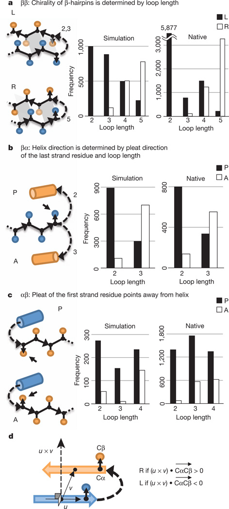
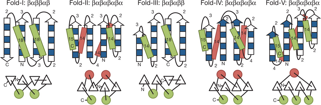
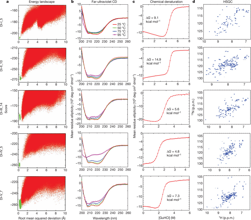
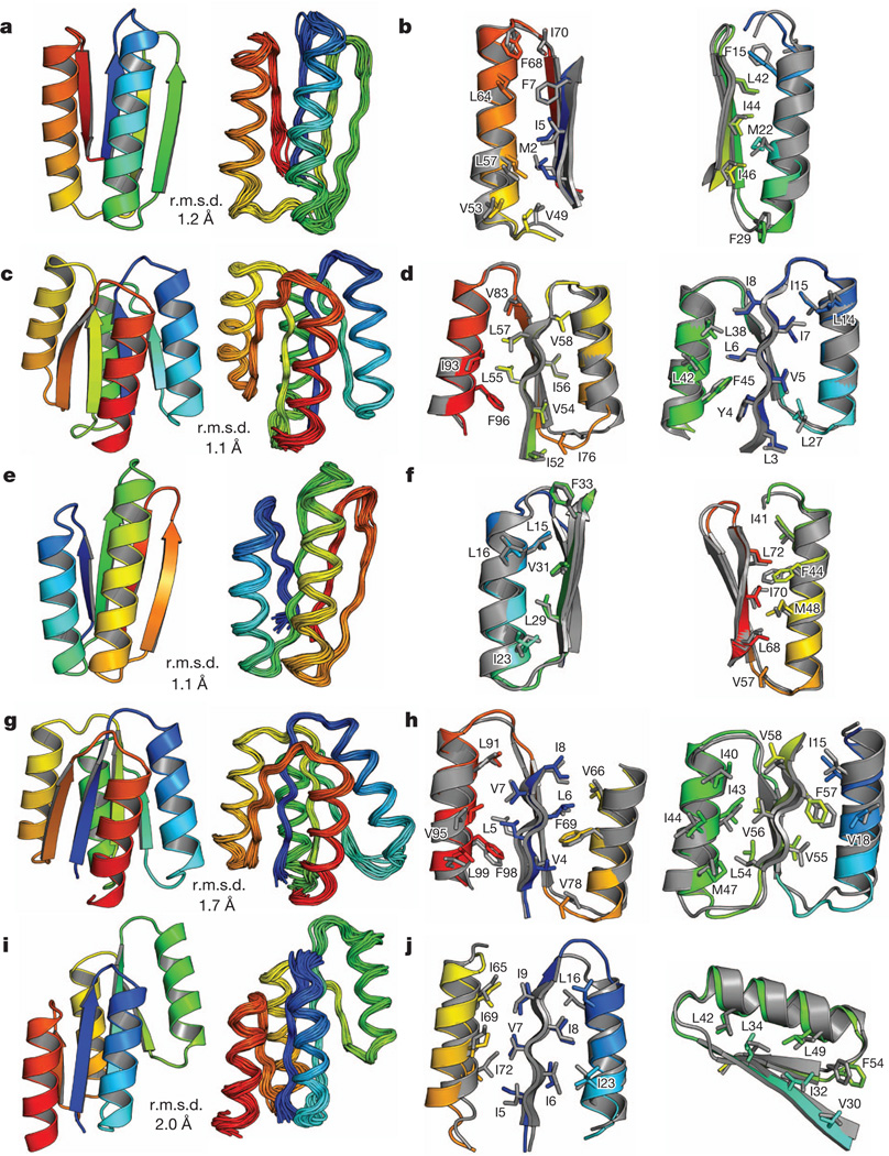
Unlike random heteropolymers, natural proteins fold into unique ordered structures. Understanding how these are encoded in amino-acid sequences is complicated by energetically unfavourable non-ideal features--for example kinked α-helices, bulged β-strands, strained loops and buried polar groups--that arise in proteins from evolutionary selection for biological function or from neutral drift. Here we describe an approach to designing ideal protein structures stabilized by completely consistent local and non-local interactions. The approach is based on a set of rules relating secondary structure patterns to protein tertiary motifs, which make possible the design of funnel-shaped protein folding energy landscapes leading into the target folded state. Guided by these rules, we designed sequences predicted to fold into ideal protein structures consisting of α-helices, β-strands and minimal loops. Designs for five different topologies were found to be monomeric and very stable and to adopt structures in solution nearly identical to the computational models. These results illuminate how the folding funnels of natural proteins arise and provide the foundation for engineering a new generation of functional proteins free from natural evolution.


Figures

Figure 1
Figure 1. Fundamental rules
a, ββ-rule. L (left-handed) and R (right-handed) ββ-units are illustrated (see Fig. 1d for chirality definition). The dependence of chirality on loop length is shown in the histograms. b, βα-rule. P (parallel) and A (antiparallel) βα-units are illustrated. The dependence of orientation (P versus A) on loop length is shown in the histograms. c, αβ-rule. d, Chirality (L versus R) of a ββ-unit. The chirality is defined on the basis of the orientation of the Cα-to-Cβ vector, CαCβ, of the strand residue preceding or following the connecting loop. u is a vector along the first strand and v is a vector from the centre of the first strand to the centre of the second strand.
Figure 2
Figure 2. Derivation of secondary structure lengths from the rules for five protein topologies
Fold-I: Ferredoxin-like fold. Fold-II: Rossmann2×2 fold. Fold-III: IF3-like fold. Fold-IV: P-loop2×2 fold. Fold-V: Rossmann3×1 fold. In the upper illustrations, numbers represent the secondary structure lengths following from the rules described in Fig. 1 and Supplementary Fig. 1. Strand lengths are represented by filled and open boxes. The filled boxes represent pleats coming out of the page, and the open boxes represent pleats going into the page. In the lower illustrations, the design topologies are represented with circles (helices) and triangles (strands) connected by solid lines (loops).
Figure 3
Figure 3. Characterization of design for each of the five folds
a, Energy landscapes obtained from Rosetta ab initio structure prediction simulations on Rosetta@home. Red points represent the lowest-energy structures obtained in independent Monte Carlo structure prediction trajectories starting from an extended chain for each sequence; the y axis shows the Rosetta all-atom energy and the x axis shows the Cα root mean squared deviation from the design model. Green points represent the lowest-energy structures obtained in trajectories starting from the design model. Less sampling around the designed minima is observed for the higher-contact-order topology, Fold-IV. b, The far-ultraviolet circular dichroism (CD) spectra at various temperatures. c, Chemical denaturations with GuHCl (square brackets denote concentration) at 220nmand 25 °C. The data were fitted to a two-state model (red solid line) to obtain the free energy of unfolding ΔG. d, Two-dimensional 1H–15N HSQC spectra at 25 °C and 600 MHz. p.p.m., parts per million.
Figure 4
Figure 4. Comparison of computational models with experimentally determined structures
a, c, e, g, i, Comparison of overall topology. Design models (left) and NMR structures (right); the Cα root mean squared deviation (r.m.s.d.) between them is indicated. b, d, f, h, j, Comparison of core side-chain packing in superpositions of design models (rainbow) and NMR structures (grey). The left and right panels show close-up views of the core packing and correspond to the left and right portions of the structures shown in a, c, e, g and i. a, b, Di-I_5 (Protein Data Bank code, 2KL8); c, d, Di-II_10 (2LV8); e, f, Di-III_14 (2LN3); g, h,Di-IV_5 (2LVB); i, j, Di-V_7 (2LTA). The design models and NMR structures are available from http://psvs-1_4-dev.nesg.org/ideal_proteins/.

Comment in

References

    1. Leopold PE, Montal M, Onuchic JN. Protein folding funnels: a kinetic approach to the sequence-structure relationship. Proc. Natl Acad. Sci. USA. 1992;89:8721–8725. - PMC - PubMed
    1. Onuchic JN, Wolynes PG, Luthey-Schulten Z, Socci ND. Toward an outline of the topography of a realistic protein-folding funnel. Proc. Natl Acad. Sci. USA. 1995;92:3626–3630. - PMC - PubMed
    1. Dill KA, Chan HS. From Levinthal to pathways to funnels. Nature Struct. Biol. 1997;4:10–19. - PubMed
    1. Hill RB, Raleigh DP, Lombardi A, DeGrado WF. De novo design of helical bundles as models for understanding protein folding and function. Acc.Chem. Res. 2000;33:745–754. - PMC - PubMed
    1. Butterfoss GL, Kuhlman B. Computer-based design of novel protein structures. Annu. Rev. Biophys. Biomol. Struct. 2006;35:49–65. - PubMed

Publication types