. 2013 Feb;131(2):558-67.
doi: 10.1093/toxsci/kfs301. Epub 2012 Nov 7.

Dioxin inhibits zebrafish epicardium and proepicardium development

Affiliations

Dioxin inhibits zebrafish epicardium and proepicardium development

Jessica Plavicki et al. Toxicol Sci. 2013 Feb.

Abstract

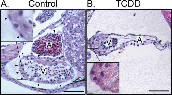
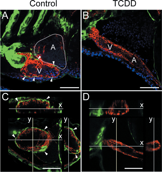
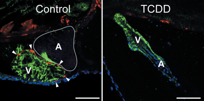
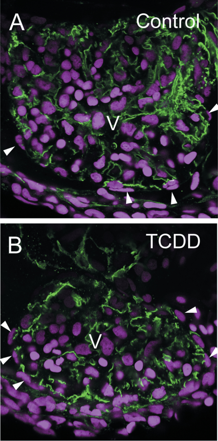
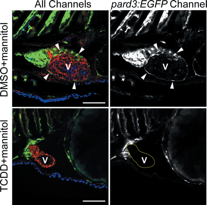
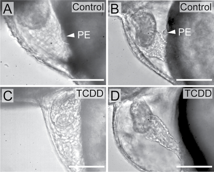
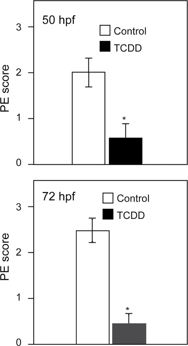
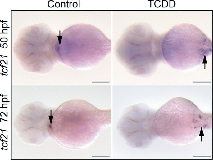
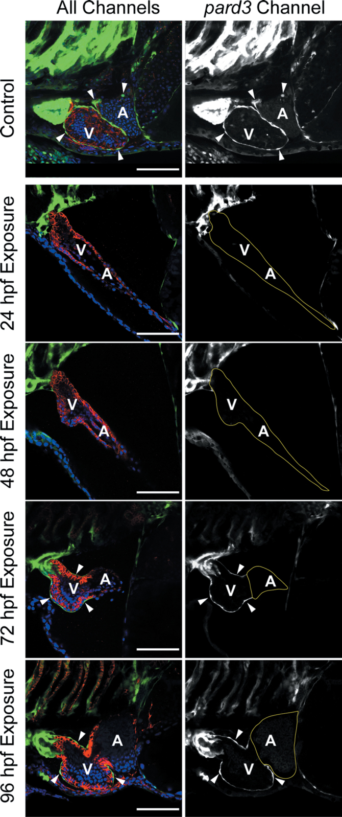
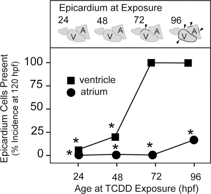
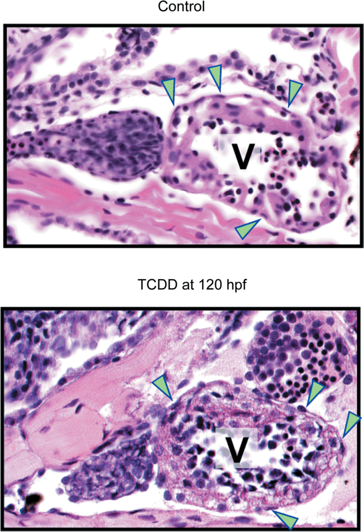
Embryonic exposure to the environmental contaminant and aryl hydrocarbon receptor agonist, 2,3,7,8-tetrachlorodibenzo-p-dioxin (TCDD, dioxin), disrupts cardiac development and function in fish, birds, and mammals. In zebrafish, the temporal window of sensitivity to the cardiotoxic effects of TCDD coincides with epicardium formation. We hypothesized that this TCDD-induced heart failure results from disruption of epicardial development. To determine whether embryonic TCDD exposure inhibits epicardium and proepicardium (PE) development in zebrafish, we used histology and fluorescence immunocytochemistry to examine the epicardium formation in fish exposed to TCDD. TCDD exposure prevented epicardium formation. Using live imaging and in situ hybridization, we found that TCDD exposure blocked the formation of the PE cluster. In situ hybridization experiments showed that TCDD exposure also prevented the expression of the PE marker tcf21 at the site where the PE normally forms. TCDD also inhibited expansion of the epicardial layer across the developing heart: Exposure after PE formation was completed prevented further expansion of the epicardium. However, TCDD exposure did not affect epicardial cells already present. Because TCDD blocks epicardium formation, but is not directly toxic to the epicardium once complete, we propose that inhibition of epicardium formation can account for the window of sensitivity to TCDD cardiotoxicity in developing zebrafish. Epicardium development is crucial to heart development. Loss of this layer during development may account for most if not all of the TCDD-induced cardiotoxicity in zebrafish.


Figures

Fig. 1.
Fig. 1.
TCDD exposure prevents epicardium formation in zebrafish. Zebrafish were exposed to vehicle (A) or TCDD (B) immediately following fertilization as described in the Materials and Methods section and collected at 120 hpf. H&E-stained sagital sections show atrium and ventricle from lateral view, with anterior to the left. A, Atrium; V, Ventricle. Scale bars: 50 μm. Vehicle control hearts are on the left and corresponding TCDD-exposed hearts are shown at right. Insets show the flattened epicardial cells in the control ventricle and the corresponding region lacking epicardial cells in the TCDD-exposed heart. Arrowheads indicate epicardial cells.
Fig. 2.
Fig. 2.
Loss of pard3 reporter expression in TCDD-exposed hearts. Zebrafish were exposed to TCDD as in Figure 1. A and B) Fish were collected at 120 hpf, and lateral confocal images of pard3:EGFP control and exposed hearts are shown. ALCAM is counterstained as red, and EGFP is indicated in green. DAPI staining shows nuclei in outer pericardium in blue. White arrowheads indicate GFP-positive epicardial cells. (C and D). X and Y orthogonal optical slices through z-stacks (z-step = 0.52 μ) showing ventricle lumen. White arrowheads indicate GFP-positive epicardial cells. Scale bars: 50 μ.
Fig. 3.
Fig. 3.
Expression of the tcf21 epicardium marker is lost in TCDD-exposed hearts. Zebrafish were exposed to TCDD immediately after fertilization and collected at 120 hpf as in the figures above. Confocal images show lateral views of hearts from the tcf21:DsRed transgenic line. Red indicates tcf21 expression; ALCAM expression is shown in green. DAPI in blue shows cell nuclei, generally at the pericardial surface. White arrowheads indicate DsRed-positive epicardial cells. Scale bars: 50 μ.
Fig. 4.
Fig. 4.
TCDD has no effect on epicardium formation in ahr2 −/− mutants. Zebrafish homozygous for loss of functional ahr2 were exposed to TCDD immediately after fertilization and collected at 120 hpf as in the figures above. Confocal images show lateral views of hearts. ALCAM expression is shown in green and delineates the cytoplasm in myocardial cells. DAPI shows cell nuclei in blue. White arrowheads indicate flattened cells lying on the surface of the heart, outside of the myocardial layer. The ventricle center is labeled as V, and rounded erythrocyte nuclei are prominent within the ventricles.
Fig. 5.
Fig. 5.
TCDD-induced pericardial edema is not linked to the loss of epicardium in TCDD-exposed larvae. Zebrafish carrying the pard3:EGFP reporter were exposed to TCDD or vehicle immediately following fertilization and moved into water with 175mM mannitol added as an osmotic support. Samples were collected at 120 hpf for confocal microscopy as described in the Materials and Methods section. Ventral views are shown with the anterior to the left. The left column of images shows all three channels of fluorescence together. ALCAM is counterstained in red, and EGFP is indicated in green. DAPI staining shows nuclei in outer pericardium in blue. White arrowheads indicate GFP-positive epicardial cells. The column of images at right shows the pard3:EGFP signal alone in white. Arrowheads show examples of pard3:EGFP-positive cells. A indicates atrium; V indicates ventricle. Scale bars: 50 μm.
Fig. 6.
Fig. 6.
TCDD exposure blocks PE development in zebrafish. Zebrafish were exposed to TCDD or vehicle immediately following fertilization as described in the Materials and Methods section. (A–D) Lateral views of hearts at 50 and 72 hpf are shown, with anterior to left in all panels. Vehicle control hearts are on the left, and corresponding TCDD-exposed hearts are shown at right. White arrowhead indicates the PE. Scale bars: 50 μm. (A) Vehicle control heart at 50 hpf. (B) Vehicle control heart at 72 hpf. (C) TCDD-exposed heart at 50 hpf. (D) TCDD-exposed heart at 72 hpf.
Fig. 7.
Fig. 7.
Scoring of PE formation. Graphs show incidence of PE formation at 50 and 72 hpf, respectively. Scoring is described in detail in Supplemental Methods. Briefly, embryos were scored using the following scale: 0, no PE; 1, slight evidence of PE; 2, moderate evidence for PE; 3, normal PE in full view. Experimenters were blind to the treatment groups scored. Asterisk indicates difference from control p < 0.01; Student’s t-test. Error bars indicate standard error.
Fig. 8.
Fig. 8.
TCDD alteration of PE and epicardium-specific marker. Zebrafish embryos were exposed to TCDD or vehicle at 24 hpf as described in the Materials and Methods section. Vehicle control hearts are on the left and corresponding TCDD-exposed hearts are shown at right. Fish were collected for in situ hybridization probing for tcf21 at either 50 or 72 hpf as indicated, and ventral views are shown with anterior end to the left. Arrows indicate regions of hybridization. Scale bars: 100 μm.
Fig. 9.
Fig. 9.
TCDD after PE formation halts further epicardium progression. Zebrafish carrying the pard3:EGFP reporter were exposed to TCDD at the indicated times. Samples were collected at 120 hpf for confocal microscopy as described in the Materials and Methods section. The left column of images shows all three channels of fluorescence together. ALCAM is counterstained in red, and EGFP is indicated in green. DAPI staining shows nuclei in outer pericardium in blue. White arrowheads indicate GFP-positive epicardial cells. The column of images at right shows the pard3:EGFP signal alone as white. The control was not exposed to TCDD and demonstrates normal epicardium formation at 120 hpf. Scale bars: 50 μm.
Fig. 10.
Fig. 10.
TCDD exposure during epicardial expansion halts further epicardium development. Zebrafish carrying the pard3:EGFP reporter were exposed to TCDD at the times indicated on the x-axis and collected at 120 hpf for staining and confocal microscopy. The control was exposed to the DMSO vehicle alone. Incidence of the appearance of GFP-positive cells on either ventricle or atrium in the confocal images was counted at 120 hpf. The schematic figures above each time point represent the normal course of epicardium formation. Chambers with at least 1 EGFP-positive cell were scored as positive. If no cells were observed, the chamber was scored as negative. For these experiments, n = 1 represents individual fish, and the n for each treatment ranged from 45 to 54. The values shown are the percentages of positive scoring individuals in the treatment group. Scoring the percentage of individuals with an all or none response produces no error bars. Instead, Fisher’s Exact Test was used, and asterisks indicate difference from control at p < 0.001.
Fig. 11.
Fig. 11.
TCDD does not remove formed epicardium. The panels show H&E sections of 168 hpf larval hearts treated with DMSO (control) or TCDD at 120 hpf. Arrowheads indicate epicardial cells. In all panels, ventral views are shown with the anterior to the left. A indicates atrium; V indicates ventricle.

References

    1. Antkiewicz D. S., Burns C. G., Carney S. A., Peterson R. E., Heideman W. (2005). Heart malformation is an early response to TCDD in embryonic zebrafish. Toxicol. Sci. 84, 368–377 - PubMed
    1. Belair C. D., Peterson R. E., Heideman W. (2001). Disruption of erythropoiesis by dioxin in the zebrafish. Dev. Dyn. 222, 581–594 - PubMed
    1. Bertazzi P. A., Bernucci I., Brambilla G., Consonni D., Pesatori A. C. (1998). The Seveso studies on early and long-term effects of dioxin exposure: A review. Environ. Health Perspect. 106,(Suppl. 2)625–633 - PMC - PubMed
    1. Brambilla G., Dellatte E., Fochi I., Iacovella N., Miniero R., di Domenico A. (2007). Depletion of selected polychlorinated biphenyl, dibenzodioxin, and dibenzofuran congeners in farmed rainbow trout (Oncorhynchus mykiss): A hint for safer fish farming. Chemosphere. 66, 1019–1030 - PubMed
    1. Carmona R., Guadix J. A., Cano E., Ruiz-Villalba A., Portillo-Sánchez V., Pérez-Pomares J. M., Muñoz-Chápuli R. (2010). The embryonic epicardium: An essential element of cardiac development. J. Cell. Mol. Med. 14, 2066–2072 - PMC - PubMed

Publication types

MeSH terms

Substances