. 2013 Jan;87(2):1115-22.
doi: 10.1128/JVI.01964-12. Epub 2012 Nov 7.

Gangliosides have a functional role during rotavirus cell entry

Affiliations

Gangliosides have a functional role during rotavirus cell entry

Miguel Angel Martínez et al. J Virol. 2013 Jan.

Abstract

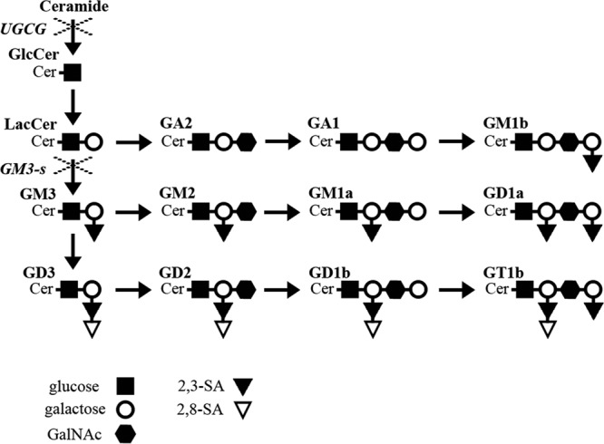
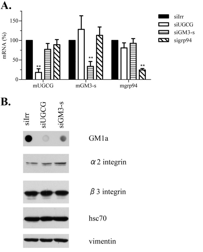
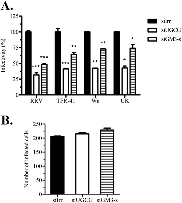
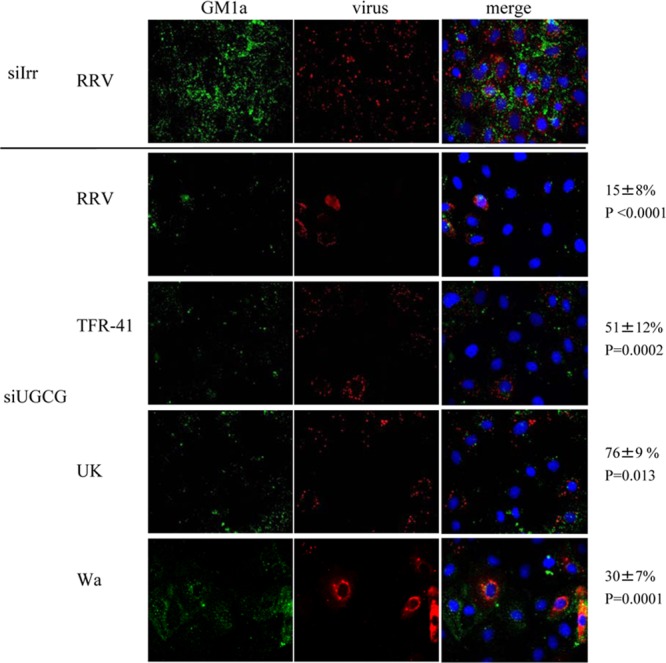
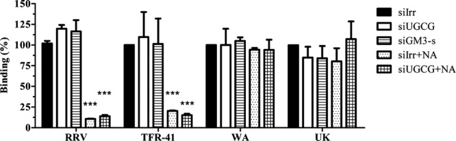
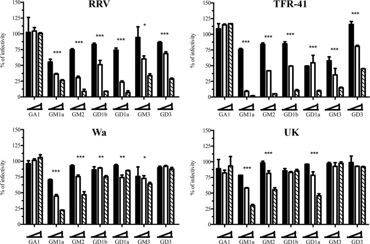
Cell entry of rotaviruses is a complex process, which involves sequential interactions with several cell surface molecules. Among the molecules implicated are gangliosides, glycosphingolipids with one or more sialic acid (SA) residues. The role of gangliosides in rotavirus cell entry was studied by silencing the expression of two key enzymes involved in their biosynthesis--the UDP-glucose:ceramide glucosyltransferase (UGCG), which transfers a glucose molecule to ceramide to produce glucosylceramide GlcCer, and the lactosyl ceramide-α-2,3-sialyl transferase 5 (GM3-s), which adds the first SA to lactoceramide-producing ganglioside GM3. Silencing the expression of both enzymes resulted in decreased ganglioside levels (as judged by GM1a detection). Four rotavirus strains tested (human Wa, simian RRV, porcine TFR-41, and bovine UK) showed a decreased infectivity in cells with impaired ganglioside synthesis; however, their replication after bypassing the entry step was not affected, confirming the importance of gangliosides for cell entry of the viruses. Interestingly, viral binding to the cell surface was not affected in cells with inhibited ganglioside synthesis, but the infectivity of all strains tested was inhibited by preincubation of gangliosides with virus prior to infection. These data suggest that rotaviruses can attach to cell surface in the absence of gangliosides but require them for productive cell entry, confirming their functional role during rotavirus cell entry.


Figures

Fig 1
Fig 1
Ganglioside biosynthesis. Schematic representation of main ganglioside synthesis. × indicates silenced enzymes UGCG (UDP-glucose:ceramide glucosyltransferase) and GM3-s (lactosyl ceramide-α-2,3–sialyl transferase 5). The key code for saccharide units composing gangliosides is shown. Ganglioside nomenclature according to IUPAC-IUB (46) was used.
Fig 2
Fig 2
siRNAs directed to enzymes involved in ganglioside biosynthesis decrease ganglioside levels, without affecting expression of other rotavirus receptors. MA104 cells were transfected with siRNAs against UGCG and GM3-s, and 72 h posttransfection parallel wells were harvested for mRNA quantification and for immunoblotting. (A) Total RNA was extracted and levels of mRNA were determined by one-step RT-PCR as described in Materials and Methods. Results are expressed as percentages relative to mRNA values obtained in cells lipofected with control siRNA (siIrr). As a negative control, the expression of endoplasmic reticulum chaperone grp94 was silenced. Means and standard deviations for results of at least three independent experiments are shown. (B) Harvested cellular proteins were resolved by SDS-PAGE, and proteins associated with rotavirus cell entry (integrin subunits α2 and β3 and hsc70 protein) were detected by immunostaining. Vimentin was used as a loading control. The result of one representative experiment of three is shown. To detect GM1a, 250 ng of total protein was applied to nitrocellulose membranes and stained using cholera toxin B subunit. Statistical analysis was done using a Student t test, and statistically significant values are shown (**, P < 0.01).
Fig 3
Fig 3
The formation of detergent-resistant membrane domains is unaffected in cells with decreased ganglioside synthesis. MA104 cells were transfected with siRNAs (siIrr and siUGCG) in 25-cm2 flasks, and 72 h posttransfection they were brought to single cell suspension and incubated or not with 10 mM MβCD. Then cells were lysed with 1% Triton X-100 at 4°C and subjected to density gradient centrifugation in discontinuous iodixanol gradients, as described in Materials and Methods. The proteins from equal volumes of individual fractions were separated by 15% SDS-PAGE, transferred to a nitrocellulose membrane, and probed with rabbit antibodies to caveolin 1 and vimentin, followed by a peroxidase-labeled secondary antibody. For GM1 detection, the samples were applied to nitrocellulose membranes with a dot blotter and developed using a horseradish peroxidase-conjugated cholera toxin B subunit. DRMs localize in the top fractions of gradient, while soluble proteins are in the bottom high-density fractions.
Fig 4
Fig 4
Inhibition of ganglioside synthesis decreases rotavirus cell entry. MA104 cells grown in 48-well plates were transfected with siRNAs (siIrr, siUGCG, and siGM3-s) and 72 h posttransfection were infected with rotavirus. In each experiment, parallel wells were harvested and ganglioside inhibition analyzed by GM1a immunoblotting as described in Materials and Methods. (A) Four different rotavirus strains (RRV, TFR-41, Wa, and UK) were inoculated for 1 h, washed, and left for 14 h for infection to proceed. Then infected cells were detected using peroxidase immunostaining with anti-rotavirus polyclonal antibodies. Results are expressed as percentages relative to irrelevant control treatment (siIrr) for each rotavirus strain. Means and standard deviations for results of at least three independent experiments are shown. (B) RRV DLPs were lipofected in ganglioside-decreased cells, and at 14 h posttransfection focus-forming units (FFUs) were detected as described previously. As a negative control, DLPs without lipofectant were inoculated (data not shown), and no FFUs were detected. Results are expressed as the number of FFUs obtained in each treatment. Means and standard deviations for results of at least three independent experiments are shown. Statistical analysis was done using a Student t test, and statistically significant values are shown as follows: ***, P < 0.001, **; P < 0.01; *, P < 0.05.
Fig 5
Fig 5
Cells with low ganglioside levels are less susceptible to rotavirus infection. MA104 cells grown in coverslips were transfected with the indicated siRNAs as described in Materials and Methods. Seventy-two hours later, cells were infected with the indicated rotavirus strain (MOI, 3), and eight hours postinfection they were fixed and processed as described in Materials and Methods. Cell membrane ganglioside GM1a was detected with Alexa 488-cholera toxin B subunit (in green); virus-infected cells were detected with polyclonal anti-NSP2 antibody and Alexa 568 anti-rabbit antibody (in red); nuclei were stained with DAPI (in blue). Images were scored independently by two persons, and scores were averaged. In the upper panel a representative staining of cells transfected with siIrr and infected with strain RRV is shown. The lower panel shows representative staining of cells transfected with siUGCG, infected with four different strains. Numbers at the right side correspond to the percentages of infected cells with low levels of ganglioside. Images acquired from at least 3 different experiments were analyzed and counted and represent at least 100 individual cells. Statistical analysis was done using a Student t test, and the values of statistical difference (P) for each virus are shown.
Fig 6
Fig 6
Neuraminidase treatment but not inhibition of ganglioside synthesis decreases cell surface binding of rotavirus strains RRV and TFR-41. MA104 cells were transfected with the indicated siRNAs, and 72 h posttransfection, the cell monolayers were incubated or not with 40 mU of neuraminidase for 1 h at 37°C. After this time, cells were blocked with BSA, and purified rotavirus TLPs were added for 1 h at 4°C, as described in Materials and Methods. Virus bound to cells was determined by an ELISA. Data are expressed as the percentage of virus bound to the cells relative to virus detected in siIrr control treatment cells. The means and standard errors of results from three independent experiments are shown. Statistical analysis was done using a Student t test, and statistically significant values are shown as follows: ***, P < 0.001; **, P < 0.01; *, P < 0.05.
Fig 7
Fig 7
Preincubation with gangliosides affects rotavirus infectivity. Four different rotavirus strains (RRV, TFR-41, Wa, and UK) were preincubated with the indicated gangliosides (GA1, GM1a, GM2, GD1b, GD1a, GM3, GD3) in three different concentrations (1.5 [black bars], 12.5 [white bars], and 50 [striped bars] μg/ml). After preincubation, the virus-ganglioside mixture was used to infect MA104 cells. Fourteen hours postinfection, cells were fixed and stained as described in Materials and Methods. The results are expressed as the percentages of infectivity relative to control (preincubation with 0 μg/ml) treatment. The means and standard errors from results of at least three independent experiments are shown. Statistical analysis was done using a Student one-way ANOVA test and a Tukey posttest, and statistically significant values for highest ganglioside concentration are shown as follows: ***, P < 0.001; **, P < 0.01; *, P < 0.05.

Similar articles

Cited by

References

    1. Lopez S, Arias CF. 2004. Multistep entry of rotavirus into cells: a Versaillesque dance. Trends Microbiol. 12:271–278 - PubMed
    1. Isa P, Arias CF, Lopez S. 2006. Role of sialic acids in rotavirus infection. Glycoconj. J. 23:27–37 - PMC - PubMed
    1. Fuentes-Panana EM, Lopez S, Gorziglia M, Arias CF. 1995. Mapping the hemagglutination domain of rotaviruses. J. Virol. 69:2629–2632 - PMC - PubMed
    1. Isa P, Lopez S, Segovia L, Arias CF. 1997. Functional and structural analysis of the sialic acid-binding domain of rotaviruses. J. Virol. 71:6749–6756 - PMC - PubMed
    1. Blanchard H, Yu X, Coulson BS, von Itzstein M. 2007. Insight into host cell carbohydrate-recognition by human and porcine rotavirus from crystal structures of the virion spike associated carbohydrate-binding domain (VP8*). J. Mol. Biol. 367:1215–1226 - PubMed

Publication types

LinkOut - more resources