. 2013 Jan 15;73(2):776-84.
doi: 10.1158/0008-5472.CAN-12-2669. Epub 2012 Nov 7.

Combination therapy targeting the Chk1 and Wee1 kinases shows therapeutic efficacy in neuroblastoma

Affiliations

Combination therapy targeting the Chk1 and Wee1 kinases shows therapeutic efficacy in neuroblastoma

Mike R Russell et al. Cancer Res. .

Abstract

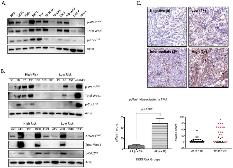
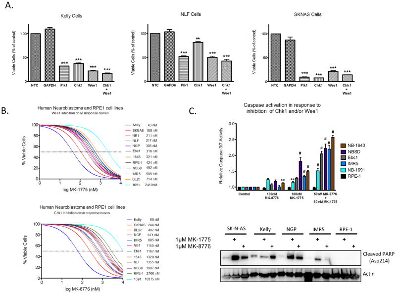
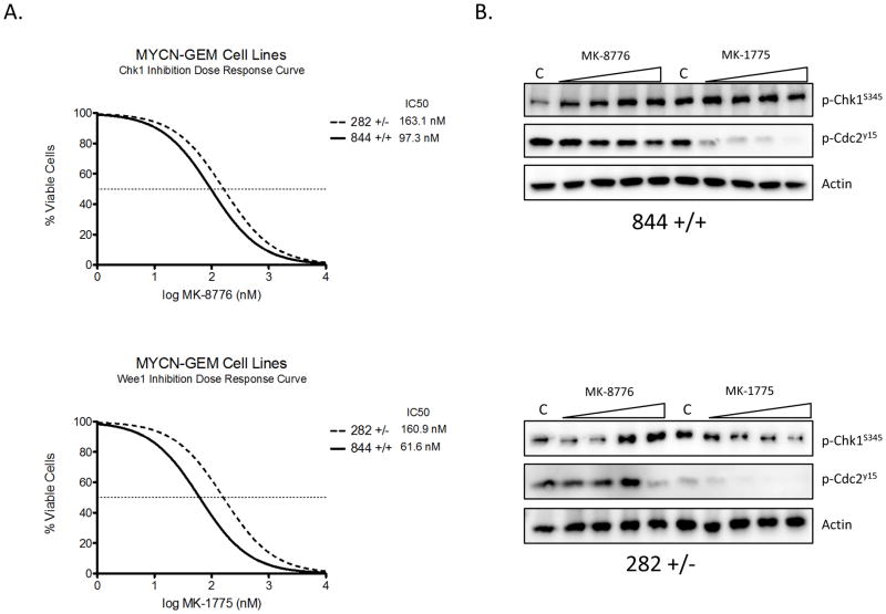
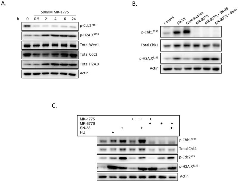
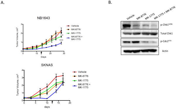
Neuroblastoma is uniquely sensitive to single-agent inhibition of the DNA damage checkpoint kinase Chk1, leading us to examine downstream effectors of this pathway and identify mitotic regulator Wee1 as an additional therapeutic target in this disease. Wee1 was overexpressed in both neuroblastoma cell lines and high-risk patient tumors. Genetic or pharmacologic abrogation of Wee1 signaling results in marked cytotoxicity in 10 of 11 neuroblastoma cell lines with a median IC(50) of 300 nmol/L for the Wee1-selective small-molecule inhibitor MK-1775. Murine tumor lines derived from mice that were either heterozygous or homozygous for MycN were particularly sensitive to single-agent inhibition of Wee1 (IC(50)s of 160 and 62 nmol/L, respectively). Simultaneous pharmacologic inhibition of Chk1 and Wee1 acted in a synergistic fashion to further impede neuroblastoma cell growth in vitro, in a manner greater than the individual inhibitors either alone or combined with chemotherapy. Combination Chk1 and Wee1 inhibition also revealed in vivo efficacy in neuroblastoma xenografts. Taken together, our results show that neuroblastoma cells depend on Wee1 activity for growth and that inhibition of this kinase may serve as a therapeutic for patients with neuroblastoma.


Conflict of interest statement

Author Conflicts of Interest: none

Figures

Figure 1
Figure 1
Wee1 kinase is highly expressed in neuroblastoma. (A) Western blot analysis of neuroblastoma cell lines demonstrates that Wee1 is highly expressed at the protein level and constitutively activated compared to non-NB lines such as DAOY medulloblastoma cells or non-transformed RPE-1 cells. Phosphorylated Wee1S642 is present in the majority of neuroblastoma lines, and often coincides with downstream phosphorylation of Cdc2Y15 (Cdk1). (B) Wee1 is also highly expressed in diagnostic patient tumor samples, with increased Wee1 activity in 67% (8 of 12) of tumors derived from high-risk patients, compared to 28.5% (2 of 7) of tumors derived from low-risk patients (C) Neuroblastoma tissue microarray (TMA) also stained positively for phospo-Wee1 (S642), confirming higher expression levels in high-risk, including MYCN amplified tumors (red triangles), as compared to low-risk samples. Representative staining for each tumor risk group is shown (top). Abbreviations: HR = high-risk, LR = low-risk, INSS = International Neuroblastoma Staging System. pWee1 score = antibody intensity (0, 1+, 2+, 3+) x the % of cells positive.
Figure 2
Figure 2
Abrogation of either Wee1 or Chk1 signaling is cytotoxic to neuroblastoma cells. (A) siRNA-mediated depletion of Chk1 or Wee1 resulted in a significant reduction in cell viability in several neuroblastoma cell lines. (B) The majority of neuroblastoma cells were sensitive to single-agent inhibition of Wee1 (MK-1775) or Chk1 (MK-8776) activity, with median IC50s of 300nM and 900nM, respectively (curve shifted <0.05 on x-axis to allow visualization where overlapped). (C) Neuroblastoma cell lines underwent apoptosis in response to Chk1 and/or Wee1 inhibition as evidenced by caspase 3/7 activation (top panel) or PARP cleavage (bottom panel) (** = p ≤0.01, *** = p ≤0.001, # = p < 0.0001).
Figure 3
Figure 3
Murine neuroblastoma lines derived from MYCN transgenic mice are sensitive to Chk1/Wee1 inhibition. (A) Cells homozygous (844) or heterozygous (282) for the MYCN oncogene were derived from MYCN-transgenic murine tumors, and were found to be sensitive to single-agent MK-1775 (bottom) and MK-8776 (top), (n=9 for each cell type). (B) Target engagement was confirmed via Western blot 6h after treatment with increasing concentrations of MK-1775 or MK-8776. Increasing phosphorylation of Chk1S345 has been shown to be a biomarker of Chk1 inhibition (15).
Figure 4
Figure 4
Dual Chk1 and Wee1 inhibition results in accumulation of DNA double-strand breaks and mitotic catastrophe. (A) Be2C cells treated with MK-1775 demonstrated an abrogation of Cdc2 activity with a concomitant increase in H2A.X phosphorylation, indicative of DNA damage. (B) Inhibition of Chk1, on the other hand, results in only marginal increases in γH2A.X, however in combination with chemotherapy MK-8776 rapidly (2h) induces double strand breaks (shown in NB-1643 cells). (C) Simultaneous inhibition of Chk1 and Wee1 (16h) resulted in robust H2A.x activation and complete abolishment of Cdc2 signaling, suggesting cells were progressing through mitosis with DNA damage (shown in NGP cells). (HU = 1mM hydroxyurea, [MK-1775] = 250nM, [MK-8776] = 500nM, [SN-38] = 100nM, Gem = 100nM gemcitabine)
Figure 5
Figure 5
Growth of neuroblastoma xenografts is significantly impaired in response to Chk1/Wee1 combinatorial therapy. (A) Mice subcutaneously implanted with neuroblastoma xenografts were treated twice daily with 30 mg/kg/dose MK-1775, MK-8776 or both for 5d x 2 weeks. Mixed linear analysis indicated a significant reduction in tumor burden in the combination treatment group in both neuroblastoma cell lines. For graphical purposes, mice removed from study from excessive tumor burden had their last measurements carried forward. Arrows indicate end of treatment. (* = p ≤ 0.05, *** = p ≤ 0.0001) (B) Target engagement was verified by resection of Ebc1 xenografts, where both Chk1 and Cdc2 activity was substantially reduced following four doses of MK-8776 or MK-1775, respectively.

References

    1. Maris JM, Hogarty MD, Bagatell R, Cohn SL. Neuroblastoma. Lancet. 2007;369(9579):2106–20. - PubMed
    1. Brodeur GM. Neuroblastoma: biological insights into a clinical enigma. Nature Rev Cancer. 2003;3:203–16. - PubMed
    1. Maris JM. Recent advances in neuroblastoma. NEJM. 2010;362:2202–11. - PMC - PubMed
    1. Hudson MM, Mertens AC, Yasui Y, Hobbie W, Chen H, et al. Health Status of Adult Long-term A Report From the Childhood Cancer Survivor Study. 2003;290:1583–1592. - PubMed
    1. Cole KA, Huggins J, Laquaglia M, Hulderman CE, Russell MR, et al. RNAi screen of the protein kinome identifies checkpoint kinase 1 (CHK1) as a therapeutic target in neuroblastoma. PNAS. 2011;108:3336. - PMC - PubMed

Publication types

MeSH terms