. 2012 Nov 7;4(159):159ra147.
doi: 10.1126/scitranslmed.3004249.

A human disease model of drug toxicity-induced pulmonary edema in a lung-on-a-chip microdevice

Affiliations

A human disease model of drug toxicity-induced pulmonary edema in a lung-on-a-chip microdevice

Dongeun Huh et al. Sci Transl Med. .

Erratum in

Abstract

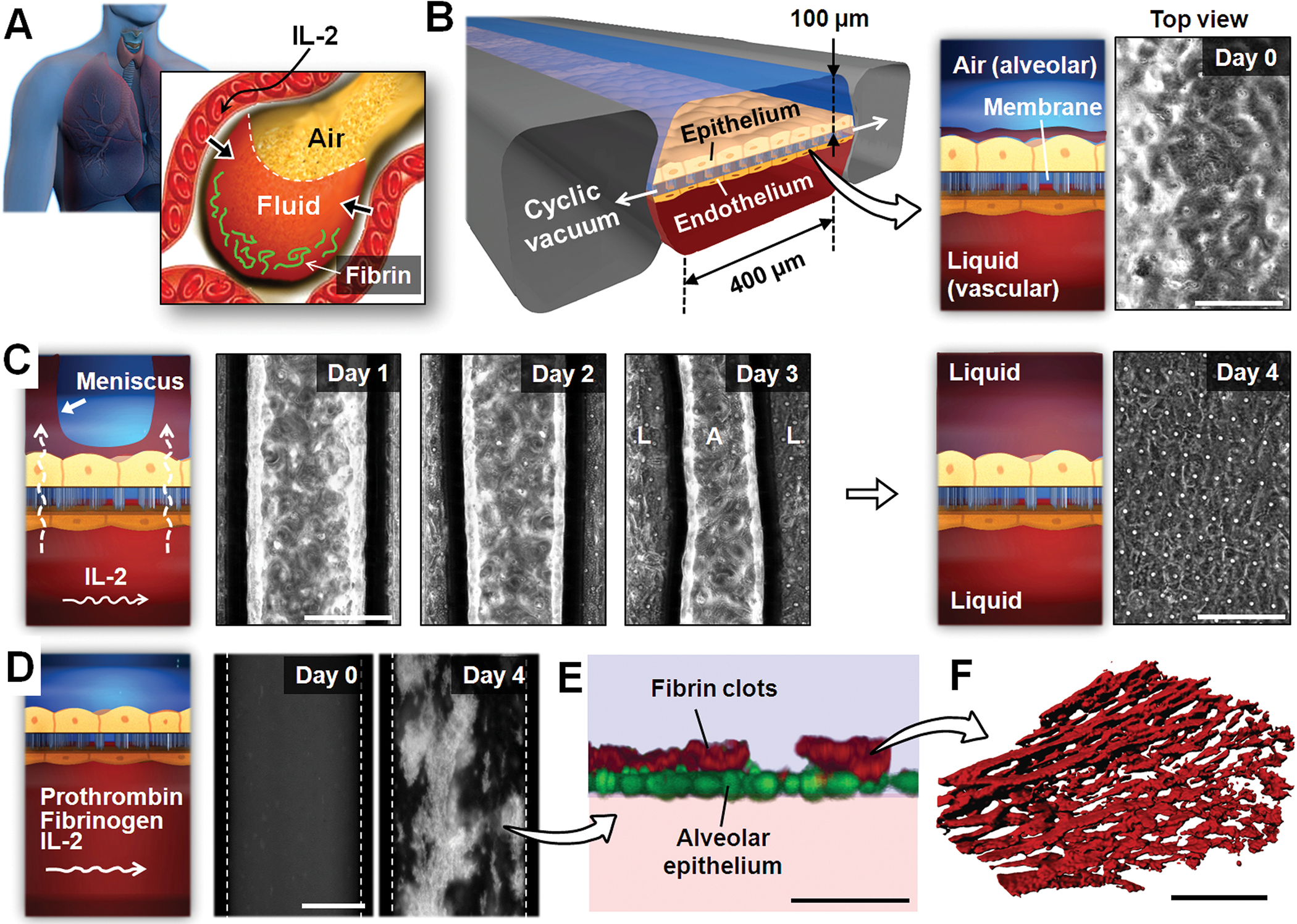
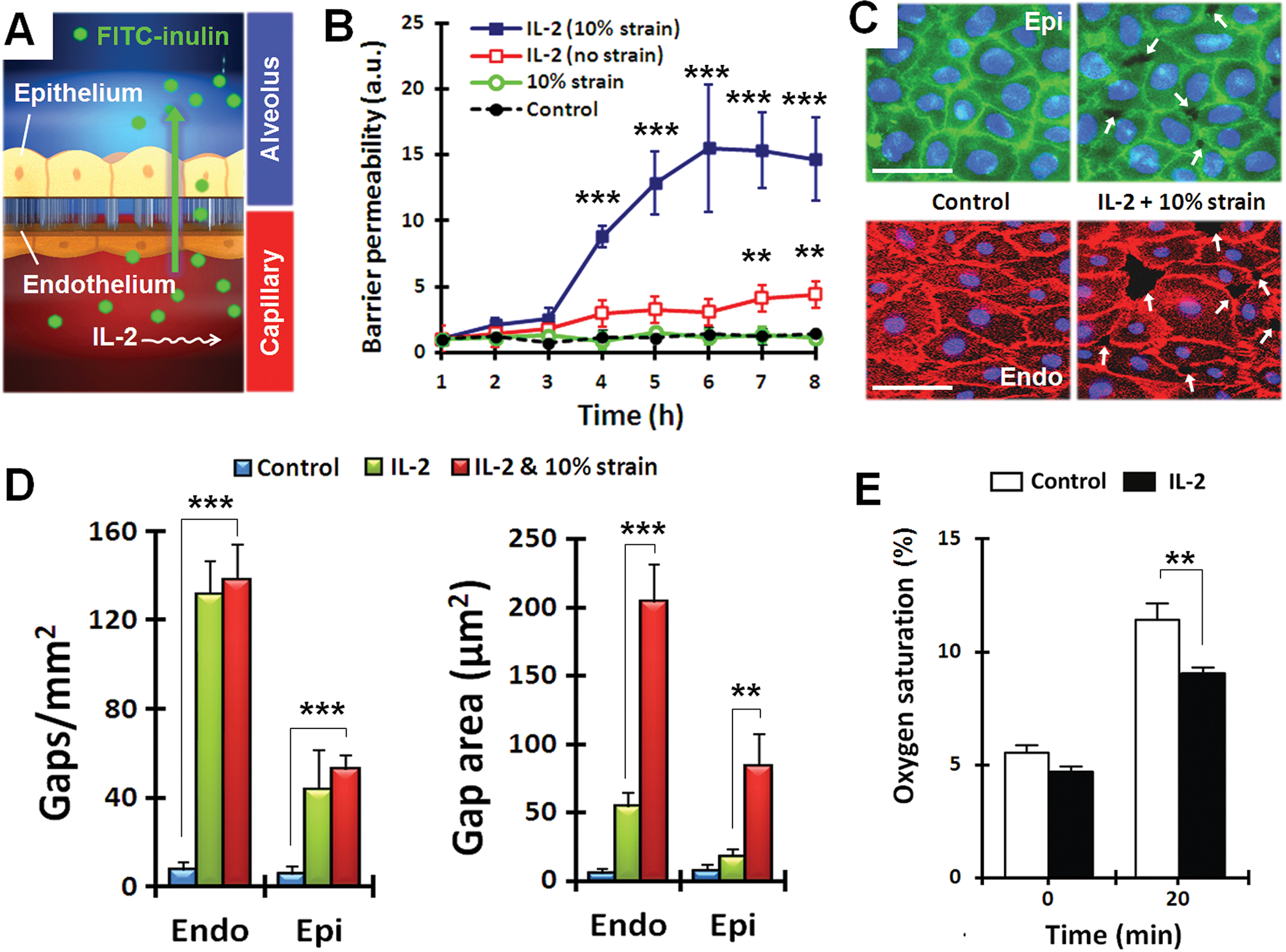
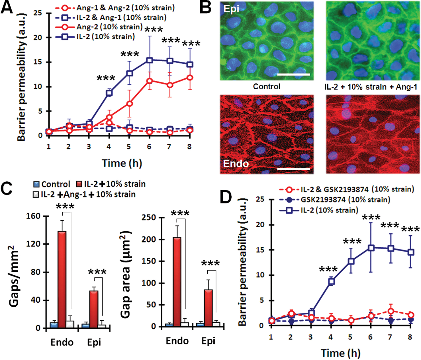
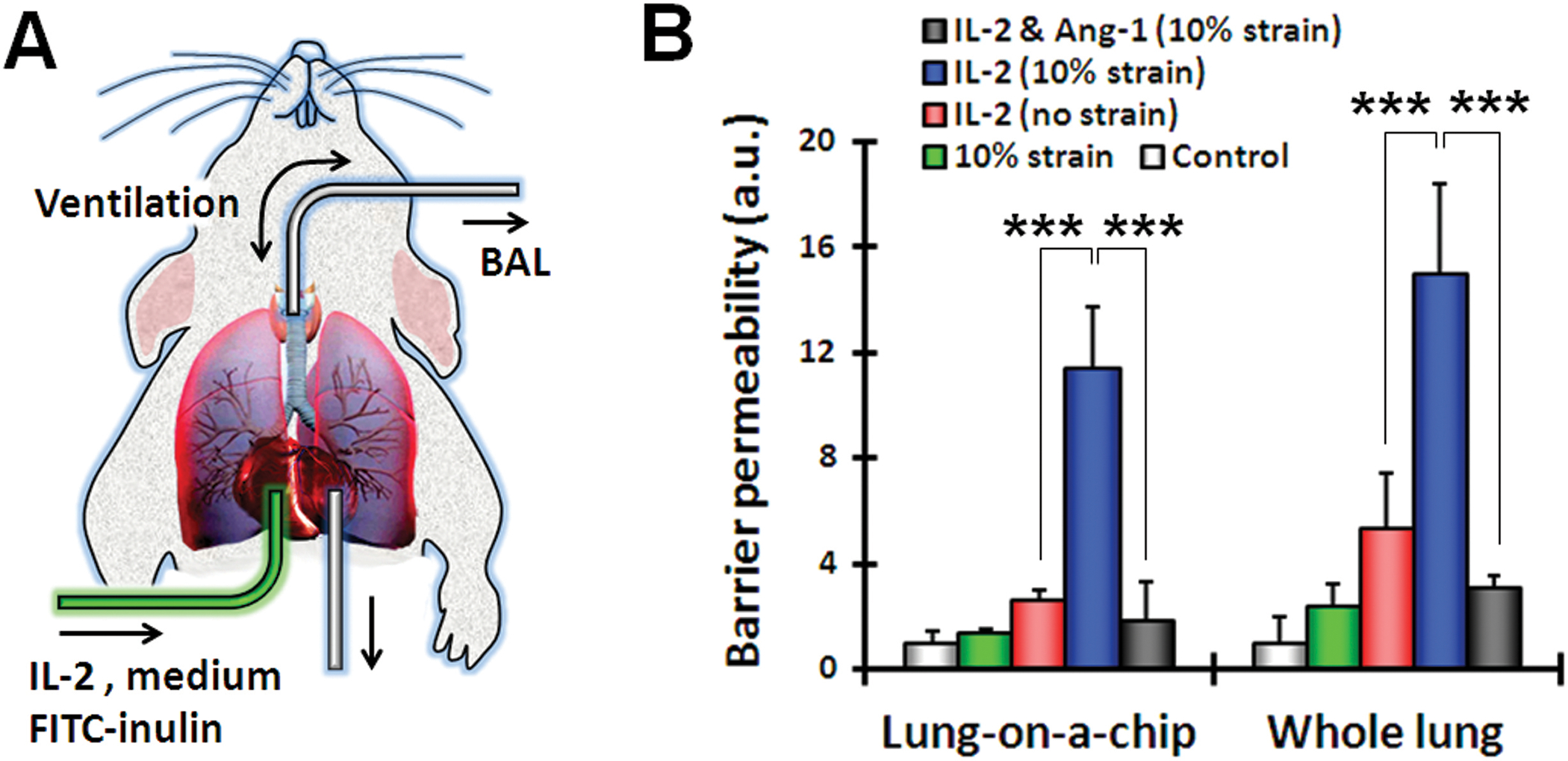
Preclinical drug development studies currently rely on costly and time-consuming animal testing because existing cell culture models fail to recapitulate complex, organ-level disease processes in humans. We provide the proof of principle for using a biomimetic microdevice that reconstitutes organ-level lung functions to create a human disease model-on-a-chip that mimics pulmonary edema. The microfluidic device, which reconstitutes the alveolar-capillary interface of the human lung, consists of channels lined by closely apposed layers of human pulmonary epithelial and endothelial cells that experience air and fluid flow, as well as cyclic mechanical strain to mimic normal breathing motions. This device was used to reproduce drug toxicity-induced pulmonary edema observed in human cancer patients treated with interleukin-2 (IL-2) at similar doses and over the same time frame. Studies using this on-chip disease model revealed that mechanical forces associated with physiological breathing motions play a crucial role in the development of increased vascular leakage that leads to pulmonary edema, and that circulating immune cells are not required for the development of this disease. These studies also led to identification of potential new therapeutics, including angiopoietin-1 (Ang-1) and a new transient receptor potential vanilloid 4 (TRPV4) ion channel inhibitor (GSK2193874), which might prevent this life-threatening toxicity of IL-2 in the future.


Conflict of interest statement

Competing interests: The authors declare no competing financial interests.

Figures

Fig. 1.
Fig. 1.
A microengineered model of human pulmonary edema. (A) IL-2 therapy is associated with vascular leakage that causes excessive fluid accumulation (edema) and fibrin deposition in the alveolar air spaces. (B) IL-2–induced pulmonary edema is modeled in a microengineered lung-on-a-chip that reproduces the lung micro-architecture and breathing-induced cyclic mechanical distortion of the alveolar-capillary interface. The top “air” portion is the alveolar channel; the bottom “liquid” portion is the vascular channel. The phase-contrast image shows a top-down view of the apical surface of the alveolar epithelium maintained at an air-liquid interface in the upper microchannel. Scale bar, 200 μm. (C) Endothelial exposure to IL-2 (1000 U/ml) causes liquid in the lower microvascular channel to leak into the alveolar chamber (days 1 to 3) and eventually fill the entire air space (day 4). The meniscus between air (A) and liquid (L) appears as dark bands in the phase contrast images. Scale bars, 200 μm. (D) During IL-2 treatment, prothrombin (100 μg/ml) and fluorescently labeled fibrinogen (2 mg/mL) introduced into the microvascular channel form fluorescent fibrin clots (white) over the course of 4 days. Dotted lines represent channel walls. Scale bar, 200 μm. (E) A fluorescence confocal microscopic image shows that the fibrin deposits (red) in (D) are found on the upper surface of the alveolar epithelium (green). Scale bar, 50 μm. (F) The clots in (D and E) are highly fibrous networks, as visualized at high magnification by confocal fluorescence microscopy. Images are representative of three independent experiments. Scale bar, 5 μm.
Fig. 2.
Fig. 2.
Quantitative analysis of pulmonary edema progression on-chip. (A) Pathological alterations of barrier function were quantified by measuring alveolar-capillary permeability to FITC-inulin (green) introduced into the microvascular channel containing IL-2. (B) Barrier permeability in response to IL-2, with and without cyclical strain. Data are means ± SEM (n = 3) and were normalized to the mean at time 0 (C) Immunostaining of epithelial occludin (green) and endothelial VE-cadherin (red) after 3 days of cyclic stretch with 10% strain without IL-2 (control) or with IL-2. White arrows indicate intercellular gaps; blue, nuclear staining. Scale bars, 30 μm. (D) Compromised barrier integrity due to IL-2 and mechanical strain assessed by quantifying the number and size of intercellular gaps. Data are means ± SEM (n = 3). (E) Quantification of O2 uptake from air in the epithelial channel by deoxygenated medium flowing through vascular channel in control and IL-2-treated lung chips. Data are means ± SEM (n = 3). For (B, D, and E), **P < 0.01, ***P < 0.001, ANOVA followed by post hoc Tukey’s multiple comparison test.
Fig. 3.
Fig. 3.
Pharmacological modulation in lung-on-a-chip pulmonary edema model. (A) Administration of Ang-1 (100 ng/ml) prevents fluid leakage caused by IL-2 (1000 U/ml) and Ang-2 (100 ng/ml) applied in conjunction with mechanical stretch. Statistical significance determined for comparison of IL-2 (10% strain) and IL-2 and Ang-1 (10% strain). (B) Immunostaining of epithelial occludin (green) and endothelial VE-cadherin (red) in cell-cell junctions after exposure to cyclic mechanical strain (10% at 0.2 Hz) without IL-2 (control) or with co-administration of IL-2 and Ang-1 for 3 days. Scale bars, 30 μm. (C) Restoration of barrier integrity by Ang-1 treatment assessed by changes in the number and size of intercellular gaps. ***P < 0.001. (D) The effect of TRPV4 inhibitor GSK2193874 (100 nM) on barrier permeability alone and in the presence of IL-2 and 10% strain. Statistical significance determined for comparison of IL-2 alone and IL-2 with GSK inhibitor. For (A, C, and E), data are means ± SEM (n = 3). For (A, C, and E), ***P < 0.001, ANOVA followed by post hoc Tukey’s multiple comparison test.
Fig. 4.
Fig. 4.
Barrier permeability in the lung-on-a-chip compared to whole mouse lung ex vivo. (A) An excised whole mouse lung was mechanically ventilated ex vivo, and the pulmonary microvasculature was perfused with culture medium containing IL-2 (1000 U/ml) and FITC-inulin (1 mg/ml). After IL-2 treatment, bronchoalveolar lavage (BAL) fluid was collected for fluorescence measurement. (B) Barrier permeability was measured in the lung-on-a-chip and in the whole animal lung model. Control represents no strain and no IL-2. Data are means ± SEM (n = 3). ***P < 0.001, ANOVA followed by post hoc Tukey’s multiple comparison test.

Comment in

References

    1. Pampaloni F, Reynaud EG, Stelzer EH, The third dimension bridges the gap between cell culture and live tissue. Nat Rev Mol Cell Biol 8, 839 (October, 2007). - PubMed
    1. Huh D, Hamilton GA, Ingber DE, From 3D cell culture to organs-on-chips. Trends Cell Biol 21, 745 (December, 2011). - PMC - PubMed
    1. Jang KJ, Suh KY, A multi-layer microfluidic device for efficient culture and analysis of renal tubular cells. Lab Chip 10, 36 (January 7, 2010). - PubMed
    1. Huh D et al. , Reconstituting organ-level lung functions on a chip. Science 328, 1662 (June 25, 2010). - PMC - PubMed
    1. Lee PJ, Hung PJ, Lee LP, An artificial liver sinusoid with a microfluidic endothelial-like barrier for primary hepatocyte culture. Biotechnol Bioeng 97, 1340 (August 1, 2007). - PubMed

Publication types

LinkOut - more resources