. 2012 Nov 7;4(159):159ra149.
doi: 10.1126/scitranslmed.3004599.

Human melanoma metastasis in NSG mice correlates with clinical outcome in patients

Affiliations

Human melanoma metastasis in NSG mice correlates with clinical outcome in patients

Elsa Quintana et al. Sci Transl Med. .

Abstract

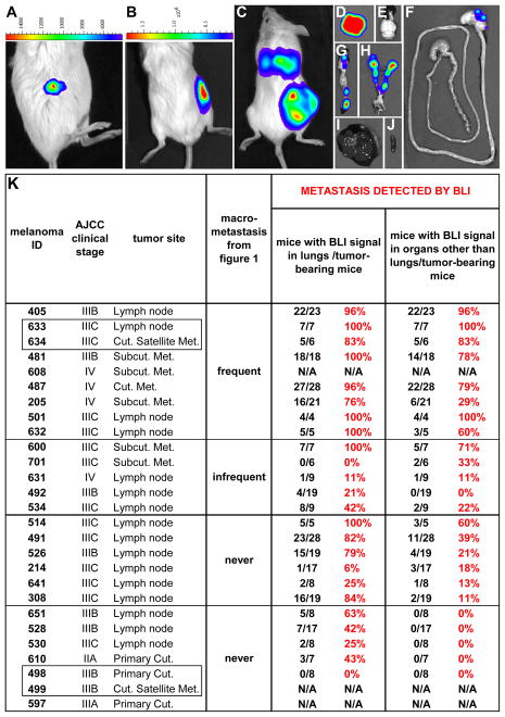
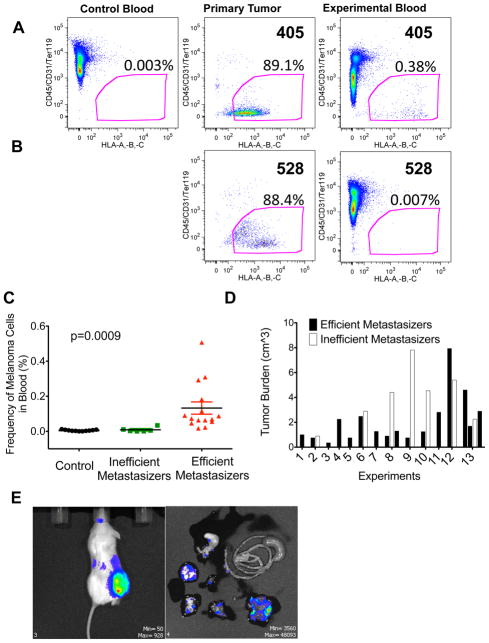
Studies of human cancer metastasis have been limited by a lack of experimental assays in which cancer cells from patients metastasize in vivo in a way that correlates with clinical outcome. This makes it impossible to study intrinsic differences in the metastatic properties of cancers from different patients. We recently developed an assay in which human melanomas readily engraft in nonobese diabetic/severe combined immunodeficient interleukin-2 receptor-γ chain null (NSG) mice. We show that melanomas from 25 patients exhibited reproducible differences in the rate of spontaneous metastasis after transplantation into NSG mice and that these differences correlated with clinical outcome in the patients. Stage IIIB/C melanomas that formed distant metastases within 22 months in patients also formed tumors that metastasized widely in NSG mice, whereas stage IIIB/C melanomas that did not form distant metastases within 22 to 50 months in patients metastasized more slowly in NSG mice. These differences in the efficiency of metastasis correlated with the presence of circulating melanoma cells in the blood of NSG mice, suggesting that the rate of entry into the blood is one factor that limits the rate of metastasis. The study of NSG mice can therefore yield information about the metastasis of human melanomas in vivo, in this case revealing intrinsic differences among stage III melanomas in their ability to circulate/survive in the blood and to metastasize.


Conflict of interest statement

Competing interests: S.J.M. is a founder and stockholder in OncoMed Pharmaceuticals. E.Q. was an employee of the University of Michigan while performing this study, but is now an employee of OncoMed Pharmaceuticals, a company that develops cancer therapeutics using xenograft assays.

Figures

Figure 1
Figure 1. Human melanomas exhibit intrinsic differences in their metastatic efficiency in NSG mice
(A) 27 melanomas from 25 patients were transplanted subcutaneously into NSG mice in doses between 1 and 50,000 cells per injection. Mice with subcutaneous tumors were monitored until the tumors grew to 1.4±0.9 cm (mean ± SD) in diameter, 21.8±10.2 weeks after transplantation. Upon necropsy, organs were visually examined for the presence of metastases. “AJCC clinical stage” is the American Joint Committee on Cancer stage of the patient at the time of melanoma removal. “Tumor site” reflects the location of the tumor in the patient. Melanomas 633/634 and 498/499 (boxed) were pairs of tumors obtained from different locations in the same patients at the same time. The metastatic behavior of each melanoma was examined in 2 to 22 independent experiments with 6 to 136 mice that formed subcutaneous tumors per melanoma (see Table S2 for more details). The rate of metastasis (%) is the percentage of mice with subcutaneous tumors that developed macrometastases. (B) Tumors that formed at the injection site as well as macrometastases in mice transplanted with cells from four patients. Metastases were confirmed as melanoma by a dermatopathologist after staining sections with hematoxylin and eosin (H&E) and S-100. Table S1 contains clinical details for each melanoma and patient. Table S2 has details regarding rates of metastasis at different cell doses for each melanoma. Table S3 shows the results for each independent experiment conducted on each melanoma. Table S4 shows the location where macrometastasis was detected for each melanoma.
Figure 2
Figure 2. Bioluminescence imaging (BLI) confirms intrinsic differences in metastatic efficiency among human melanomas in NSG mice
Luciferase-GFP+ melanoma cells from different patients exhibited intrinsic differences in metastatic efficiency revealed by BLI in NSG mice. BLI of an NSG mouse 2 days (A) and 45 days (B) after subcutaneous transplantation of 100 luciferase-GFP+ cells from melanoma 205, a heavily pigmented melanoma (see Fig. 1B). (C) BLI of an NSG mouse 13 weeks after subcutaneous transplantation of 100 luciferase-GFP+ cells from melanoma 487. BLI of individual organs dissected from the mouse in (C) revealed metastases in the lungs (D), stomach (F), pancreas (G), kidneys, adrenal glands and ovaries (H), but not in the brain (E), liver (I) or spleen (J). The maximum luminescence shown in red in panels C and D is 31x106 photons/second/cm2/steradian and in panels E–J is 6.2x10⁶ photons/second/cm2/steradian. (K) A summary for each melanoma of the percentage of mice with subcutaneous tumors that developed metastases detected by BLI. Supplementary Table 4 summarizes the locations in which metastases were detected by BLI. Melanomas 608, 499, and 597 did not undergo BLI.
Figure 3
Figure 3. Metastatic efficiency in NSG mice correlated with disease progression in patients
(A) Side-by-side comparison of distant metastasis in patients versus metastasis in mice for all 22 patients from whom follow-up clinical data were available after tumor banking. Most of the melanomas were lymph node metastases from stage IIIB/C patients at the time of banking. The melanomas were ranked by clinicians in terms of the aggressiveness of disease in patients after banking, based primarily on the rate of metastasis, from 514 at the top (most aggressive) to 498/499 at the bottom (least aggressive). The latest AJCC stage (or stage at time of death) and the time (in months) of survival after tumor banking are indicated in the “most recent survival and staging data” column. The patients with melanomas 514 to 492 formed distant metastases (stage IV disease) and died 3.4 to 40.6 months after melanoma banking. The patient who donated melanoma 600 remained alive at last follow-up, 33.2 months after melanoma banking but had progressed to stage IV disease with brain metastasis. All of the patients who progressed to stage IV disease did so within 22 months of tumor banking. The patients with melanomas 651 to 498/499 did not form distant metastases or progress to stage IV disease. Patient 528 died 40.7 months after melanoma banking with lung cancer and advanced chronic obstructive pulmonary disease but no evidence of melanoma at the time of death. See Table S1 for clinical details. All 16 of the melanomas that formed distant metastases in patients (stage IV disease) also metastasized to multiple organs in NSG mice (see Fig. 2K). Of the 6 melanomas that did not form distant metastases in patients, 5 also did not metastasize widely in mice (no metastasis or only micrometastases in the lungs, no metastasis to other organs; see Fig. 2K). (B) Side by side comparison of brain metastasis in patients and in NSG mice injected subcutaneously with the same melanomas. (C) Kaplan-Meier survival curves for all patients with at least 30 months of follow-up after tumor banking. Patients with melanomas that did not metastasize widely in mice lived significantly longer than all patients or than patients whose melanomas metastasized widely in mice (log-rank test, P<0.05).
Figure 4
Figure 4. The frequency of circulating melanoma cells in the blood of xenografted NSG mice correlates with metastatic efficiency
Blood was collected from untransplanted control or melanoma-transplanted experimental NSG mice by cardiac puncture. Live nucleated blood cells were analysed for HLA+ melanoma cells by flow cytometry. Blood from mice bearing an efficiently metastasizing melanoma (A; melanoma 405) and an inefficiently metastasizing melanoma (B; melanoma 528). HLA+ cells were not detected in the blood of untransplanted control mice (A, <0.01% background staining) or in the blood of mice bearing inefficiently metastasizing melanomas (B, <0.01% background staining) but were detected in the blood of mice bearing efficiently metastasizing melanomas (A, 0.02–0.51%). Note that melanoma 528 (B) was analysed side-by-side with melanoma 405 (A) such that the negative control in panel (A) applied to both analyses. (C) The frequency of HLA+ melanoma cells in the blood of all mice. The difference between mice bearing efficiently-metastasizing (n=15 mice transplanted with melanomas 405, 481, and 633) and inefficiently metastasizing melanomas (n=7 mice transplanted with melanomas 528 and 651) was statistically significant (p=0.0023 by Anova comparing efficient metastasizers to both other treatments; p=0.0004 by post-hoc pairwise Mann-Whitney t-tests). (D) Several of the mice with inefficiently metastasizing melanomas had large tumor burdens and yet still did not have detectable levels of circulating melanoma cells in their blood (see C). (E) NSG mice were injected subcutaneously with 100 HLA+/GFP+ cells from the blood of NSG mice with subcutaneous melanomas. Palpable subcutaneous tumors developed within 2 months after injection. Mice were analysed by BLI for metastases when the subcutaneous tumor reached 2cm in diameter. Widespread metastasis was observed, just as in the primary melanomas from which the circulating melanoma cells derived.

References

    1. Quintana E, Shackleton M, Foster HR, Fullen DR, Sabel MS, Johnson TM, Morrison SJ. Phenotypic heterogeneity among tumorigenic melanoma cells from patients that is reversible and not hierarchically organized. Cancer cell. 2010 Nov 16;18:510–523. - PMC - PubMed
    1. Quintana E, Shackleton M, Sabel MS, Fullen DR, Johnson TM, Morrison SJ. Efficient tumour formation by single human melanoma cells. Nature. 2008 Dec 4;456:593–598. - PMC - PubMed
    1. Balch CM, Gershenwald JE, Soong SJ, Thompson JF, Atkins MB, Byrd DR, Buzaid AC, Cochran AJ, Coit DG, Ding S, Eggermont AM, Flaherty KT, Gimotty PA, Kirkwood JM, McMasters KM, Mihm MC, Jr, Morton DL, Ross MI, Sober AJ, Sondak VK. Final version of 2009 AJCC melanoma staging and classification. J Clin Oncol. 2009 Dec 20;27:6199–6206. - PMC - PubMed
    1. Jeffs AR, Glover AC, Slobbe LJ, Wang L, He S, Hazlett JA, Awasthi A, Woolley AG, Marshall ES, Joseph WR, Print CG, Baguley BC, Eccles MR. A gene expression signature of invasive potential in metastatic melanoma cells. PLoS One. 2009;4:e8461. - PMC - PubMed
    1. John T, Black MA, Toro TT, Leader D, Gedye CA, Davis ID, Guilford PJ, Cebon JS. Predicting clinical outcome through molecular profiling in stage III melanoma. Clin Cancer Res. 2008 Aug 15;14:5173–5180. - PubMed

Publication types

Substances