. 2013 Feb 15;22(4):685-95.
doi: 10.1093/hmg/dds475. Epub 2012 Nov 6.

The frontotemporal lobar degeneration risk factor, TMEM106B, regulates lysosomal morphology and function

Affiliations

The frontotemporal lobar degeneration risk factor, TMEM106B, regulates lysosomal morphology and function

Owen A Brady et al. Hum Mol Genet. .

Abstract

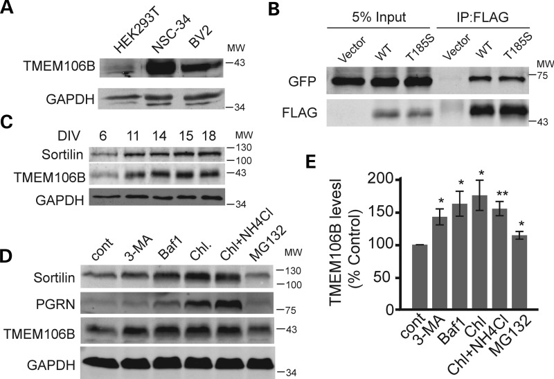
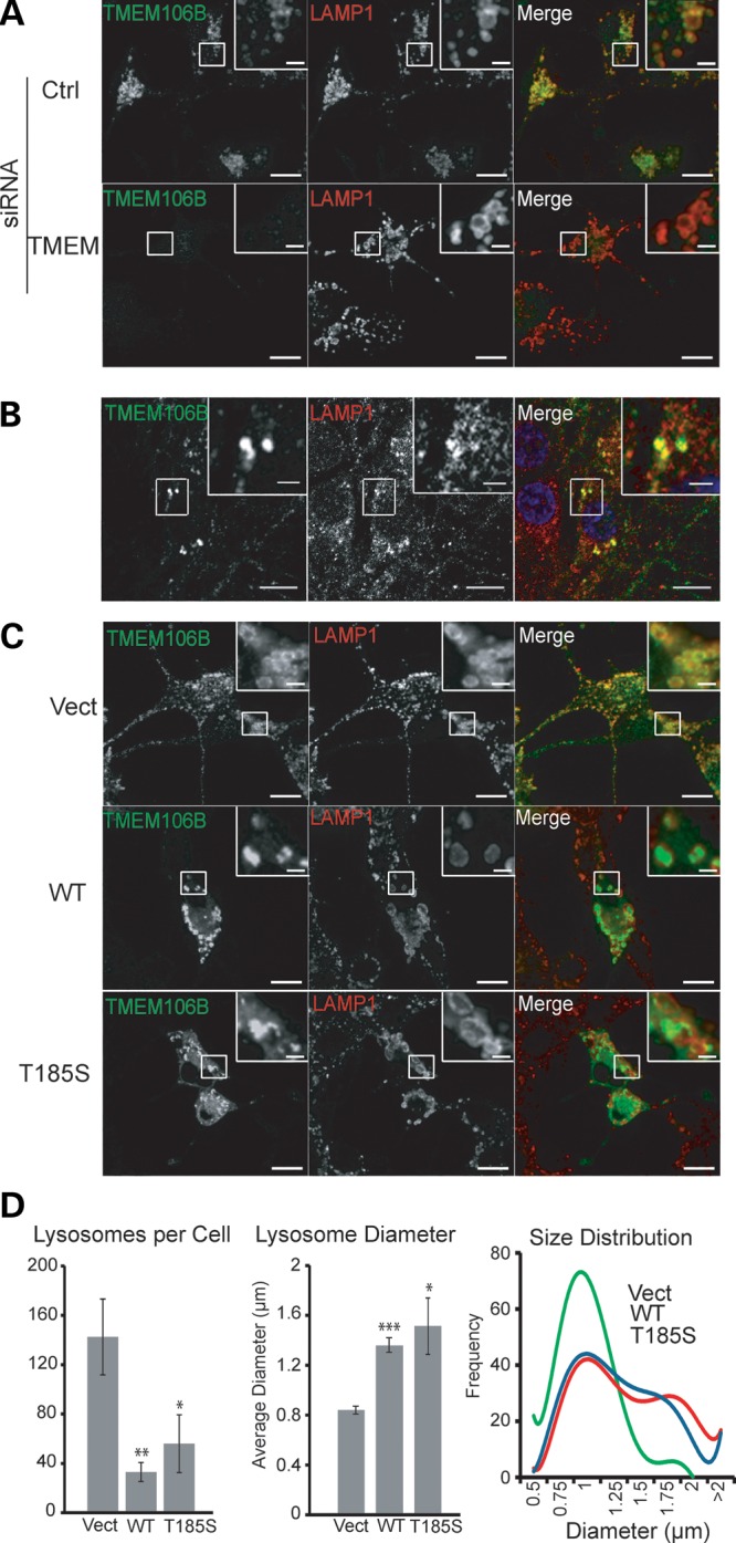
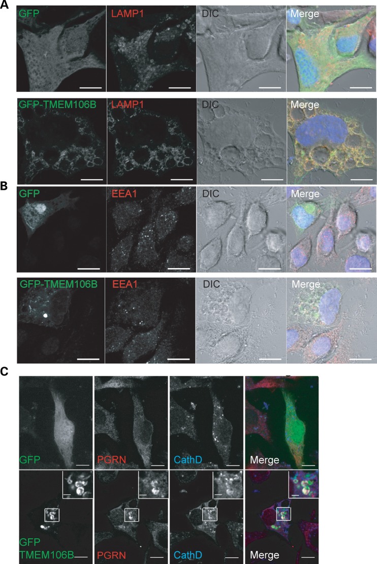
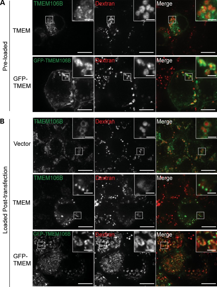
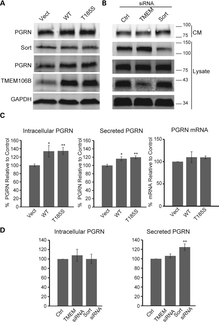
Haploinsufficiency of Progranulin (PGRN), a gene encoding a secreted glycoprotein, is a major cause of frontotemporal lobar degeneration with ubiquitin (FTLD-U) positive inclusions. Single nucleotide polymorphisms in the TMEM106B gene were recently discovered as a risk factor for FTLD-U, especially in patients with PGRN mutations. TMEM106B is also associated with cognitive impairment in amyotrophic lateral sclerosis patients. Despite these studies, little is known about TMEM106B at molecular and cellular levels and how TMEM106B contributes to FTLD. Here, we show that TMEM106B is localized in the late endosome/lysosome compartments and TMEM106B levels are regulated by lysosomal activities. Ectopic expression of TMEM106B induces morphologic changes of lysosome compartments and delays the degradation of endocytic cargoes by the endolysosomal pathway. Furthermore, overexpression of TMEM106B correlates with elevated levels of PGRN, possibly by attenuating lysosomal degradation of PGRN. These results shed light on the cellular functions of TMEM106B and the roles of TMEM106B in the pathogenesis of FTLD-U with PGRN mutations.


Figures

Figure 1.
Figure 1.
Expression of TMEM106B in different cell types. (A) Cell lysates from HEK293T, NSC-34 and BV-2 were loaded and blotted with anti-TMEM106B and anti-glyceraldehyde 3-phosphate dehydrogenase (GAPDH) antibodies. (B) TMEM106B forms homodimers. FLAG-tagged TMEM106B wild type (WT) and T185S variants were cotransfected with GFP-TMEM106B WT into HEK293T cells. Anti-FLAG antibodies were used to immunoprecipitate FLAG-TMEM106B. IP products were blotted for GFP and FLAG as indicated. (C) TMEM106B expression in rat cortical neurons. E17 rat cortical neurons were isolated and allowed to differentiate for indicated days. Cell lysates were prepared and blotted for sortilin, TMEM106B and GAPDH. Cortical neurons were treated with 2 µm Ara-C after DIV6 to inhibit the growth of glial cells. (D) N2A cells were treated with 5 mm 3-MA, 50 nm Baf1, 15 mm NH4Cl + 100 µm chloroquine or 10 µm MG-132 for 14 h as indicated. Cell lysates were prepared and blotted for TMEM106B and GAPDH. (E) Quantification of data shown in (D), n = 4, ±SEM, * P<0.05, ** P < 0.01 Student's t-test.
Figure 2.
Figure 2.
TMEM106B localizes to late endosomes/lysosomes. (A) Colocalization of endogenous TMEM106B with LAMP1-positive vesicles. N2A cells were fixed and stained with polyclonal anti-TMEM106B and monoclonal anti-LAMP1 antibodies. siRNA knockdown of TMEM106B confirms the specificity of our anti-TMEM106B antibody. (B) Colocalization of endogenous TMEM106B with LAMP1 in DIV15 cortical neurons. (C) Overexpression of TMEM106B induces enlarged LAMP1-positive vesicles. Vector control, pCMV-TMEM106B WT and T185S transfected N2A cells were stained with anti-TMEM106B and anti-LAMP1 antibodies. Green fluorescence exposure time was reduced for cells overexpressing TMEM106B to highlight differences in expression levels when compared with nearby non-transfected cells. Scale bars: 10 µm (2 µm in the inset). (D) Quantification of average number of lysosomes per cell and mean lysosome diameter ±SEM along with best fit curves of lysosome size histograms from cells imaged in (C). A minimum of 200 lysosomes were measured from six randomly selected cells in each condition. * P < 0.05, ** P < 0.01, ***P < 0.001 Student's t-test.
Figure 3.
Figure 3.
GFP-TMEM106B overexpression results in LAMP1-positive vacuoles in N2A cells. (A) GFP-TMEM106B expression in N2A cells results in enlarged vacuoles that are rimmed by TMEM106B and LAMP1. (B) GFP-TMEM106B-induced vacuoles are EEA1 negative. (C) Colocalization of PGRN with GFP-TMEM106B in some of the vacuoles. Scale bar: 10 µm (2 µm in the inset).
Figure 4.
Figure 4.
The fluid-phase marker, dextran, accumulates in TMEM106B-positive vesicles. (A) N2A cells preloaded with dextran were transfected with TMEM106B and GFP-TMEM106B. Cells were fixed and stained for 24 h post transfection. Untagged and GFP-TMEM106B can be seen on the surface of dextran-containing vesicles. (B) Dextran-labeled endosomes are capable of fusion with TMEM106B enlarged lysosomes. N2A cells were transfected with vector control, TMEM106B and GFP-TMEM106B. After 28 h, cells were loaded with dextran for 16 h, washed and chased 4 h in growth medium. Cells were fixed and stained 48 h post transfection. Scale bars: 10 µm (2 µm in the inset).
Figure 5.
Figure 5.
GFP-TMEM106B overexpression results in defects in EGFR degradation. (A) T98G cells transfected with vector control or GFP-TMEM106B were serum starved and stimulated with EGF in the presence of cycloheximide for indicated times. Levels of EGFR, phosphorylated ERK1/2 and GAPDH were quantified by western blots. A representative image of three experiments is shown. (B) Quantification of EGFR levels relative to loading control for experiment in (A). n = ±SEM.
Figure 6.
Figure 6.
Regulation of PGRN levels by TMEM106B. (A) Western blot analysis of N2A cells overexpressing TMEM106B WT and T185S. Transfected cells were changed to serum-free medium 24 h after transfection. After another 24 h, lysates and CM were collected. CM were further concentrated using TCA precipitation. (B) Western blot analysis of N2A cells transfected with control siRNA pool or siRNA pools against TMEM106B and sortilin. Transfected cells were changed to serum-free medium 48 h after siRNA transfection. After another 24 h, lysates and CM were collected. CM were further concentrated using TCA precipitation. (C) Overexpression of TMEM106B in N2A cells leads to increased intracellular and secreted PGRN levels as measured by western blot or ELISA, respectively (n = 5). PGRN levels were normalized to the mean of two vector transfected controls. No change in PGRN mRNA levels was detected via qPCR (n = 3). (D) Knockdown of TMEM106B in N2A cells has no effect on intracellular or secreted PGRN levels as measured by western blot or ELISA, respectively (n = 6). Sortilin knockdown leads to increased levels of PGRN in the media. * P < 0.05, **P < 0.01, Student's t-test.

References

    1. Neary D., Snowden J., Mann D. Frontotemporal dementia. Lancet Neurol. 2005;4:771–780. - PubMed
    1. Ratnavalli E., Brayne C., Dawson K., Hodges J.R. The prevalence of frontotemporal dementia. Neurology. 2002;58:1615–1621. - PubMed
    1. Baker M., Mackenzie I.R., Pickering-Brown S.M., Gass J., Rademakers R., Lindholm C., Snowden J., Adamson J., Sadovnick A.D., Rollinson S., et al. Mutations in progranulin cause tau-negative frontotemporal dementia linked to chromosome 17. Nature. 2006;442:916–919. - PubMed
    1. Cruts M., Gijselinck I., van der Zee J., Engelborghs S., Wils H., Pirici D., Rademakers R., Vandenberghe R., Dermaut B., Martin J.J., et al. Null mutations in progranulin cause ubiquitin-positive frontotemporal dementia linked to chromosome 17q21. Nature. 2006;442:920–924. - PubMed
    1. Gass J., Cannon A., Mackenzie I.R., Boeve B., Baker M., Adamson J., Crook R., Melquist S., Kuntz K., Petersen R., et al. Mutations in progranulin are a major cause of ubiquitin-positive frontotemporal lobar degeneration. Hum. Mol. Genet. 2006;15:2988–3001. - PubMed

Publication types

MeSH terms