. 2012 Dec;139(24):4561-70.
doi: 10.1242/dev.087247. Epub 2012 Nov 7.

In vivo evidence for the crucial role of SF1 in steroid-producing cells of the testis, ovary and adrenal gland

Affiliations

In vivo evidence for the crucial role of SF1 in steroid-producing cells of the testis, ovary and adrenal gland

F William Buaas et al. Development. 2012 Dec.

Abstract

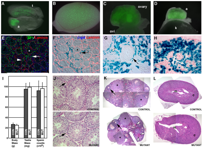
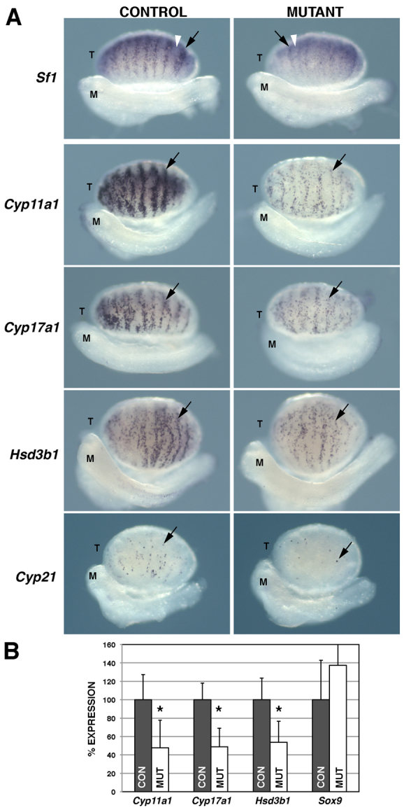
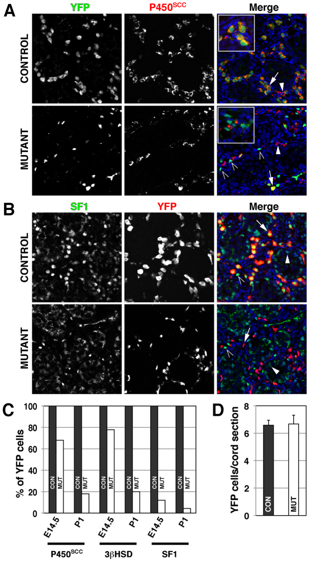
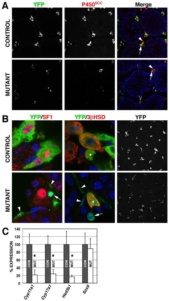
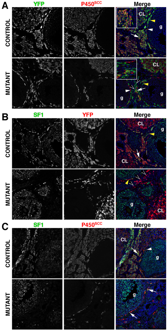
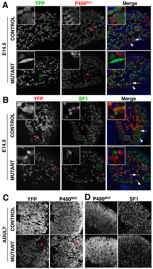
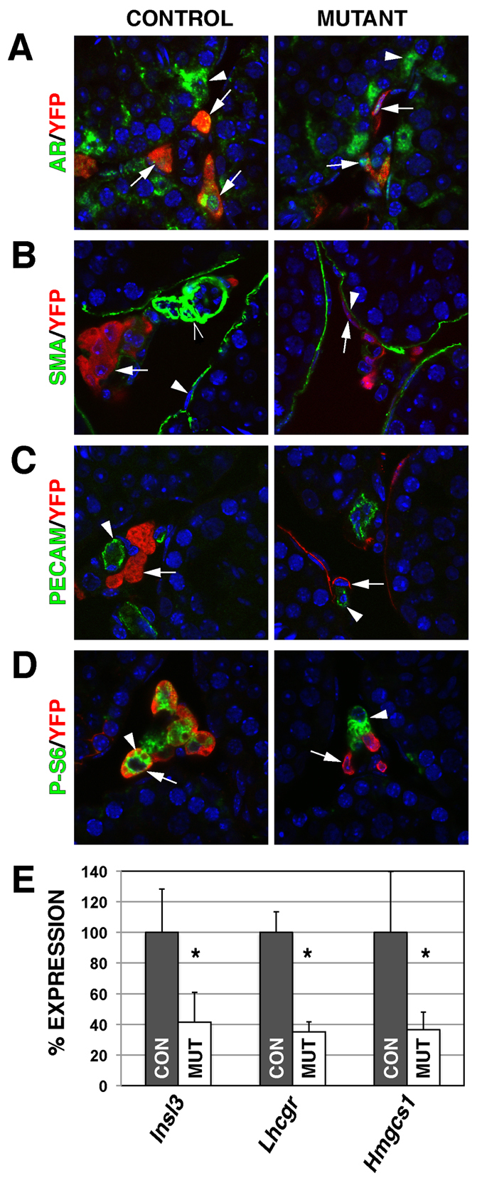
Adrenal and gonadal steroids are essential for life and reproduction. The orphan nuclear receptor SF1 (NR5A1) has been shown to regulate the expression of enzymes involved in steroid production in vitro. However, the in vivo role of this transcription factor in steroidogenesis has not been elucidated. In this study, we have generated steroidogenic-specific Cre-expressing mice to lineage mark and delete Sf1 in differentiated steroid-producing cells of the testis, the ovary and the adrenal gland. Our data show that SF1 is a regulator of the expression of steroidogenic genes in all three organs. In addition, Sf1 deletion leads to a radical change in cell morphology and loss of identity. Surprisingly, sexual development and reproduction in mutant animals were not compromised owing, in part, to the presence of a small proportion of SF1-positive cells. In contrast to the testis and ovary, the mutant adult adrenal gland showed a lack of Sf1-deleted cells and our studies suggest that steroidogenic adrenal cells during foetal stages require Sf1 to give rise to the adult adrenal population. This study is the first to show the in vivo requirements of SF1 in steroidogenesis and provides novel data on the cellular consequences of the loss of this protein specifically within steroid-producing cells.


Figures

Fig. 1.
Fig. 1.
Reporter gene expression and deletion of Sf1 in mouse foetal and adult steroidogenic cells. (A-D) YFP fluorescence from Cyp11a1-Cre;R26RYFP E14.5 testis (t) and adrenal (a) (A), adult testis (B), adult ovary and adjacent oviduct (ovi) (C), and adult adrenal (a) juxtaposed to the kidney (k) (D). (E) Double immunofluorescence staining on E14.5 wild-type testis of laminin surrounding the testicular cords, and SF1 in the interstitial region (arrow) and in Sertoli cells (arrowhead), within cords (asterisk). (F-H) β-Gal staining of sections from Cyp11a1-Cre;R26RlacZ tissues in E14.5 testicular interstitial cells (F, arrow), presumptive theca (black arrow) and stroma (white arrowhead) in the adult ovary (G) and E16.5 adrenal gland (H). (I) Body mass, testis mass and epididymal sperm counts from mutant (MUT) and control (CON) adult (2- to 3-month-old) male mice. Error bars represent s.e.m. (J-L) PAS-stained sections of adult control and mutant testes (J), ovaries (K) and adrenal glands (L). (J) Presumptive Leydig cells (arrows) based on position and morphology. (K) Follicles (black arrows) and corpora lutea (asterisks). Increased PAS-positive cells in mutant stroma (white arrowheads). (L) Medulla (m) and cortical (c) regions of adrenal glands.
Fig. 2.
Fig. 2.
The steroidogenic gene expression pathway is reduced in Cyp11a1-Cre;Sf1FI/FI foetal testes. (A) Whole mount in situ hybridization of representative E14.5-15 testes from control and mutant mouse embryos were analysed for the expression of steroidogenic genes as indicated. Anterior is to the left with the testis (T) positioned above the mesonephros (M). Steroidogenic Leydig cells (black arrows) reside between seminiferous tubules, which contain Sf1-expressing Sertoli cells (white arrowhead). (B) Quantitative RT-PCR on RNA from E14.5 control (CON) and mutant (MUT) testes for steroidogenic genes as indicated. *P<0.05. Error bars represent s.e.m.
Fig. 3.
Fig. 3.
Foetal Leydig cells require SF1 for steroidogenesis. (A) Protein expression of the nuclear and cytoplasmic YFP (green) and mitochondrial P450SCC (red) in control and mutant E14.5 testes (blue, DAPI). YFP-positive cells in controls are always positive for P450SCC (arrow and magnified inset). P450SCC single-positive cells were also present (filled arrowhead). Mutant testes have fewer double-positive cells (arrow) with the continued presence of P450SCC single positives (filled arrowhead). YFP single positives (unfilled arrowheads) are present throughout the interstitium. Mutant YFP cells show reduced P450SCC and a flattened morphology (inset). (B) Protein expression of nuclear SF1 (green) and nuclear and cytoplasmic YFP (red) in control and mutant E14.5 testes. Control YFP cells show SF1 expression (arrow) whereas Sertoli cells in the testicular cords express SF1 but no YFP (filled arrowhead). SF1-positive interstitial cells, not expressing YFP, are infrequent in control and mutants (unfilled arrowheads). Mutant interstitial YFP cells (arrow) do not express SF1, exhibit a reduced size and flattened morphology. SF1 expression in Sertoli cells is unaffected (filled arrowhead). (C) Frequency of YFP cells expressing P450SCC, 3βHSD and SF1 in E14.5 and P1 control (CON) and mutant (MUT) testes (n=2 control and mutant, two sections per sample and >65 YFP cells counted per section). (D) Quantification of E14.5 testis sections for the number of YFP cells per testis cord. Error bars represent s.e.m.
Fig. 4.
Fig. 4.
Adult Leydig cells require SF1 for steroidogenesis. (A) Protein expression of nuclear and cytoplasmic YFP (green) and mitochondrial P450SCC (red) in control and mutant adult testes (blue, DAPI). YFP-positive cells in controls are always positive for P450SCC (arrow). Leydig cells not targeted by the Cre recombinase show P450SCC expression but no YFP (filled arrowhead). Mutant testes also contain YFP-positive cells with no P450SCC expression (unfilled arrowhead). (B) YFP/SF1 (left) and YFP/3βHSD (middle) protein expression in control and mutant testes. Control interstitial YFP cells show nuclear SF1 expression whereas YFP mutant Leydig cells have lost SF1 expression (arrow and arrowheads) and display peritubular (arrowheads) or interstitial (arrow) positions. Mutant Leydig cells (middle panel) lose 3βHSD expression (arrow and arrowheads) whereas non-SF1-deleted cells remain steroidogenic (asterisk). Low magnification (20×) of the YFP adult Leydig cells (right- hand panels) reveals mutant Leydig cells that are broadly distributed throughout the testes and show interstitial and peritubular positions. (C) Quantitative RT-PCR analyses of steroidogenic Leydig and Sertoli cell-specific genes in control (CON) and mutant (MUT) testes. *P<0.05. Error bars represent s.e.m.
Fig. 5.
Fig. 5.
Theca and stromal cells of the adult ovary require SF1 for steroidogenesis. (A) Protein expression of nuclear and cytoplasmic YFP (green) and mitochondrial P450SCC (red) in control and mutant adult ovaries (blue, DAPI). YFP-positive cells in controls are always positive for P450SCC (arrow). Double-positive theca cells (yellow arrowhead) surround the growing follicle with occasional non-Cre targeted theca cells (white arrowhead). YFP/P450SCC double positives reside in the corpora lutea (CL). Mutant ovaries contain cells with all the expression profiles described above. Additionally, YFP single positives (unfilled arrowheads) are present throughout the mutant interstitium. Mutant YFP cells show reduced P450SCC and a flattened morphology (insets). YFP/P450SCC cells are unaffected in mutant CL. (B) Protein expression of nuclear SF1 and YFP in control and mutant adult ovaries. Control theca (yellow arrowhead) and stromal (white arrow) YFP cells express SF1 but the cells of the CL do not. Granulosa (g) cells express SF1 but not YFP. Mutant YFP stromal (unfilled arrowhead) and theca (yellow arrowhead) do not express SF1, exhibit a reduced size and flattened morphology. SF1 expression in mutant granulosa cells is unaffected. (C) Protein expression of SF1 and P450SCC in control and mutant adult ovaries. Control P450SCC stromal (arrow) and theca (arrowhead) cells express SF1. P450SCC-positive theca and stromal cells continue to express SF1 in mutant ovaries (arrows).
Fig. 6.
Fig. 6.
Adrenal defects in Cyp11a1-Cre;Sf1Fl/Fl mice. (A) Protein expression of nuclear and cytoplasmic YFP and mitochondrial P450SCC in control and mutant E14.5 adrenal glands. In control adrenal glands, all YFP-expressing cells are P450SCC positive (arrow and inset), but some P450SCC-expressing cells have no YFP (arrowhead). Mutant adrenal glands contain cells expressing only YFP (unfilled arrowhead and inset), indicative of reduced P450SCC expression. (B) SF1 and YFP protein expression in control and mutant E14.5 adrenal glands. Control and mutant adrenal glands exhibit cells positive for both YFP and nuclear SF1 (arrow and inset), whereas some remain negative for YFP (arrowhead and inset). Additionally, a large number of cells in mutant adrenal glands express only YFP (unfilled arrowhead and inset), indicating Cre-mediated deletion of SF1. (C) YFP and P450SCC protein expression in control and mutant adult adrenal glands. In control adrenal glands, most cells co-express YFP and P450SCC, although a few express only P450SCC. P450SCC expression was found throughout the cortex of adult mutant adrenal glands. Unlike the control adrenal gland, a number of P450SCC-positive cells in the mutant did not express YFP (red arrowhead). (D) P450SCC and SF1 expression in control and mutant adult adrenal glands in adjacent sections.
Fig. 7.
Fig. 7.
Properties of SF1-deficient adult Leydig cells. (A) Control Leydig and Sertoli cells express nuclear-localized androgen receptor (AR) (arrows and arrowhead, respectively). Mutant Leydig cells express AR (arrows), and AR-expressing Sertoli cells (arrowhead) are unaffected. (B) Myoid epithelial cells (arrowhead) and endothelial cells (unfilled arrowhead) of the microvasculature express smooth muscle actin (SMA). Control and mutant Leydig cells (red, arrows) do not express SMA. (C) Control and mutant Leydig cells (arrows) do not express the endothelial marker PECAM (arrowheads). (D) Control Leydig cells express YFP (arrow) and high levels of P-S6 (arrowheads) whereas mutant Leydig cells (arrows) no longer express P-S6. A Leydig cell that escaped Cre-mediated recombination continues to express P-S6 (arrowhead). (E) Quantitative RT-PCR analyses of non-steroidogenic Leydig cell genes in adult control (CON) and mutant (MUT) testes. *P<0.05. Error bars represent s.e.m.

References

    1. Buaas F. W., Val P., Swain A. (2009). The transcription co-factor CITED2 functions during sex determination and early gonad development. Hum. Mol. Genet. 18, 2989–3001 - PubMed
    1. Davidoff M. S., Middendorff R., Enikolopov G., Riethmacher D., Holstein A. F., Müller D. (2004). Progenitor cells of the testosterone-producing Leydig cells revealed. J. Cell Biol. 167, 935–944 - PMC - PubMed
    1. Duggavathi R., Volle D. H., Mataki C., Antal M. C., Messaddeq N., Auwerx J., Murphy B. D., Schoonjans K. (2008). Liver receptor homolog 1 is essential for ovulation. Genes Dev. 22, 1871–1876 - PMC - PubMed
    1. Falender A. E., Lanz R., Malenfant D., Belanger L., Richards J. S. (2003). Differential expression of steroidogenic factor-1 and FTF/LRH-1 in the rodent ovary. Endocrinology 144, 3598–3610 - PubMed
    1. Ferraz-de-Souza B., Lin L., Achermann J. C. (2011). Steroidogenic factor-1 (SF-1, NR5A1) and human disease. Mol. Cell. Endocrinol. 336, 198–205 - PMC - PubMed

Publication types

MeSH terms