. 2012 Nov 7;32(45):15779-90.
doi: 10.1523/JNEUROSCI.3557-12.2012.

Aversive stimuli differentially modulate real-time dopamine transmission dynamics within the nucleus accumbens core and shell

Affiliations

Aversive stimuli differentially modulate real-time dopamine transmission dynamics within the nucleus accumbens core and shell

Aneesha Badrinarayan et al. J Neurosci. .

Abstract

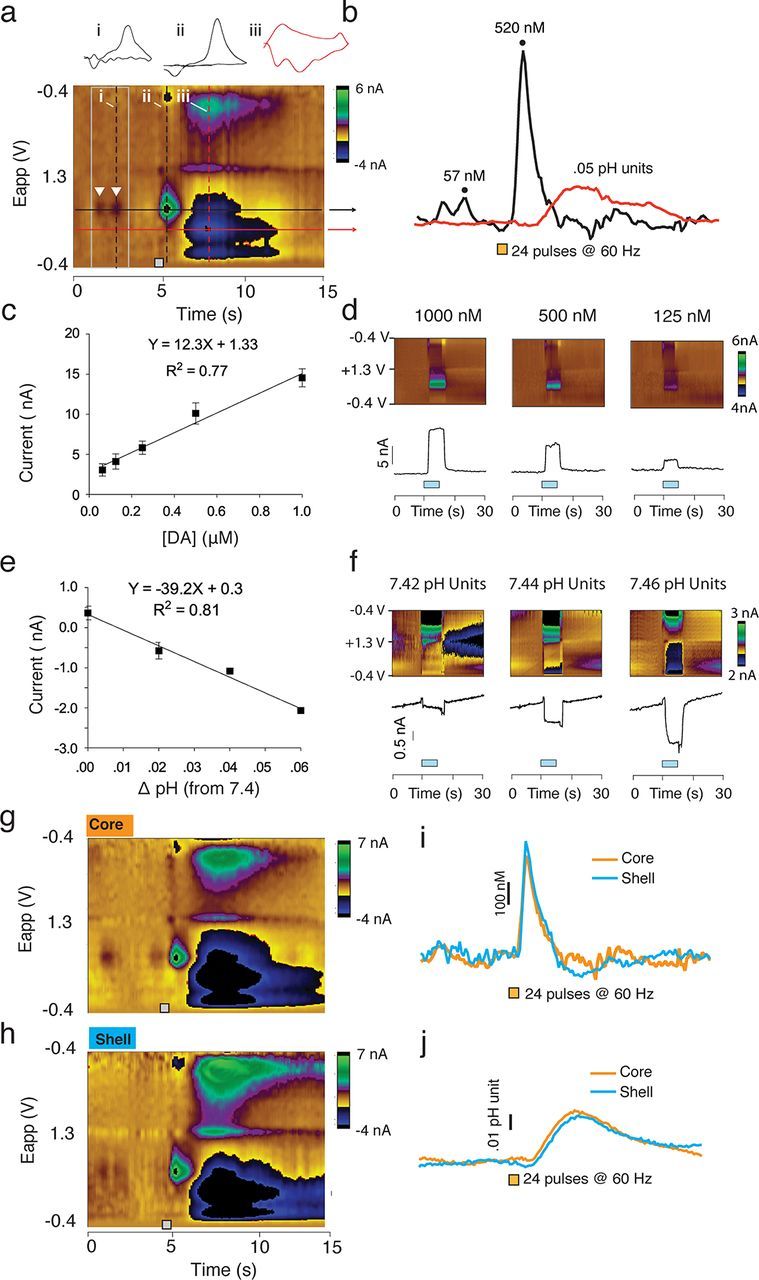
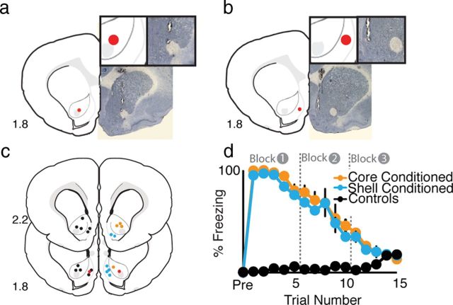
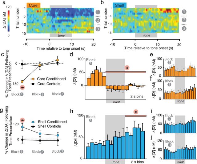
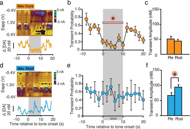
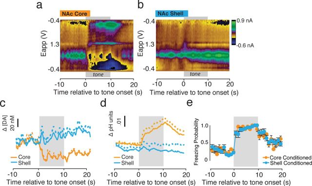
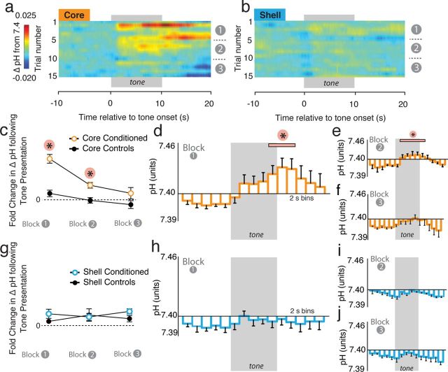
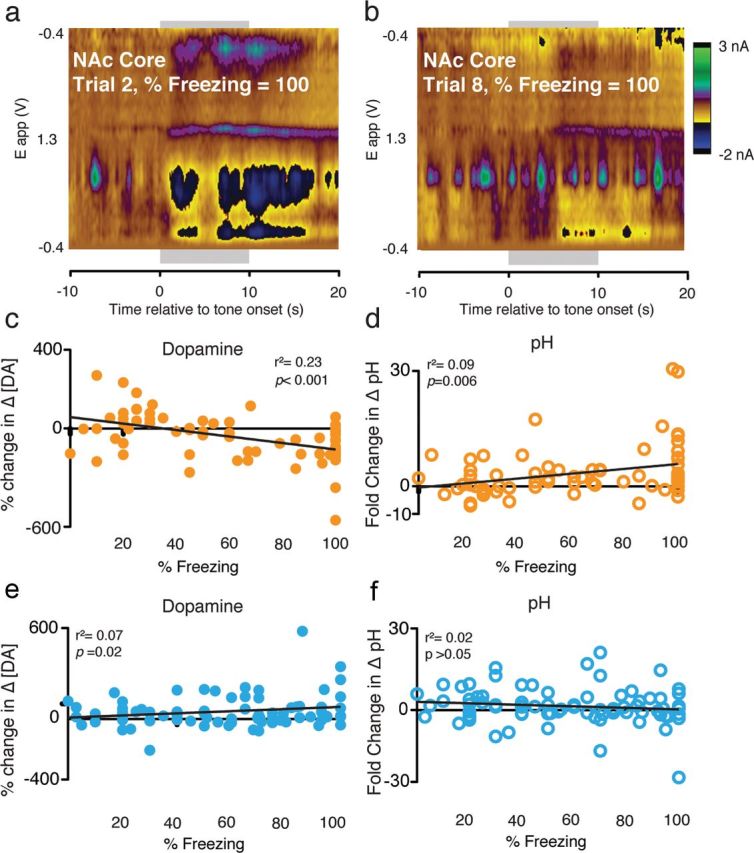
Although fear directs adaptive behavioral responses, how aversive cues recruit motivational neural circuitry is poorly understood. Specifically, while it is known that dopamine (DA) transmission within the nucleus accumbens (NAc) is imperative for mediating appetitive motivated behaviors, its role in aversive behavior is controversial. It has been proposed that divergent phasic DA transmission following aversive events may correspond to segregated mesolimbic dopamine pathways; however, this prediction has never been tested. Here, we used fast-scan cyclic voltammetry to examine real-time DA transmission within NAc core and shell projection systems in response to a fear-evoking cue. In male Sprague Dawley rats, we first demonstrate that a fear cue results in decreased DA transmission within the NAc core, but increased transmission within the NAc shell. We examined whether these changes in DA transmission could be attributed to modulation of phasic transmission evoked by cue presentation. We found that cue presentation decreased the probability of phasic DA release in the core, while the same cue enhanced the amplitude of release events in the NAc shell. We further characterized the relationship between freezing and both changes in DA as well as local pH. Although we found that both analytes were significantly correlated with freezing in the NAc across the session, changes in DA were not strictly associated with freezing while basic pH shifts in the core more consistently followed behavioral expression. Together, these results provide the first real-time neurochemical evidence that aversive cues differentially modulate distinct DA projection systems.


Figures

Figure 1.
Figure 1.
Changes in [DA] and pH values are characterized based on stimulation both in vivo and in vitro. a, Representative color plot showing current changes observed at the recording electrode plotted against the applied voltage and time. Analytes (DA and pH) are identified based on specific oxidation-reduction profiles. The holding potential applied to the carbon-fiber electrode (−0.4 V) was ramped to +1.3 V and back to 0.4 V at a rate of 10 Hz. This example shows two spontaneous DA transients, indicated by white inverted triangles. Electrical stimulation (24 pulses, 60 Hz) of the VTA results in a near instantaneous surge in [DA] and a more delayed and long-lasting basic shift in pH. Vertical dashed lines on the color plot indicate the time point of representative cyclic voltammograms, shown above. Horizontal black (DA) and red (pH) lines indicate the applied voltage at the point of maximum oxidation, used to identify neurochemical species. b, Traces for Δ[DA] and pH units were obtained by using principal components analysis. Δ[DA] for the first transient observed in a and the stimulation is shown, as well as peak change in pH units. c, Calibration curve for DA. Known concentrations of DA were used to determine the average current evoked in a standard set of electrodes. The slope of the resulting trend line was then used as the calibration factor for conversion of current to DA concentration. d, Representative color plots and current traces recorded in vitro during the application of specific DA concentrations. Blue bars indicate infusion of DA. e, Calibration curve for pH. Current changes were again recorded for known basic shifts in pH. The resulting slope was again used as the calibration factor. f, Representative color plots and current traces recorded in vitro during the application of basic pH shifts. Blue bars indicate the duration of the infusion. g–j, Representative color plots and traces of electrical stimulation (24 pulses, 60 Hz) of DA afferents to the NAc core (g, i, j) and shell (h–j). These plots indicate similar DA and pH responses within the core and shell.
Figure 2.
Figure 2.
Histology and fear expression during recording sessions. a, Example of histological verification of recording site in the NAc core (b) and within the NAc shell. c, Diagrammatic representation of electrode placements for all animals within the NAc. Core-conditioned placements are indicated in orange (n = 5), Shell-Conditioned placements are indicated in cyan (n = 6), and placements for control animals are shown in black (n = 11). Although the placements are distributed throughout both cerebral hemispheres here for clarity, recordings from all animals were taken from the same hemisphere. Red indicates the examples shown in a and b. d, Percentage freezing during fear expression for core-conditioned, shell-conditioned, and control animals. There was no difference between conditioned groups in fear expression. Unconditioned rats did not freeze during CS presentation. All conditioned rats showed robust freezing that attenuated as the session progressed. By the end of the session, conditioned rats showed no more freezing than control rats.
Figure 3.
Figure 3.
Fear cues cause decreased DA transmission in the NAc core but enhanced DA transmission in the NAc shell. a, b, Heatmaps depicting trial-by-trial Δ[DA] across all 15 cue presentations in the core (n = 5) and shell (n = 6), respectively. Tone presentation was immediately followed by a dramatic decrease in [DA] within the NAc core, but within the shell, [DA] was gradually increased following cue offset. In both regions, the effect attenuated as the session progressed. c, Percentage change in mean [DA] in the NAc core in conditioned versus unconditioned animals in response to CS presentations. A significant decrease in [DA] was observed in response to CS presentations during the first five tone block. d–f, Δ[DA] within the NAc core relative to CS presentation across each block of trials. [DA] was calculated in 2 s bins. Tone presentation causes an immediate and significant decrease in [DA] during block 1 (d) that attenuated during subsequent blocks (e, f). g, Percentage change in mean [DA] in the NAc shell in conditioned versus unconditioned animals in response to CS presentations. A significant increase in [DA] was observed in response to the CS during the first five tone block. h–j, Δ[DA] within the NAc shell relative to CS presentation across each block of trials. Tone presentation resulted in a gradual increase in [DA] that was significant relative to pretone [DA] following cue offset during block 1 (h). There was no significant change following tone presentation in block 2 (i) or 3 (j). *p < 0.05.
Figure 4.
Figure 4.
Changes in [DA] in response to the fear-evoking CS are attributable to changes in the probability and amplitude of DA release events. a, d, Representative transient traces. White inverted triangles indicate transients. The panel underneath each color plot shows the converted [DA] traces. b, e, Transient probability calculated during 2 s bins for block 1 within the NAc core (i.e. averaged over first five tones for n = 5; 25 traces all together) and shell (i.e. averaged over first five tones for n = 6; 30 traces). Tone onset caused a significant decrease in transient probability that lasted the duration of the tone presentation, but returned to pretone levels following tone offset in the NAc core. Transient probability did not change with tone presentation in the NAc shell. c, f, Average transient amplitude before and following cue onset in the NAc core and shell. Cue onset did not alter transient amplitude in the NAc core, but increased transient amplitude in the NAc shell. *p < 0.05.
Figure 5.
Figure 5.
Cue onset is accompanied by basic pH shifts within the NAc core. a, b, Average color plots for the first block of CS presentations in the NAc core (n = 5; a) and shell (n = 6; b) were generated to illustrate the raw changes in neurochemical activity associated with tone presentation (gray bar). Within the NAc core, cue onset is accompanied by a delayed but lasting basic pH shift. No visible pH shifts were detected in the shell. c, Quantification of changes in [DA] in core and shell following cue onset during block 1. Cue presentation altered both core and shell [DA]. d, Quantification of pH changes during block 1 within the NAc core and shell. Cue presentation results in a significant basic pH shift within the NAc core, but does not cause any change in pH within the NAc shell. e, Within-trial probability of freezing across block 1 for both core-conditioned and shell-conditioned rats. Cue onset causes rapid increases in freezing probability that last throughout the tone presentation and attenuate slowly following tone-offset.
Figure 6.
Figure 6.
Fear cues cause basic shifts in pH within the NAc core but not within the NAc shell. a, b, Heatmaps depicting trial-by-trial pH shifts across all 15 cue presentations in the core (n = 5) and shell (n = 6), respectively. Tone presentation was followed by a basic pH shift within the NAc core, but not within the shell. The basic shift within the NAc core remained prominent through blocks 1 and 2 and attenuated during block 3. c, Fold-change in average pH in the NAc core in conditioned versus unconditioned animals in response to CS presentations. A significant increase in pH was observed in response to CS presentations during the first and second five tone blocks. d–f, ΔpH units within the NAc core relative to CS presentation across each block of trials. pH was calculated in 2 s bins. Cue presentation causes a significant increase in pH during block 1 (d) and 2 (e) that attenuated during the final blocks (f). g, Fold-change in mean pH in the NAc shell in conditioned versus unconditioned animals in response to CS presentations. There was no difference in pH changes within any block between conditioned and unconditioned animals. h–j, ΔpH within the NAc shell relative to CS presentation across each block of trials. Tone presentation did not significantly alter pH shifts in the NAc shell during blocks 1 (h), 2 (i) or 3 (j). *p < 0.05.
Figure 7.
Figure 7.
Analysis of the relationship between locomotor activity and neurochemical changes. a, b, Representative traces from a single animal that displayed identical freezing behavior on two trials during block 1 (a) and block 2 (b) illustrate that [DA] does not strictly follow locomotor activity. c, Quantification of the correlation between percentage change in [DA] and freezing in the NAc core shows a significant correlation between behavioral expression and changes in dopamine. d, Quantification of the correlation between pH and freezing revealed a modest but significant relationship in the NAc core. e, f, Conversely, no relationships were observed between changes in [DA] (e) or pH (f) and freezing within the NAc shell.

References

    1. Adhikari A, Topiwala MA, Gordon JA. Synchronized activity between the ventral hippocampus and the medial prefrontal cortex during anxiety. Neuron. 2010;65:257–269. - PMC - PubMed
    1. Aragona BJ, Cleaveland NA, Stuber GD, Day JJ, Carelli RM, Wightman RM. Preferential enhancement of dopamine transmission within the nucleus accumbens shell by cocaine is attributable to a direct increase in phasic dopamine release events. J Neurosci. 2008;28:8821–8831. - PMC - PubMed
    1. Aragona BJ, Day JJ, Roitman MF, Cleaveland NA, Wightman RM, Carelli RM. Regional specificity in the real-time development of phasic dopamine transmission patterns during acquisition of a cue-cocaine association in rats. Eur J Neurosci. 2009;30:1889–1899. - PMC - PubMed
    1. Arbuthnott GW, Wickens J. Space, time and dopamine. Trends Neurosci. 2007;30:62–69. - PubMed
    1. Arcediano F, Escobar M, Miller RR. Bidirectional associations in humans and rats. J Exp Psychol Anim Behav Process. 2005;31:301–318. - PubMed

Publication types

LinkOut - more resources