. 2012 Nov 7;32(45):15886-901.
doi: 10.1523/JNEUROSCI.4505-11.2012.

Neurodevelopmental role for VGLUT2 in pyramidal neuron plasticity, dendritic refinement, and in spatial learning

Affiliations

Neurodevelopmental role for VGLUT2 in pyramidal neuron plasticity, dendritic refinement, and in spatial learning

Hongbo He et al. J Neurosci. .

Abstract

The level and integrity of glutamate transmission during critical periods of postnatal development plays an important role in the refinement of pyramidal neuron dendritic arbor, synaptic plasticity, and cognition. Presently, it is not clear how excitatory transmission via the two predominant isoforms of the vesicular glutamate transporter (VGLUT1 and VGLUT2) participate in this process. To assess a neurodevelopmental role for VGLUT2 in pyramidal neuron maturation, we generated recombinant VGLUT2 knock-out mice and inactivated VGLUT2 throughout development using Emx1-Cre(+/+) knock-in mice. We show that VGLUT2 deficiency in corticolimbic circuits results in reduced evoked glutamate transmission, release probability, and LTD at hippocampal CA3-CA1 synapses during a formative developmental period (postnatal days 11-14). In adults, we find a marked reduction in the amount of dendritic arbor across the span of the dendritic tree of CA1 pyramidal neurons and reduced long-term potentiation and levels of synaptic markers spinophilin and VGLUT1. Loss of dendritic arbor is accompanied by corresponding reductions in the number of dendritic spines, suggesting widespread alterations in synaptic connectivity. Conditional VGLUT2 knock-out mice exhibit increased open-field exploratory activity yet impaired spatial learning and memory, endophenotypes similar to those of NMDA receptor knock-down mice. Remarkably, the impairment in learning can be partially restored by selectively increasing NMDA receptor-mediated glutamate transmission in adult mice by prolonged treatment with d-serine and a d-amino acid oxidase inhibitor. Our data indicate that VGLUT2 expression is pivotal to the proper development of mature pyramidal neuronal architecture and plasticity, and that such glutamatergic deficiency leads to cognitive malfunction as observed in several neurodevelopmental psychiatric disorders.


Figures
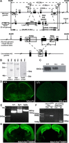
Figure 1.
Figure 1.
VGLUT2 gene targeting and inactivation of VGLUT2 in Emx1-Cre+/+/VGLUT2fx/fx mice. A, Top, A targeting construct engineered with LoxP sites flanking exon 2 of the Slc17a6 gene encoding VGLUT2 was inserted into the endogenous VGLUT2 locus by homologous recombination. Wild-type genome DNA schematic surrounding exons (E)1–3 is shown. The locations of Probe 1 and Probe 2 that recognize wild-type sequence are indicated. Approximate sites of insertion of vector targeting sequences (*, LoxP-Frt, and LoxP) in wild-type genome are shown. The vector cassette (pVBFRTCKR01) with the thymidine kinase promoter (TK) and I-CeuI vector linearization site are shown. The selectable marker (Neo, neomycin gene) is flanked by Frt sites. Exon 2 (E2), including this Frt region, is flanked by LoxP sites. Relative distances between sites in relation to the wild-type genome are marked and indicated in kilobase pairs (kb). New restriction sites (NsiI, NdeI, BstEII) introduced in the downstream LoxP site of the conditional allele are indicated (arrow), and the predicted sizes of fragments following restriction in the wild-type genome and conditional allele are shown. A, Bottom, Expanded view of floxed conditional VGLUT2 allele in flp+ mice whereby excision of the neomycin gene (Neo) has taken place, leaving one Frt site remaining in genome. Note: additional DNA sequences are added to intronic regions flanking exon 2 of VGLUT2 (shaded). B, Southern blot screening of ES cell genomic DNA from wild-type (Wt) and recombinant (rec) sample using NdeI and NsiI with probe 2 and BstEII with probe 1. In the normal genome, NsiI digestion gives rise to a 6.0 kb fragment and NdeI digestion gives rise to a 5.9 kb fragment hybridizing to probe 2. Instead, homologous recombination reveals bands of 4.5 kb and 4.8 kb following NsiI and NdeI digestion, respectively. BstEII digestion gives an 8.9 kb band in the recombinant (rec). C, Loss of VGLUT2 protein expression in VGLUT2Δ/Δ mice. Western blot of enriched whole brain vesicles prepared from E16-E19 wild-type (+/+), heterozygote (Δ/+), and unconditional VGLUT2 knock-out (Δ/Δ) mice. D, Immunostaining of wild-type mouse brain (+/+, left) shows VGLUT2 immunoreactivity absent from the unconditional VGLUT2 knock-out (Δ/Δ, right). E, Genotypes of wild-type (+/+), heterozygotes (fx/+), and homozygous (fx/fx) mice were verified by PCR using DNA extracted from tail samples and PCR amplification, revealing an 857 bp fragment in the floxed VGLUT2 conditional allele and a 763 bp fragment in the wild-type allele. F, VGLUT2 mRNA is inactivated by excision of exon 2 in cortical/hippocampal neurons in vitro from Emx1-Cre+/+/VGLUT2fx/fx mice. In VGLUT2fx/fx control cultures, a 414 bp fragment can be amplified using cRNA primers in exon 1 and exon 3, whereas only a 165 bp fragment was amplified from Emx1-Cre+/+/VGLUT2fx/fx cultures. Con, Control; GZ, gabazine. G, EGFP autofluorescence in coronal brain sections from Emx1-Cre+/+/Z-EG and Emx-Cre+/−/Z-EG mice shows the vast majority of EGFP+ neurons are found in cortical/hippocampal regions, demonstrating the specificity of recombination in vivo.
Figure 2.
Figure 2.
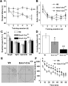
Impairment of spatial learning yet increased novelty–exploratory activity in Emx1-Cre+/+/VGLUT2fx/fx mice. A, We used the Morris water maze to assess spatial learning. Comparison of the latency among Emx1-Cre+/+/VGLUT2fx/fx, Emx1-Cre+/−/VGLUT2fx/fx, and control VGLUT2fx/fx (Wt) mice shows impairment of spatial learning in Emx1-Cre+/+/VGLUT2fx/fx mice. A similar decrease in escape latency occurred for Emx1-Cre+/−/VGLUT2fx/fx mice and Wt mice during training. Data are presented as the mean ± SEM (n = 14–21 in each group; males and females); **p < 0.001; repeated-measures ANOVA with a LSD post hoc test. B, Comparison of swimming speed during training among Emx1-Cre+/+/VGLUT2fx/fx, Emx1-Cre+/−/VGLUT2fx/fx, and control mice showed no differences between groups. C, Control mice spent significantly more time in the quadrant where the platform was previously located in a probe test for spatial reference memory. *p < 0.05; ANOVA. In contrast, Emx1-Cre+/+/VGLUT2fx/fx and Emx1-Cre+/−/VGLUT2fx/fx mice showed no preference for any quadrant. D, During the probe test, control mice crossed the platform more often than Emx1-Cre+/−/VGLUT2fx/fx mice and Emx1-Cre+/+/VGLUT2fx/fx mice; *p < 0.05, **p < 0.01, significant difference from control mice; ANOVA. E, Swimming traces during the probe test. F, Emx1-Cre+/+/VGLUT2fx/fx mice retain higher forward locomotion level throughout a 1 h period compared to control mice using a novelty exploration test in an open-field chamber. Error bars represent SEM; *p < 0.05; Student's t test.
Figure 3.
Figure 3.
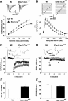
VGLUT2-deficiency results in reduced dendritic arbor and reduced number of spines in adult CA1 hippocampal pyramidal neurons. A, Golgi–Cox impregnated neurons and their corresponding digital camera lucida reconstructions. Scale bar represents 50 μm for whole neurons and 10 μm for spine images. Wt, Wild type. B, Basal dendrites of Emx1-Cre+/+/VGLUT2fx/fx mice show a significant reduction in arbor over most of the dendritic tree. Apical dendrites show incremental decreases in arbor over the tree. C, Basal and apical dendrites have significantly reduced total arbor and decreased average segment length. D, Both the basal and apical dendrites show reductions in the number of spines. E, Spine density remains unchanged across groups. Results for each group are obtained from 30 neurons among 6 animals. Error bars represent SEM; *p < 0.05, **p < 0.01; two-way repeated-measures ANOVA with a Scheffé post hoc or a Student's t test.
Figure 4.
Figure 4.
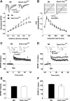
VGLUT2-deficiency results in reduced basal transmission, increased paired-pulse ratio, and reduced LTD in young CA3-CA1 connections. A, Representative fEPSP waveforms recorded at in the stratum radiatum of the CA1 region from hippocampal CA3–CA1 connections in young (P11–14) control VGLUT2fx/fx (Wt, wild type) and Emx1-Cre+/+/VGLUT2fx/fx mice. Input–output function (Wt, 29 recordings/6 animals; Emx1-Cre+/+/VGLUT2fx/fx, 18 recordings/5 animals). Stimulus intensity was normalized to the maximum intensity. B, Representative fEPSP traces. fEPSPs were induced by a paired-pulse protocol with varying interpulse intervals. Paired-pulse ratio (fEPSP2/fEPSP1) in control and Emx1-Cre+/+/VGLUT2fx/fx mice (Wt, 19 recordings/4 animals; Emx1-Cre+/+/VGLUT2fx/fx, 25 recordings/5 animals). C, Representative traces of fEPSPs recorded before (1) and after (2) LFS by stimulating the Schaffer collateral pathway in acute hippocampal slices from young (P11–14) animals. Time courses of fEPSP slopes are also shown. D, Representative traces of fEPSPs recorded before and after LFS by stimulating the Schaffer collateral pathway in acute hippocampal slices from adult (P45–60) animals. Time courses of fEPSP slopes are also shown. E, Mean values of the potentiation of fEPSPs averaged from 55 to 60 min following LFS (Wt, 11 recordings/5 animals; Emx1-Cre+/+/VGLUT2fx/fx, 10 recordings/5 animals). F, Mean values of the potentiation of fEPSPs averaged from 55 to 60 min following TBS (Wt, 7 recordings/4 animals; Emx1-Cre+/+/VGLUT2fx/fx, 10 recordings/4 animals). Error bars represent SEM; **p < 0.01; ANOVA.
Figure 5.
Figure 5.
Basal synaptic transmission and LTP are reduced in CA3–CA1 synapses in adult Emx1-Cre+/+/VGLUT2fx/fx mice. A, Representative fEPSP waveforms recorded at in the stratum radiatum of the CA1 region following Schaffer collateral stimulation in adult (P45–60) control VGLUT2fx/fx (Wt, Wild type) and Emx1-Cre+/+/VGLUT2fx/fx mice. Input–output function is shown for Wt (15 recordings/4 animals) and Emx1-Cre+/+/VGLUT2fx/fx (18 recordings/4 animals) mice. B, Representative fEPSP waveforms. Paired-pulse ratio (fEPSP2/fEPSP1) is shown for adult Wt (13 recordings/4 animals) and Emx1-Cre+/+/VGLUT2fx/fx (15 recordings/4 animals) mice. C, Representative traces of fEPSPs recorded before (1) and after (2) TBS by stimulating CA3-CA1 connections in acute hippocampal slices from young (P11–14) animals. Time course of fEPSP slopes is also shown. D, Representative traces of fEPSPs recorded before and after TBS by stimulating Schaffer collateral pathway in acute hippocampal slices from adult (P45–60) animals. Time course of fEPSP slopes are also shown. E, Mean values of the potentiation of fEPSPs averaged from 55 to 60 min following TBS (Wt, 8 recordings/4 animals; Emx1-Cre+/+/VGLUT2fx/fx, 12 recordings/4 animals). F, Mean values of the potentiation of fEPSPs averaged from 55 to 60 min following TBS (Wt, 16 recordings/5 animals; Emx1-Cre+/+/VGLUT2fx/fx, 16 recordings/5 animals). Error bars represent SEM; *p < 0.05; ANOVA.
Figure 6.
Figure 6.
Reduced hippocampal expression of VGLUT1 and spinophilin in adult Emx1-Cre+/+/VGLUT2fx/fx mice. A, Western blot of vesicle-enriched membranes (milligram membrane protein) from the following: (1) VGLUT1 (2 μg); (2) VIAAT (4 μg); (3) spinophilin (50 μg); (4) GAD67 (50 μg); (5) PSD95 (50 μg); and (6) β-actin (4 μg). Total hippocampal protein extracts were prepared from three mice in each group. B, Quantitation of bands by densitometry. Results are expressed as a ratio of VGLUT1, VIAAT, spinophilin, and GAD67 against β-actin or against PSD95, which did not differ between adult Emx1-Cre+/+/VGLUT2fx/fx and control VGLUT2fx/fx mice. Error bars represent SEM; *p < 0.05; Student's t test.
Figure 7.
Figure 7.
Representative examples of VGLUT1, VGLUT2, and VIAAT expression in the hippocampus and cerebral cortex of adult Emx1-Cre+/+/VGLUT2fx/fx, Emx1-Cre+/−/VGLUT2fx/fx, and control VGLUT2fx/fx mice. AC, These micrographs show robust immunofluorescence signals for VGLUT1 in the hippocampus. Note: although cell nuclei are unlabeled, an intact structure of the basic trisynaptic circuit is evident in conditional VGLUT2 knock-out mice. Rad, Striatum radiatum; grDG, supragranular region of dentate gyrus; Lmol, stratum lacunosum-molecular; DGmol, molecular layer of the dentate gyrus. Scale bar, 100 μm. DL, Distributions of VGLUT1 and VGLUT2 immunoreactivities in area S1 of the neocortex. As expected, the highest levels of VGLUT2 expression are seen in layer IV of area S1 barrel subfield, followed by layers II/III and the border between layers V and VI in control VGLUT2fx/fx (Wt, Wild type) mice. Similar immunostaining for VGLUT2 is observed in control and Emx1-Cre+/−/VGLUT2fx/fx mice. However, VGLUT2 immunofluorescence is noticeably reduced in layer IV and absent in layer III in Emx1-Cre+/+/VGLUT2fx/fx mice. Scale bar, 100 μm. MO, VGLUT2 expression is retained in the supragranular region (gr) of the dentate gyrus in Emx1-Cre+/+/VGLUT2fx/fx mice, where it colocalizes with VIAAT. Scale bar, 20 μm.
Figure 8.
Figure 8.
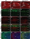
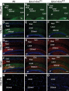
Representative examples of VGLUT1 and VGLUT2 expression in the hippocampus of Emx1-Cre+/+/VGLUT2fx/fx, Emx1-Cre+/−/VGLUT2fx/fx, and VGLUT2fx/fx mice at P18. A–C, Selective reduction of VGLUT2 expression in hippocampal subfields in conditional VGLUT2 knock-out mice. VGLUT2 immunofluorescence is strong in the supragranular region (gr) of the dentate gyrus, the stratum lacunosum-moleculare (Lmol), and the molecular layer of the dentate gyrus (DGmol) in VGLUT2fx/fx (Wt, Wild type) mice. Note: loss of VGLUT2 expression in DGmol in both Emx1-Cre lines. Note: Emx1-Cre+/− mice retain more expression of VGLUT2 in Lmol than Emx1-Cre+/+ mice. Note: high VGLUT2 expression in the supragranular region of the dentate gyrus and in the thalamus (Th) across all genotypes. Scale bar, 100 μm. D–L, Higher power micrographs reveal VGLUT2 (green), VGLUT1 (red), and merged images, together with the DAPI nuclear labeling (blue) Scale bar, 100 μm. M–O, Deconvolution microscopy and projection images show a clear reduction in the number of VGLUT2 puncta in the Lmol fields from Emx1-Cre+/−VGLUT2fx/fx mice and their absence in Emx1-Cre+/+/VGLUT2fx/fx mice. Scale bar, 20 μm.
Figure 9.
Figure 9.
d-Serine administration, together with d-amino acid oxidase inhibition, enhances learning in adult Emx1-Cre+/+/VGLUT2fx/fx mice. Adult Emx1-Cre+/+/VGLUT2fx/fx mice (P60–80, males/females) were treated with daily injections of saline, clozapine (1 mg/kg), or d-serine (600 mg/kg) and the DAAO inhibitor (compound 8; 50 mg/kg) for 2 weeks in the home cage and then each day 1 h before training or testing. A, The doses of these compounds used did not adversely affect locomotor activity at 1 or 6 h postinjection in an open-field chamber. B, In the Morris water maze test there was no significant improvement in the escape latency between the saline-injected and clozapine-injected groups during 7 d of training. However, the animals in the d-serine/compound 8-injected group had significantly lower latency and time to reach the hidden platform; *p < 0.05; repeated-measures ANOVA with a LSD post hoc test. C, No significant preference for the quadrant where the platform was previously located in the probe test was observed for all groups. D, A comparison of the number of times the mice crossed the location where the platform was previously hidden revealed that d-serine/compound 8-treated mice showed significant improvement when compared to saline and clozapine-treated animals. Error bars represent SEM; *p < 0.05; ANOVA.

Similar articles

Cited by

References

    1. Amitai Y. Thalamocortical synaptic connections: efficacy, modulation, inhibition and plasticity. Rev Neurosci. 2001;12:159–173. - PubMed
    1. Asaka Y, Jugloff DG, Zhang L, Eubanks JH, Fitzsimonds RM. Hippocampal synaptic plasticity is impaired in the Mecp2-null mouse model of Rett syndrome. Neurobiol Dis. 2006;21:217–227. - PubMed
    1. Atkinson L, Batten TF, Moores TS, Varoqui H, Erickson JD, Deuchars J. Differential co-localisation of the P2X7 receptor subunit with vesicular glutamate transporters VGLUT1 and VGLUT2 in rat CNS. Neuroscience. 2004;123:761–768. - PubMed
    1. Balschun D, Moechars D, Callaerts-Vegh Z, Vermaercke B, Van Acker N, Andries L, D'Hooge R. Vesicular glutamate transporter VGLUT1 has a role in hippocampal long-term potentiation and spatial reversal learning. Cereb Cortex. 2010;20:684–693. - PubMed
    1. Bergeron R, Meyer TM, Coyle JT, Greene RW. Modulation of N-methyl-d-aspartate receptor function by glycine transport. Proc Natl Acad Sci U S A. 1998;95:15730–15734. - PMC - PubMed

Publication types

MeSH terms

Substances

LinkOut - more resources