. 2012 Nov 7;32(45):15934-45.
doi: 10.1523/JNEUROSCI.3227-12.2012.

BMP4 sufficiency to induce choroid plexus epithelial fate from embryonic stem cell-derived neuroepithelial progenitors

Affiliations

BMP4 sufficiency to induce choroid plexus epithelial fate from embryonic stem cell-derived neuroepithelial progenitors

Momoko Watanabe et al. J Neurosci. .

Abstract

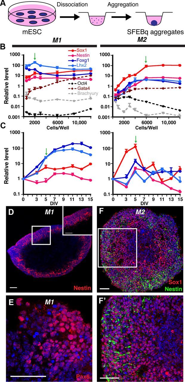
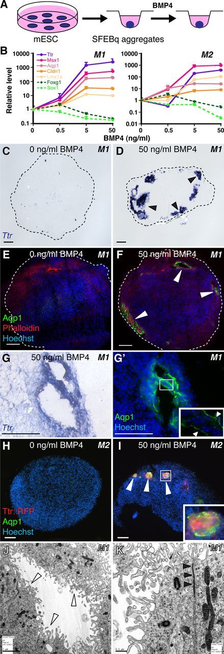
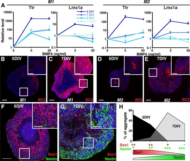
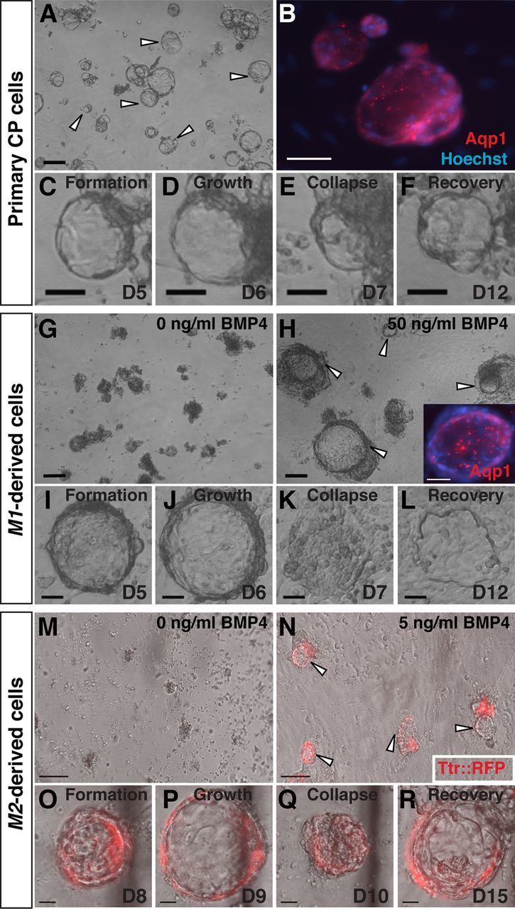
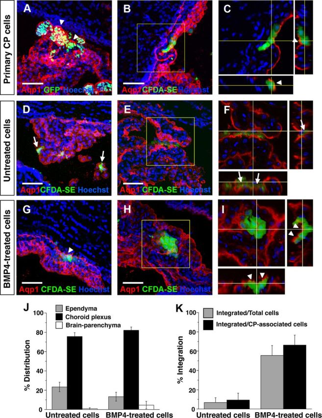
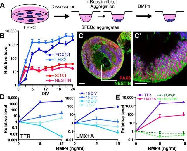
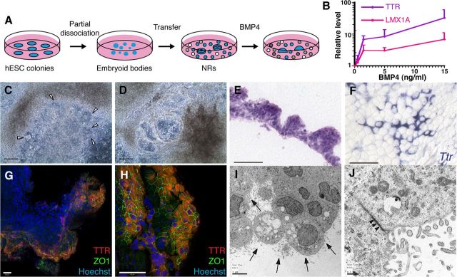
Choroid plexus epithelial cells (CPECs) have essential developmental and homeostatic roles related to the CSF and blood-CSF barrier they produce. Accordingly, CPEC dysfunction has been implicated in many neurological disorders, such as Alzheimer's disease, and transplant studies have provided proof-of-concept for CPEC-based therapies. However, such therapies have been hindered by the inability to expand or generate CPECs in culture. During development, CPECs differentiate from preneurogenic neuroepithelial cells and require bone morphogenetic protein (BMP) signaling, but whether BMPs suffice for CPEC induction is unknown. Here we provide evidence for BMP4 sufficiency to induce CPEC fate from neural progenitors derived from mouse embryonic stem cells (ESCs). CPEC specification by BMP4 was restricted to an early time period after neural induction in culture, with peak CPEC competency correlating to neuroepithelial cells rather than radial glia. In addition to molecular, cellular, and ultrastructural criteria, derived CPECs (dCPECs) had functions that were indistinguishable from primary CPECs, including self-assembly into secretory vesicles and integration into endogenous choroid plexus epithelium following intraventricular injection. We then used BMP4 to generate dCPECs from human ESC-derived neuroepithelial cells. These findings demonstrate BMP4 sufficiency to instruct CPEC fate, expand the repertoire of stem cell-derived neural derivatives in culture, and herald dCPEC-based therapeutic applications aimed at the unique interface between blood, CSF, and brain governed by CPECs.


Figures

Figure 1.
Figure 1.
Efficient neural induction from mouse ESCs. A, Schematic of neural differentiation using the SFEBq method (Eiraku et al., 2008). B, Cells/well optimization for neural induction (qRT-PCR, normalized to starting mouse ESCs); 2000 and 6000 cells/well (green arrows) yield optimal gene expression profiles for the M1 and M2 mouse ESC lines, respectively. C, Temporal analysis of 2000-cell (M1) or 6000-cell (M2) aggregates (qRT-PCR, normalized to mouse ESCs). Plateaus for the four NEC markers are seen in M1 aggregates, whereas they peak, then decrease in M2 aggregates. Green arrows designate the time point at which most CPEC differentiations were initiated. D–F', Nestin, Pax6, and Sox1 ICC of 7-day M1 aggregates and 5-day M2 aggregates reveal efficient neural induction. Scale bars, 50 μm.
Figure 2.
Figure 2.
BMP4 sufficiency and concentration dependence for CPEC differentiation from mouse ESC-derived neural progenitors. A, Schematic of the differentiation method. Neural aggregates are treated for 5–7 DIV with BMP4. B, qRT-PCR of 5-day M1 and M2 aggregates treated with BMP4 (normalized to no BMP4 controls). CPEC markers are strongly induced, while neural progenitor markers Sox1 and Foxg1 are downregulated in a BMP4 concentration-dependent manner. C–G', ISH and ICC of sectioned M1 aggregates. Ttr and Aqp1 expression are detected in peripheral regions of BMP4-treated aggregates (arrowheads; n = 11/13 and 7/7 aggregates, respectively), but not in aggregates without BMP4 (n = 0/6 and 0/3 aggregates). Expression often occurs in vesicles that coexpress Ttr and Aqp1 (adjacent sections in G and G') and localize Aqp1 to the apical (luminal) surface (G', arrowheads) as seen in CPECs in vivo. H–I, ICC of sectioned M2 aggregates. Ttr::RFP (native fluorescence) and Aqp1 expression (arrowheads) are BMP4-dependent (n = 6/8 and 0/6 aggregates with and without BMP4, respectively), and colocalize in cell clusters toward the periphery of aggregates (I). J, K, Electron microscopy of M1-derived BMP4-treated aggregates. Vesicle-lining cells have extensive microvilli (white arrowheads), rare cilia, and juxtalumenal tight junctions (black arrowheads) characteristic of CPECs in vivo. Scale bars: C–I, 50 μm; J, 2 μm; K, 0.5 μm.
Figure 3.
Figure 3.
Temporally restricted CPEC competency correlates with NEC rather than RG fate. A, SFEBq aggregates treated with BMP4 for 7 DIV (M1) or 5 DIV (M2) (qRT-PCR normalized to no BMP4 controls). Five-day M1 and M2 aggregates show much stronger CPEC gene induction than 7-, 9-, or 11-day aggregates. B–E, RC2 ICC of sectioned M1 and M2 aggregates. Five-day aggregrates exhibit low RC2 expression levels characteristic of NECs, while strong RC2 expression typical of RG is present throughout 7-day aggregates. F–G, Sox1 and Nestin ICC of sectioned M2 aggregates. Five-day aggregates exhibit the strong Sox1 and weak Nestin patterns characteristic of NECs, whereas 7-day aggregates display the opposite RG-like pattern. H, Scoring of Sox1/Nestin staining patterns in M2 aggregates. Scale bars, 100 μm.
Figure 4.
Figure 4.
Vesicle self-assembly and secretion by primary and derived CPECs (ICC, phase contrast, and fluorescence microscopy; the same vesicle is shown in C–F, I–L, or O–R). A–F, Primary CPECs. CPECs from dissociated primary CP self-assemble into vesicles on Matrigel (A, arrowheads) that are Aqp1-positive (B). Due to their secretory activity, these vesicles enlarge (C, D), collapse upon treatment with the secretion inhibitors acetazolamide and ouabain (E), then regrow upon inhibitor withdrawal (F). G–L, M1-derived cells. M1 aggregates treated with 50 ng/ml BMP4 (n = 6/6 cultures), but not controls (n = 0/6 cultures), formed Aqp1-positive vesicles after dissociation and plating on Matrigel (G, H). Like primary CPEC vesicles, M1-derived vesicles expand (I, J), collapse upon secretion inhibitor treatment (K), and recover after inhibitor withdrawal (L). M–R, M2-derived cells. M2 aggregates treated with 5 ng/ml BMP4 (n = 3/3 cultures), but not controls (n = 0/3 cultures), form TTR::RFP-expressing vesicles (N, arrowheads) that expand, collapse upon secretion inhibitor treatment, and regrow after inhibitor withdrawal (O–R) in a fashion indistinguishable from that of primary CPECs. Scale bars: A–N, 100 μm; O–R, 50 μm.
Figure 5.
Figure 5.
Engraftment of endogenous CP by primary and derived CPECs following intraventricular injection. A–C, Primary CP cell injections, single or collapsed z-stack images of ipsilateral telencephalic choroid plexus 2 days after injection. Injected H2B-GFP (A) or CFDA-SE-labeled CP cells (B) preferentially associate with host CP, which expresses Aqp1 (red). Confocal projections reveal labeled cells on the nuclear side of the continuous Aqp1-positive apical border (C, arrowheads), indicating integration of injected cells into host CP with appropriate apicobasal polarity. D–F, Mouse ESC-derived (M1) aggregates, no BMP4 treatment, 2 d after injection. Dye-labeled cells are associated with ipsilateral host CP, but remain on the ventricular side of the Aqp1-expressing apical border (F, arrows), indicating a failure to engraft host CP. G–I, Mouse ESC-derived (M1) aggregates, with BMP4 treatment and Ara-C enrichment, 1–2 d after injection. Like the primary cells, most of the dye-labeled cells associate with host CP and are appropriately polarized on the nuclear side of the endogenous Aqp1 border (G, I, arrowheads), indicating host CP engraftment. J, K, Quantification of distributions and integration of injected BMP4-treatedand-untreatedcells. Cells from both groups are found mostly in association with CP (J), but the BMP4-treated cells integrate into host CP with much greater efficiency (K), consistent with their dCPEC identity (see Results for numbers; error bars represent SEM.). Scale bars, 50 μm.
Figure 6.
Figure 6.
BMP4 sufficiency to induce CPEC markers from human ESC-derived neural progenitors. A, Schematic of the differentiation method. The human ESCs form multiple and less uniform SFEBq aggregates than mouse ESCs. B, Temporal analysis of neural induction in human SFEBq aggregates (9000 human ESCs/well, qRT-PCR normalized to human ESCs). Dramatic increases in forebrain (LHX2, FOXG1) and generic neural progenitor gene expression (SOX1, NESTIN) occur by 6 DIV and are stably maintained thereafter. C, ICC of 9000 cells/well human SFEBq aggregates at 21 DIV for PAX6 and NESTIN demonstrate efficient neural induction. D, Temporal-dependent and BMP4 concentration-dependent induction of CPEC genes in human SFEBq aggregates (9000 cells/well, qRT-PCR normalized to no BMP4 controls). Eighteen-day aggregates show marked BMP4-mediated upregulation of TTR and LMX1A, while little to no induction is seen in 9-, 12-, or 15-day aggregates. TTR and LMX1A baselines also increased with aggregation time (∼64× for Ttr; 4× for Lmx1a), but these baseline changes were much less than the BMP4-mediated changes in 18-day aggregates (>1000× for Ttr; >100× for Lmx1a). E, CPEC and neural progenitor markers, 18-day aggregates (qRT-PCR normalized to no BMP4 controls). BMP4 induces CPEC markers TTR, LMX1A, and OTX2 at the expense of neural progenitor markers FOXG1 and NESTIN. Scale bars, 50 μm.
Figure 7.
Figure 7.
CPEC differentiation from human ESC-derived NRs. A, Schematic of the NR differentiation method (Zhang et al., 2001). After 16 DIV, NR-containing cultures are treated for 20 DIV with BMP4. B, BMP4 effects on CPEC markers in H9-derived NR cultures (qRT-PCR normalized to no BMP4 controls). Similar induction curves are seen for CPEC genes LMX1A and TTR. C, D, Phase contrast images of H1-derived NR cultures (same field in C and D). In the presence of BMP4, NRs (arrowheads) transform into phase-bright vesicular structures, which eventually form flat epithelial sheets. E, H&E-stained cross section of an H1-derived BMP4-treated colony. The peripheral NR-containing region consists of cells with apical cytoplasm that form a papillary-like tissue reminiscent of endogenous CP. F, ISH of an H9-derived BMP4-treated colony. Strong Ttr expression is evident in epithelial cells toward the colony periphery. G, H, ICC of an H1-derived BMP4-treated colony. Extensive colocalization of TTR and ZO1 occurs at the colony periphery (G and H from two different fields). I, J, Electron microscopy of an H1-derived BMP4-treated colony. Abundant epithelial cells demonstrate extensive lumenal microvilli (arrows), rare cilia, and juxtalumenal tight junctions (arrowheads). Scale bars: C, D, 100 μm; E–H, 50 μm; I, 5 μm; J, 0.5 μm.

References

    1. Buxbaum JN, Ye Z, Reixach N, Friske L, Levy C, Das P, Golde T, Masliah E, Roberts AR, Bartfai T. Transthyretin protects Alzheimer's mice from the behavioral and biochemical effects of Abeta toxicity. Proc Natl Acad Sci U S A. 2008;105:2681–2686. - PMC - PubMed
    1. Caviness VS, Jr, Takahashi T, Nowakowski RS. Numbers, time and neocortical neuronogenesis: a general developmental and evolutionary model. Trends Neurosci. 1995;18:379–383. - PubMed
    1. Chauhan AN, Lewis PD. A quantitative study of cell proliferation in ependyma and choroid plexus in the postnatal rat brain. Neuropath Appl Neurobiol. 1979;5:303–309. - PubMed
    1. Cheng X, Hsu CM, Currle DS, Hu JS, Barkovich AJ, Monuki ES. Central roles of the roof plate in telencephalic development and holoprosencephaly. J Neurosci. 2006;26:7640–7649. - PMC - PubMed
    1. Chodobski A, Szmydynger-Chodobska J. Choroid plexus: target for polypeptides and site of their synthesis. Microsc Res Tech. 2001;52:65–82. - PubMed

Publication types

Substances