. 2012 Nov 7;32(45):15946-51.
doi: 10.1523/JNEUROSCI.0831-12.2012.

Differential feedback modulation of center and surround mechanisms in parvocellular cells in the visual thalamus

Affiliations

Differential feedback modulation of center and surround mechanisms in parvocellular cells in the visual thalamus

Helen E Jones et al. J Neurosci. .

Abstract

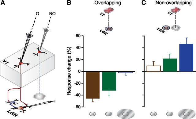
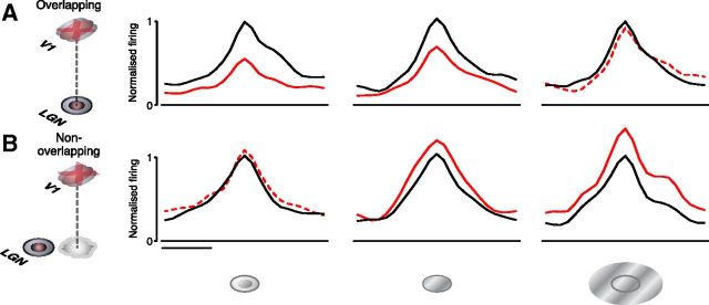
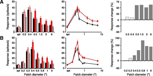
Many cells in both the central visual system and other sensory systems exhibit a center surround organization in their receptive field, where the response to a centrally placed stimulus is modified when a surrounding area is also stimulated. This can follow from laterally directed connections in the local circuit at the level of the cell in question but could also involve more complex interactions. In the lateral geniculate nucleus (LGN), the cells relaying the retinal input display a concentric, center surround organization that in part follows from the similar organization characterizing the retinal cells providing their input. However, local thalamic inhibitory interneurons also play a role, and as we examine here, feedback from the visual cortex too. Here, we show in the primate (macaque) that spatially organized cortical feedback provides a clear and differential influence serving to enhance both responses to stimulation within the center of the receptive field and the ability of the nonclassical surround mechanism to attenuate this. In short, both center and surround mechanisms are influenced by the feedback. This dynamically sharpens the spatial focus of the receptive field and introduces nonlinearities from the cortical mechanism into the LGN.


Figures

Figure 1.
Figure 1.
Differential effects of focal inactivation of corticothalamic feedback by iontophoretic application of GABA on overlapping and nonoverlapping LGN cell locations. A, Schematic summary of experimental method. Drug inactivation electrode is shown inserted in either an overlapping (O) or nonoverlapping (NO) retinotopic location with respect to that of the LGN. B, C, Population summary histograms plot the mean percentage change in LGN cell responses for overlapping (B, n = 14) and nonoverlapping (C, n = 12) recording sites for smaller than optimal (left), optimal (middle), and larger than optimal (right) stimuli, across our sample. Error bars indicate SEM. Significant response changes (see Results) denoted by filled bars, nonsignificant changes by open bars.
Figure 2.
Figure 2.
Population peristimulus time histograms (PSTHs) documenting differential effects of focal inactivation of corticothalamic feedback by iontophoretic application of GABA on overlapping (A) and nonoverlapping (B) LGN cell locations. A, B, Wrapped PSTHs of the population averaged responses to smaller than optimal (left), optimal (middle), and larger than optimal (right) diameter stimuli during control (black) and V1 inactivation (red) conditions. Significant response changes (see Results) during V1 inactivation denoted by solid red lines, nonsignificant changes denoted by dotted lines. To compare PSTHs across neurons, individual histograms were shifted by the neuron's latency so that the bin containing the maximal responses was aligned. For each stimulus condition, responses for each neuron were normalized to the control peak response. Histograms were averaged for each group and smoothed. Scale bar, 100 ms.
Figure 3.
Figure 3.
Inactivation of retinotopically overlying V1 feedback reduces responses of LGN cells to smaller than optimal and optimal diameter stimuli. A–C, Three representative examples of LGN cell responses for cells whose receptive fields retinotopically overlapped the cortical inactivation site. Records show responses, plotted as both bar histograms (left) and tuning curves (middle), to gratings of varying diameter before (black), during (red), and after (gray) focal GABA inactivation in V1. Responses during V1 inactivation that differed significantly from control levels (see Materials and Methods) are marked by solid red fill (left) and filled symbols (middle). Error bars indicate SEM. Histograms to the right plot the percentage change in response during V1 inactivation, normalized with respect to control values. Significant changes, solid fill; nonsignificant changes, crosshatched. Spt, Spontaneous activity level.
Figure 4.
Figure 4.
Inactivation of retinotopically displaced V1 feedback enhances responses of LGN cells to optimal and larger than optimal diameter stimuli. A, B, Two representative examples of LGN cell responses for cells with receptive fields that did not overlap the cortical inactivation site. Records show responses, plotted as both bar histograms (left) and tuning curves (middle), to gratings of varying diameter before (black), during (red), and after (gray) focal GABA inactivation in V1. Responses during V1 inactivation that differed significantly from control levels (see Materials and Methods) are marked by solid red fill (left) and filled symbols (middle). Error bars indicate SEM. Histograms to the right plot the percentage change in response during V1 inactivation, normalized with respect to control values. Significant changes, solid fill; nonsignificant changes, crosshatched.

References

    1. Alitto HJ, Usrey WM. Origin and dynamics of extraclassical suppression in the lateral geniculate nucleus of the macaque monkey. Neuron. 2008;57:135–146. - PMC - PubMed
    1. Andolina IM, Jones HE, Wang W, Sillito AM. Corticothalamic feedback enhances stimulus response precision in the visual system. Proc Natl Acad Sci U S A. 2007;104:1685–1690. - PMC - PubMed
    1. Briggs F, Usrey WM. Parallel processing in the corticogeniculate pathway of the macaque monkey. Neuron. 2009;62:135–146. - PMC - PubMed
    1. Briggs F, Usrey WM. Corticogeniculate feedback and visual processing in the primate. J Physiol. 2011;589:33–40. - PMC - PubMed
    1. Cudeiro J, Sillito AM. Spatial frequency tuning of orientation-discontinuity-sensitive corticofugal feedback to the cat lateral geniculate nucleus. J Physiol. 1996;490:481–492. - PMC - PubMed

Publication types

LinkOut - more resources