

Gene expression patterns in the histopathological classification of epithelial ovarian cancer
- PMID: 23136613
- PMCID: PMC3490392
- DOI: 10.3892/etm_00000030
Gene expression patterns in the histopathological classification of epithelial ovarian cancer
Abstract
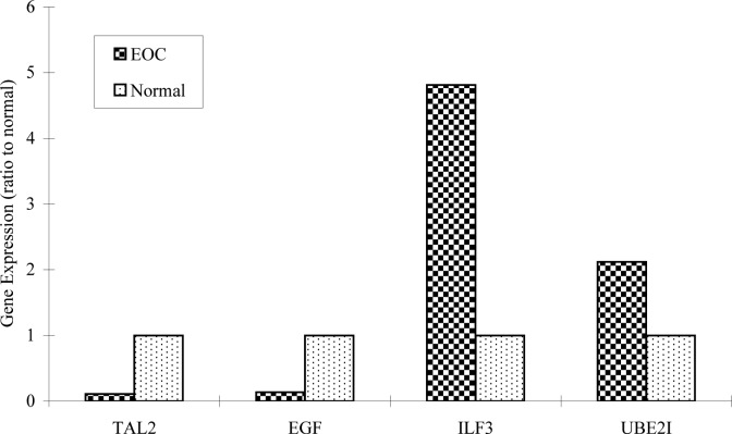
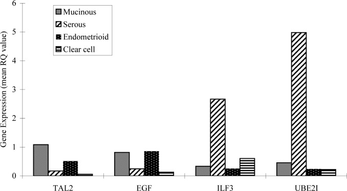
The purpose of this study was to screen cancer-related genes and to identify histopathological gene expression patterns as potential biomarkers in human epithelial ovarian cancer (EOC). Fifty genes were screened by reverse-transcription polymerase chain reaction assay with cDNA from 83 EOC tissues and 48 normal ovarian specimens of ovarian cancer patients and evaluated by gel electrophoresis analysis. Twenty expressed genes were assessed by real-time relative-quantity (RQ)-PCR in 30 EOC specimens for gene signature study. Four genes, TAL2, EGF, ILF3 and UBE2I, were investigated for gene expression patterns in histopathological classification of EOC. RQ-value (Ct, ΔCt, ΔΔCt, RQ and gene expression plots) was generated by ABI 7500 Fast System SDS Software (version 1.4). SPSS 15.0 software was used for statistical analysis. Using real-time RQ-PCR, we found that TAL2, EGF, ILF3 and UBE2I demonstrated distinct expression patterns in histological types of epithelial ovarian cancer. The expression of ILF3 and UBE2I in tumors was significantly higher than in normal tissue, with extremely high expression in serous carcinomas compared to mucinous, endometrium and clear cell carcinomas. In addition, ILF3 and UBE2I were overexpressed in advanced stage and advanced grade ovarian cancer, compared to early stage or well-differentiated ovarian cancer. This is the first report of TAL2 and ILF3 expression in the normal human ovary and epithelial ovarian cancer. Our results indicate that overexpression of ILF3 and UBE2I in advanced stage and advanced grade suggest that these two genes may play an important role in tumorigenesis/tumor progression and pathological differentiation of the disease. Notably, ILF3 plays a role in DNA binding activity and transcriptional and post-transcriptional regulation; UBE2I is required in ubiquitination and sumoylation and is involved in DNA repair and apoptosis of cells. Further investigations to reveal the molecular mechanisms related to the activation of ILF3 and UBE2I in the development of EOC are warranted.
Figures
References
-
- Baranova A, Gowder S, Naouar S, et al. Expression profile of ovarian tumors distinct signature of Sertoli-Leydig cell tumor. Int J Gynecol Cancer. 2006;16:1963–1972. - PubMed
-
- Yu JJ. Unlocking the molecular mechanisms of DNA repair and platinum drug resistance in cancer chemotherapy. Curr Drug Ther. 2009;4:19–28.
-
- Bell DA. Origins and molecular pathology of ovarian cancer. Mod Pathol. 2005;18:19–32. - PubMed
-
- Cecco LD, Marchionni L, Gariboldi M, et al. Gene expression profiling of advanced ovarian cancer characterization of a molecular signature involving fibroblast growth factor 2. Oncogene. 2004;23:8171–8183. - PubMed
Grants and funding
LinkOut - more resources
Full Text Sources
Miscellaneous