

Ultrasound Characteristics of Patients with Urinary Stress Incontinence with or without Genital Prolapse
- PMID: 23136629
- PMCID: PMC3490089
- DOI: 10.4111/kju.2012.53.10.691
Ultrasound Characteristics of Patients with Urinary Stress Incontinence with or without Genital Prolapse
Abstract
Purpose: The study purpose was to evaluate the clinical and ultrasound characteristics of women with urinary stress incontinence (USI) with or without genital prolapse (GP).
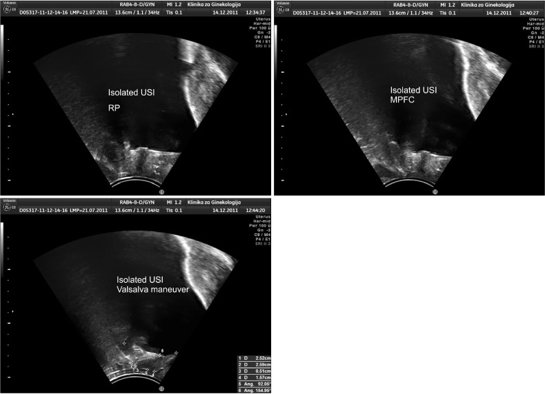
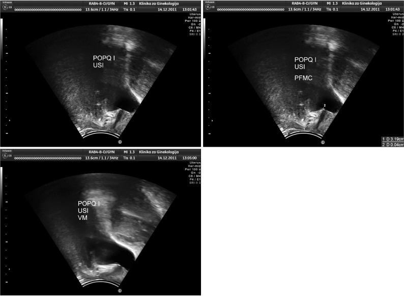
Materials and methods: A total of 268 patients who underwent ultrasound perineal evaluation were divided into two groups: isolated USI (n=132) and USIGP (n=136) with USI/GP stage I/II. The latter group was additionally divided into two subgroups: USIGP(A) (n=78) with USI/GP stage I and USIGP(B) (n=58) with USI/GP stage II.
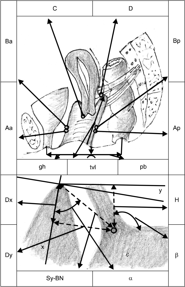
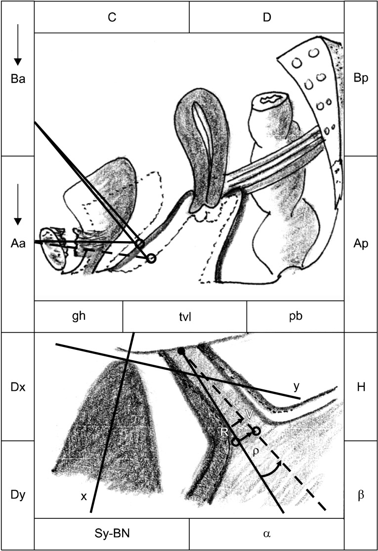
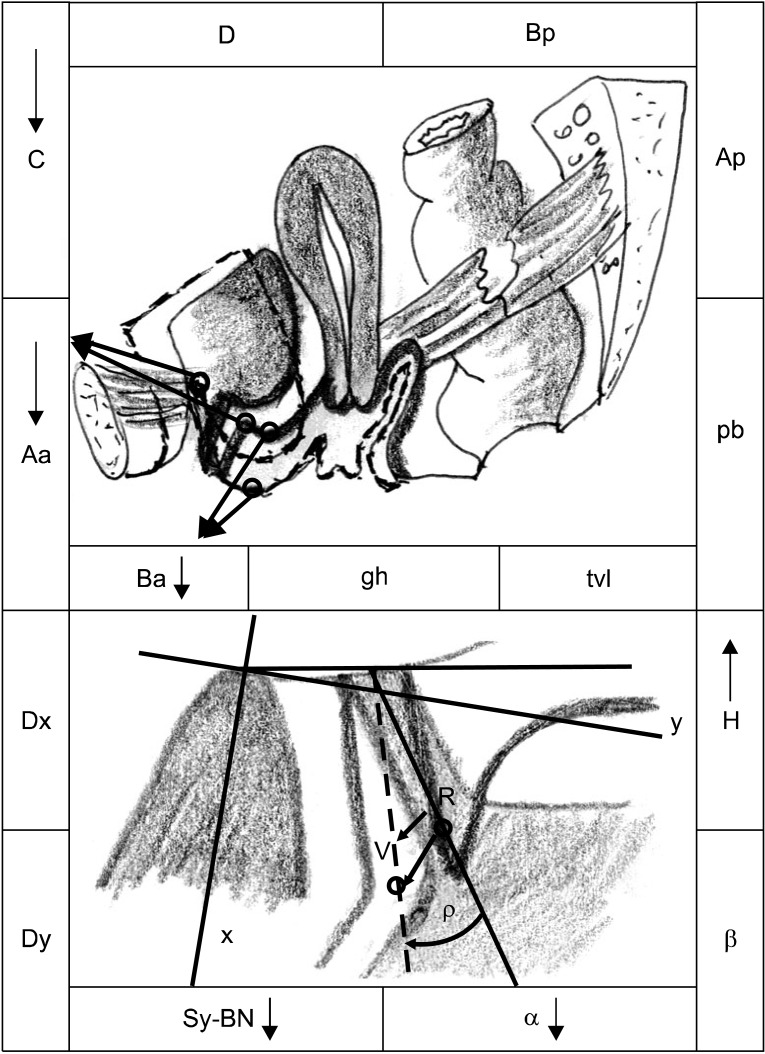
Results: Point Aa (pelvic organ prolapse quantification system), which is the projection of the bladder neck (BN) on the anterior vaginal wall, was situated higher in the rest position (RP) but moved lower during a Valsalva maneuver (VM) in the USI group than in the USIGP group (p<0.05). The ultrasound parameters α-angle and the distance Sy-BN (symphisis-bladder neck) decreased, whereas distance H increased, in the USIGP group during VM. The ultrasound parameters that gave the best insight into the range of BN movements were as follows: distance R→V and angle of rotation (ρ), which were significantly higher in the USI group than in the USIGP group during VM.
Conclusions: According to the clinical and ultrasound findings, we can conclude that the BN is situated higher during the RP but moved lower during a VM in patients with isolated USI compared with those with concomitant USI/GP, which could be explained by the cystocele-immobilizing effect on the BN during the VM in the latter group but also by the deteriorated pubo-urethral ligaments in the former group.
Keywords: POP-Q; Pelvic organ prolapsed; Ultrasonography; Urinary incontinence stress.
Conflict of interest statement
The authors have nothing to disclose.
Figures
Similar articles
-
Outcome Assessment of the Marshall Coughing Test during Cervix Reposition Maneuver in Women with Urinary Stress Incontinence with/without Genital Prolapse.ISRN Urol. 2012;2012:109858. doi: 10.5402/2012/109858. Epub 2012 Feb 20. ISRN Urol. 2012. PMID: 22523712 Free PMC article.
-
[Bologna procedure in stress urinary incontinence with stage III cystocele (with or without vaginal hysterectomy)].Prog Urol. 1999 Feb;9(1):81-7. Prog Urol. 1999. PMID: 10212956 French.
-
A new modification of the POPQ system--its effectiveness in the diagnosis of supravaginal elongation of the uterine cervix in cases with genital prolapse.Bratisl Lek Listy. 2008;109(7):307-12. Bratisl Lek Listy. 2008. PMID: 18792485
-
What is normal bladder neck anatomy?Int Urogynecol J. 2016 Jun;27(6):945-50. doi: 10.1007/s00192-015-2916-1. Epub 2015 Dec 23. Int Urogynecol J. 2016. PMID: 26700104
-
Utility of 2D-ultrasound in pelvic floor muscle contraction and bladder neck mobility assessment in women with urinary incontinence.J Gynecol Obstet Hum Reprod. 2020 Jan;49(1):101629. doi: 10.1016/j.jogoh.2019.101629. Epub 2019 Sep 6. J Gynecol Obstet Hum Reprod. 2020. PMID: 31499282 Review.
Cited by
-
Transperineal ultrasonography in stress urinary incontinence: The significance of urethral rotation angles.Arab J Urol. 2016 Mar;14(1):66-71. doi: 10.1016/j.aju.2015.11.003. Epub 2015 Dec 29. Arab J Urol. 2016. PMID: 26966596 Free PMC article.
-
The role of transperineal ultrasound in the evaluation of stress urinary incontinence cases.Int Braz J Urol. 2022 Jan-Feb;48(1):70-77. doi: 10.1590/S1677-5538.IBJU.2020.1100. Int Braz J Urol. 2022. PMID: 34528775 Free PMC article.
References
-
- Schaer G, Koelbl H, Voigt R, Merz E, Anthuber C, Niemeyer R, et al. Recommendations of the German Association of Urogynecology on functional sonography of the lower female urinary tract. Int Urogynecol J Pelvic Floor Dysfunct. 1996;7:105–108. - PubMed
-
- Bump RC, Mattiasson A, Bo K, Brubaker LP, DeLancey JO, Klarskov P, et al. The standardization of terminology of female pelvic organ prolapse and pelvic floor dysfunction. Am J Obstet Gynecol. 1996;175:10–17. - PubMed
-
- Moher D, Schulz KF, Altman DG. The CONSORT statement: revised recommendations for improving the quality of reports of parallel-group randomised trials. Lancet. 2001;357:1191–1194. - PubMed
-
- Abrams P, Blaivas JG, Stanton SL, Andersen JT The International Continence Society Committee on Standardisation of Terminology. The standardisation of terminology of lower urinary tract function. Scand J Urol Nephrol Suppl. 1988;114:5–19. - PubMed
-
- Chen HY, Huang YL, Hung YC, Chen WC. Evaluation of stress urinary incontinence by computer-aided vector-based perineal ultrasound. Acta Obstet Gynecol Scand. 2006;85:1259–1264. - PubMed
LinkOut - more resources
Full Text Sources
