. 2013 Jan;13(1):56-66.
doi: 10.1111/j.1600-6143.2012.04316.x. Epub 2012 Nov 8.

Targeting TIM-1 on CD4 T cells depresses macrophage activation and overcomes ischemia-reperfusion injury in mouse orthotopic liver transplantation

Affiliations

Targeting TIM-1 on CD4 T cells depresses macrophage activation and overcomes ischemia-reperfusion injury in mouse orthotopic liver transplantation

Y Zhang et al. Am J Transplant. 2013 Jan.

Abstract

Hepatic injury due to cold storage followed by reperfusion remains a major cause of morbidity and mortality after orthotopic liver transplantation (OLT). CD4 T cell TIM-1 signaling costimulates a variety of immune responses in allograft recipients. This study analyzes mechanisms by which TIM-1 affects liver ischemia-reperfusion injury (IRI) in a murine model of prolonged cold storage followed by OLT. Livers from C57BL/6 mice, preserved at 4°C in the UW solution for 20 h, were transplanted to syngeneic recipients. There was an early (1 h) increased accumulation of TIM-1+ activated CD4 T cells in the ischemic OLTs. Disruption of TIM-1 signaling with a blocking mAb (RMT1-10) ameliorated liver damage, evidenced by reduced sALT levels and well-preserved architecture. Unlike in controls, TIM-1 blockade diminished OLT expression of Tbet/IFN-γ, but amplified IL-4/IL-10/IL-22; abolished neutrophil and macrophage infiltration/activation and inhibited NF-κB while enhancing Bcl-2/Bcl-xl. Although adoptive transfer of CD4 T cells triggered liver damage in otherwise IR-resistant RAG(-/-) mice, adjunctive TIM-1 blockade reduced Tbet transcription and abolished macrophage activation, restoring homeostasis in IR-stressed livers. Further, transfer of TIM-1(Hi) CD4+, but not TIM-1(Lo) CD4+ T cells, recreated liver IRI in RAG(-/-) mice. Thus, TIM-1 expressing CD4 T cells are required in the mechanism of innate immune-mediated hepatic IRI in OLTs.


Conflict of interest statement

Disclosure: The authors of this manuscript have no conflicts of interest to disclose as described by the American Journal of Transplantation.

Figures

Figure 1
Figure 1
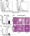
B6 livers subjected to ex-vivo cold storage (20 h) were transplanted into syngeneic recipients. By 1 h, CD4 T cells, which infiltrated OLTs were (a) activated, evidenced by CD69 staining; and (b) expressed TIM-1 (representative of 4 experiments). By 6 h after OLT, the hepatocellular function was analyzed by (c) sALT levels, (d) liver histology (representative H&E staining; magnification x100 and x400), and (e) Suzuki’s histological score (*p<0.05, **p<0.01, n=8–10/group).
Figure 2
Figure 2
Quantitative RT-PCR-assisted detection of cytokine/transcription factors in OLTs (6 h post-transplant after 20 h of cold ischemia): (a) IFN-γ, Tbet; (b) IL-4, IL-10; (c) FoxP3, and (d) IL-17, IL-22, RORγt). Data normalized to HPRT gene expression (*p<0.05, **p<0.01, n=4–6/group).
Figure 3
Figure 3
Neutrophils and macrophages in OLTs following administration of anti-TIM-1 mAb or control Ig (6 h post-transplant after 20 h of cold ischemia). (a) MPO levels (n=4–6/group); immunohistochemical staining of (b) Ly-6G+ neutrophils; and (c) CD68+ macrophages; (d) immunofluorescence double staining of TIM-4-expressing macrophages (TIM-4+CD68+ cells -head arrow). Green – macrophage; red – TIM-4; blue – DAPI nuclear stain. Results scored semi-quantitatively by averaging number of positively-stained cells/field (400x magnification). Representative of 4–6 mice/group (b, c) and 2 mice/group (d).
Figure 4
Figure 4
Measurement of TIM-4 expression and associated cytokine/chemokine programs in OLTs by quantitative RT-PCR (6 h post-transplant after 20 h of cold ischemia): (a) TIM-4; (b) CXCL-1, CCL-2, CXCL-10; and (c) TNF-α, IFN-β, IL-1β, and IL-6). Data normalized to HPRT gene expression (*p<0.05, **p<0.01, n=4–6/group).
Figure 5
Figure 5
Necrosis/apoptosis events in OLTs (6 h post-transplant after 20 h of cold ischemia). (a, b) TUNEL-assisted detection of hepatic necrosis/apoptosis (dark arrows; magnification x100 and x400); (c) caspase-3 activity; (d) Western blot-assisted detection of Bcl-2/Bcl-xl; p-IκBα, and β-actin (**p<0.01, n=4–6/group).
Figure 6
Figure 6
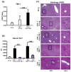
TIM-1+ CD4 T cells are required to trigger liver IRI in RAG−/− mice. Intrahepatic TIM-1 levels were elevated in livers subjected to 90 min of warm ischemia in RAG−/− mice repopulated with syngeneic CD4 T cells (a). Adjunctive infusion of anti-TIM-1 mAb (day -2) restored IR-liver resistance in RAG−/− mice repopulated with CD4 T cells, as evidenced by (b) decreased sALT levels; and (c) preserved liver histology (representative H&E; magnification x100 and x400) (n=4–6/group). Adoptive transfer of ConA-activated TIM-1Hi but not TIM-1Lo CD4+ T cells (−1 h) mediated IR-liver damage in RAG−/− mice, as shown by (b) sALT levels; and (c) liver histology. (**p<0.01), n=2/group).
Figure 7
Figure 7
TIM-1+ CD4 T cells mediate IR-triggered (a) Tbet transcription; and promote (b) TNF-α, IL-6, and CXCL-10 expression in RAG−/− recipients adoptively transferred with either syngeneic CD4+ T cells or ConA-activated TIM-1Hi cells. (**p<0.01, *p<0.05, n=4–6/group).

Similar articles

Cited by

References

    1. Zhai Y, Busuttil RW, Kupiec-Weglinski JW. Liver ischemia and reperfusion injury: new insights into mechanisms of innate-adaptive immune-mediated tissue inflammation. Am J Transplant. 2011;11:1563–1569. - PMC - PubMed
    1. Zwacka RM, Zhang Y, Halldorson J, et al. CD4(+) T-lymphocytes mediate ischemia/reperfusion-induced inflammatory responses in mouse liver. J Clin Invest. 1997;100:279–289. - PMC - PubMed
    1. Caldwell CC, Tschoep J, Lentsch AB. Lymphocyte function during hepatic ischemia/reperfusion injury. J Leukoc Biol. 2007;82:457–464. - PubMed
    1. Shen X, Wang Y, Gao F, et al. CD4 T cells promote tissue inflammation via CD40 signaling without de novo activation in a murine model of liver ischemia/reperfusion injury. Hepatology. 2009;50:1537–1546. - PMC - PubMed
    1. Shen XD, Ke B, Zhai Y, et al. Stat4 and Stat6 signaling in hepatic ischemia/reperfusion injury in mice: HO-1 dependence of Stat4 disruption-mediated cytoprotection. Hepatology. 2003;37:296–303. - PubMed

Publication types

Substances