Review
. 2013 Oct;1834(10):2176-86.
doi: 10.1016/j.bbapap.2012.10.015. Epub 2012 Nov 5.

Platelet-derived growth factors and their receptors: structural and functional perspectives

Affiliations
Review

Platelet-derived growth factors and their receptors: structural and functional perspectives

Po-Han Chen et al. Biochim Biophys Acta. 2013 Oct.

Abstract

The four types of platelet-derived growth factors (PDGFs) and the two types of PDGF receptors (PDGFRs, which belong to class III receptor tyrosine kinases) have important functions in the development of connective tissue cells. Recent structural studies have revealed novel mechanisms of PDGFs in propeptide loading and receptor recognition/activation. The detailed structural understanding of PDGF-PDGFR signaling has provided a template that can aid therapeutic intervention to counteract the aberrant signaling of this normally silent pathway, especially in proliferative diseases such as cancer. This review summarizes the advances in the PDGF system with a focus on relating the structural and functional understandings, and discusses the basic aspects of PDGFs and PDGFRs, the mechanisms of activation, and the insights into the therapeutic antagonism of PDGFRs. This article is part of a Special Issue entitled: Emerging recognition and activation mechanisms of receptor tyrosine kinases.

Keywords: Growth factor; Propeptide recognition; Receptor activation; Receptor tyrosine kinase; Signal transduction.


Figures

Fig. 1
Fig. 1
The four types PDGFs and their domain compositions. The starting numbers of specific domains/segments in the coding sequences are marked above the boundaries. Shown are all numbers for human PDGFs. The proprotein convertase-recognition sequences are indicated and positions of cleavage are marked with a black triangle. Abbreviations used: SP: signal peptide; PRO: the pro-sequence preceding the growth factor domain; Cys-Knot, the cysteine-knot growth factor domain that is responsible for receptor recognition; CUB, the Complement subcomponents C1r/C1s, Urchin EGF-like protein, and Bone marrow protein 1-like domain, which can be considered as part of the pro-sequence in PDGF-C and PDGF-D, but PDGF-C and PDGF-D also have sequences homologous to the PDGF-A and PDGF-B pro-sequences.
Fig. 2
Fig. 2
The cystine-knot growth-factor domain of PDGFs. Shown is the ribbon model of a PDGF-A dimer, as selected from PDB ID 3MJK. The two protomers in the dimer are colored in green and cyan respectively. The disulfide bridges are depicted as orange balls and sticks. Note that three intermolecular disulfide bridges are concentrated at one end of each protomer, and inter-molecular disulfide bridges are located at the dimerization interface. The N-terminal segment of each protomer, depicted as a coil, runs vertical to the β-strands of the other protomer.
Fig. 3
Fig. 3
The association between propeptide and mature growth factor domain in PDGF-A. (A) The pro-peptide of PDGF-A is cleaved from mature PDGF-A, but remains tightly associated in gel filtration. The fractions from the elution peak are shown. (B) Two residues in mature PDGF-A, W120 and N134, play organizing roles in the association of the propeptide with the mature domain. (C) Ribbon model of the propeptide-loaded PDGF-A dimer, with the mature domains colored in green and cyan, whereas the propeptides colored in pink and magenta. (D) The hydrophobic interaction pattern between PDGF-A propeptide and mature domain. The backbone of the propeptide is shown as ribbons, and the backbone of the mature domains is shown as coils. The interacting sidechains are shown as sticks, colored according to their backbones. (E) Sequence comparison between all four types of PDGFs shows that the two helices used in the interactions with mature growth factors domains are a common feature, and the hydrophobic residues for interactions are preserved.
Fig. 4
Fig. 4
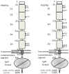
The two types of PDGFRs and their domain compositions. The starting numbers of the specific domains/segments in the coding sequences are marked left to the boundaries. Shown are all numbers for human PDGFRs. The positions of N-linked glycosylations are also marked. The lipid bilayer is represented by two straight lines. Note that D1 and D2 are an integral module, and the intracellular kinase domain is a split domains with an insert between N-terminal and C-terminal lobes.
Fig. 5
Fig. 5
PDGF:PDGFR recognition. (A) The surface model of the PDGF-B in complex with the D1-D3 domains of PDGFRβ. PDGF-B protomers are colored in green and cyan, and PDGFRβ is colored in magenta and orange. The N-linked glycans are colored in gray. (B) The recognition involves the dimeric seam of PDGF-B, extending two arms clamping the D2-D3 boundary of PDGFRβ. Both PDGF-B and PDGFRβ are shown as tubes, and the interacting parts are shown as thicker tubes than the rest. (C) PDGF:PDGFR recognition is reminiscent of VEGF:VEGFR recognition. Shown is the ribbon model of VEGF-C in complex with the D2-D3 domains of VEGFR2. Despite roughly equivalent structural elements involved, there are major differences including the interface chemistry, the domain orientations, and the length of the L1 loops.
Fig. 6
Fig. 6
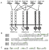
The specificity in PDGF:PDGFR recognition. (A) The biochemically defined interactions between PDGF homodimers/heterodimers and the PDGFR homodimers/heterodimers. Note that there is no proof for pre-associated PDGFR dimers, therefore the receptor dimers are just a result of ligand-driven clustering. (B) Sequence comparison of PDGFs with the residues involved in PDGF-B:PDGFRβ interaction highlighted. This comparison shows that the hydrophobic residues used for the core of the PDGF:PDGFR interface are preserved, but the hydrophilic residues at the periphery of the interface have significant variations. (D) Sequence comparison between the PDGFRs fragments used at the interface, with the PDGFRβ residues in ligand recognition highlighted. This comparison also shows that the hydrophobic nature of the ligand-recognition surface is preserved. (B) and (C) also show changes from aromatic residues to branched residues, or from larger to smaller residues, particularly between PDGF-A and PDGF-B, and between PDGFRα and PDGFRβ.
Fig. 7
Fig. 7
The activation of PDGFRs is dependent on the homotypic interactions at the membrane-proximal region of the extracellular segment. (A) Schematic model of PDGFR activation by PDGF-B. The dimeric PDGF ligand binds the D2-D3 boundary of PDGFRs, enabling or forcing the interaction between PDGFR D4 domains. The precise positioning of D4-D5 domains lead to transmembrane and juxtamembrane changes, ultimately leading to the activations of the tyrosine kinase domain. (B) The PDGFR D4-D4 homotypic interaction is likely analogous to the KIT D4- D4 interaction. Shown is the part of the structure of stem cells factor (SCF) in complex with KIT D1-D5. The D4 domains from two copies of receptors are colored in orange and pink respectively. The inter-receptor salt bridges are shown as sticks. (C) PDGFR D4-D4 interaction is also likely to be analogous to that of the D7 domains of VEGFRs, the class of RTKs evolutionarily related to PDGFRs. (D) Sequence comparison of the segment that provides inter-domain interactions for PDGFRs, KIT, and VEGFR2. Note that arginine is always required at one position, but the other position can have either glutamate or aspartate, which are different in length and could result in the difference in the interaction strength depending on the contexts.

References

    1. Kohler N, Lipton A. Platelets as a source of fibroblast growth-promoting activity. Exp Cell Res. 1974;87:297–301. - PubMed
    1. Ross R, Glomset J, Kariya B, Harker L. A platelet-dependent serum factor that stimulates the proliferation of arterial smooth muscle cells in vitro. Proc Natl Acad Sci U S A. 1974;71:1207–1210. - PMC - PubMed
    1. Andrae J, Gallini R, Betsholtz C. Role of platelet-derived growth factors in physiology and medicine. Genes Dev. 2008;22:1276–1312. - PMC - PubMed
    1. Ostman A. PDGF receptors-mediators of autocrine tumor growth and regulators of tumor vasculature and stroma. Cytokine Growth Factor Rev. 2004;15:275–286. - PubMed
    1. Lemmon MA, Schlessinger J. Cell signaling by receptor tyrosine kinases. Cell. 2010;141:1117–1134. - PMC - PubMed

Publication types

MeSH terms

Substances