Quantification of nucleolar channel systems: uniform presence throughout the upper endometrial cavity
- PMID: 23137760
- PMCID: PMC4074880
- DOI: 10.1016/j.fertnstert.2012.10.027
Quantification of nucleolar channel systems: uniform presence throughout the upper endometrial cavity
Abstract
Objective: To determine the prevalence of nucleolar channel systems (NCSs) by uterine region, applying continuous quantification.
Design: Prospective clinical study.
Setting: Tertiary care academic medical center.
Patient(s): Forty-two naturally cycling women who underwent hysterectomy for benign indications.
Intervention(s): NCS presence was quantified by a novel method in six uterine regions-fundus, left cornu, right cornu, anterior body, posterior body, and lower uterine segment (LUS)-with the use of indirect immunofluorescence.
Main outcome measure(s): Percentage of endometrial epithelial cells (EECs) with NCSs per uterine region.
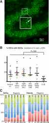
Result(s): NCS quantification was observer independent (intraclass correlation coefficient 0.96) and its intrasample variability low (coefficient of variation 0.06). Eleven of 42 hysterectomy specimens were midluteal, ten of which were analyzable with nine containing >5% EECs with NCSs in at least one region. The percentage of EECs with NCSs varied significantly between the LUS (6.1%; interquartile range [IQR] 3.0-9.9) and the upper five regions (16.9%; IQR 12.7-23.4), with fewer NCSs in the basal layer of the endometrium (17 ± 6%) versus the middle (46 ± 9%) and luminal layers (38 ± 9%) of all six regions.
Conclusion(s): NCS quantification during the midluteal phase demonstrates uniform presence throughout the endometrial cavity, excluding the LUS, with a preference for the functional luminal layers. Our quantitative NCS evaluation provides a benchmark for future studies and further supports NCS presence as a potential marker for the window of implantation.
Copyright © 2013 American Society for Reproductive Medicine. Published by Elsevier Inc. All rights reserved.
Figures

Similar articles
-
Premature formation of nucleolar channel systems indicates advanced endometrial maturation following controlled ovarian hyperstimulation.Hum Reprod. 2013 Dec;28(12):3292-300. doi: 10.1093/humrep/det358. Epub 2013 Sep 18. Hum Reprod. 2013. PMID: 24052503 Free PMC article.
-
Status of nucleolar channel systems in uterine secretions accurately reflects their prevalence-a marker for the window of implantation-in simultaneously obtained endometrial biopsies.Fertil Steril. 2018 Jan;109(1):165-171. doi: 10.1016/j.fertnstert.2017.10.005. Epub 2017 Nov 23. Fertil Steril. 2018. PMID: 29175063
-
Timing the window of implantation by nucleolar channel system prevalence matches the accuracy of the endometrial receptivity array.Fertil Steril. 2014 Nov;102(5):1477-81. doi: 10.1016/j.fertnstert.2014.07.1254. Epub 2014 Sep 17. Fertil Steril. 2014. PMID: 25241377 Clinical Trial.
-
Cyclic changes in the fine structure of the epithelial cells of human endometrium.Int Rev Cytol. 1975;42:127-72. doi: 10.1016/s0074-7696(08)60980-8. Int Rev Cytol. 1975. PMID: 172466 Review.
-
Ultrastructural changes in human endometrium at different phases of the menstrual cycle and their functional significance.Gynecol Obstet Invest. 1983;15(4):193-212. doi: 10.1159/000299412. Gynecol Obstet Invest. 1983. PMID: 6341179 Review.
Cited by
-
Presence of endometrial nucleolar channel systems at the time of frozen embryo transfer in hormone replacement cycles with successful implantation.F S Sci. 2021 Feb;2(1):80-87. doi: 10.1016/j.xfss.2021.01.001. Epub 2021 Jan 10. F S Sci. 2021. PMID: 35156063 Free PMC article.
-
Progesterone Threshold Determines Nucleolar Channel System Formation in Human Endometrium.Reprod Sci. 2014 Jul;21(7):915-920. doi: 10.1177/1933719113519177. Epub 2014 Jan 23. Reprod Sci. 2014. PMID: 24458483 Free PMC article.
-
Premature formation of nucleolar channel systems indicates advanced endometrial maturation following controlled ovarian hyperstimulation.Hum Reprod. 2013 Dec;28(12):3292-300. doi: 10.1093/humrep/det358. Epub 2013 Sep 18. Hum Reprod. 2013. PMID: 24052503 Free PMC article.
References
-
- Cornillie FJ, Lauweryns JM, Brosens IA. Normal human endometrium. An ultrastructural survey. Gynecol Obstet Invest. 1985;20:113–29. - PubMed
-
- Dubrauszky V, Pohlmann G. Strukturveränderungen am Nukleolus von Korpusendometriumzellen während der Sekretionsphase. Naturwissenschaften. 1960;47:523–4.
-
- Spornitz UM. The functional morphology of the human endometrium and decidua. Adv Anat Embryol Cell Biol. 1992;124:1–99. - PubMed
-
- Clyman MJ. A new structure observed in the nucleolus of the human endometrial epithelial cell. Am J Obstet Gynecol. 1963;86:430–2. - PubMed
Publication types
MeSH terms
Grants and funding
LinkOut - more resources
Full Text Sources
Other Literature Sources

