Clinical Trial
. 2012 Dec;43(12):3258-65.
doi: 10.1161/STROKEAHA.112.673400. Epub 2012 Nov 8.

Early change in ferumoxytol-enhanced magnetic resonance imaging signal suggests unstable human cerebral aneurysm: a pilot study

Affiliations
Clinical Trial

Early change in ferumoxytol-enhanced magnetic resonance imaging signal suggests unstable human cerebral aneurysm: a pilot study

David Hasan et al. Stroke. 2012 Dec.

Abstract

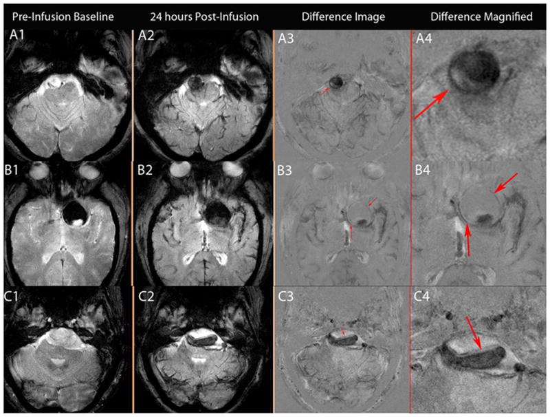
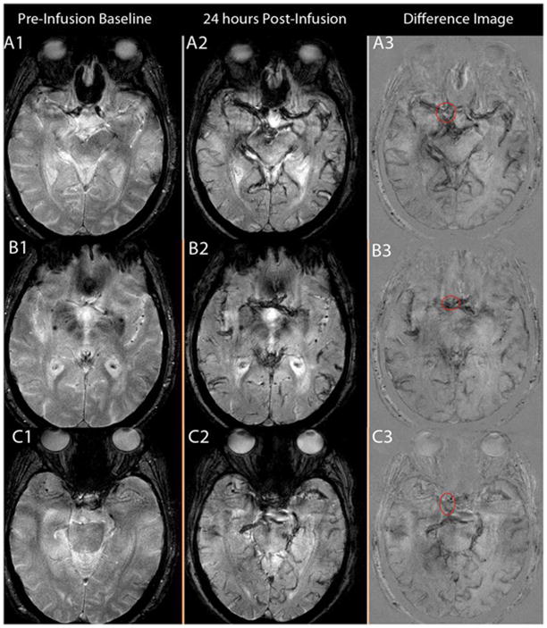
Background and purpose: The clinical significance of early (ie, within the first 24 hours) uptake of ferumoxytol by macrophages in the wall of human cerebral aneurysms is not clear. The purpose of this study was to determine whether early uptake of ferumoxytol suggests unstable cerebral aneurysm.

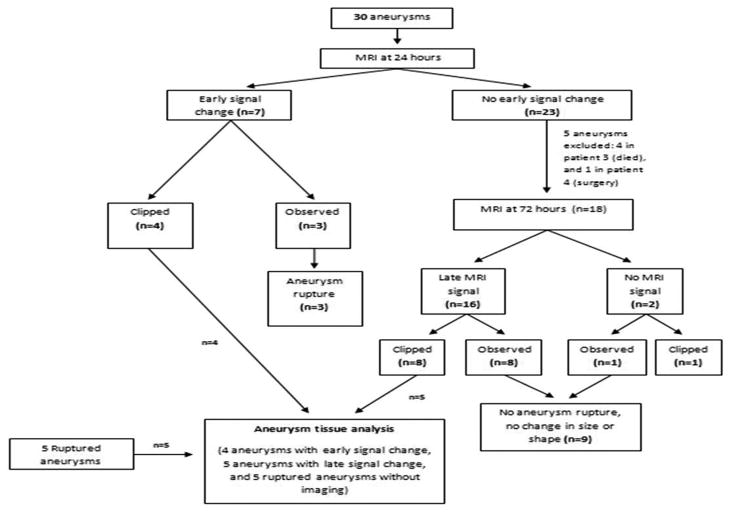
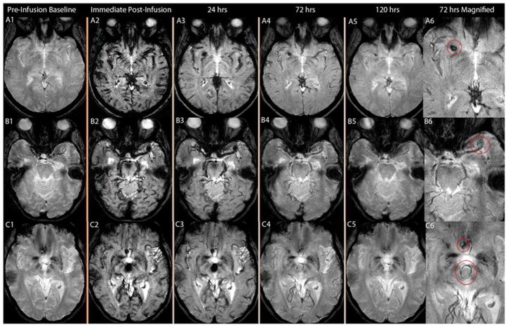
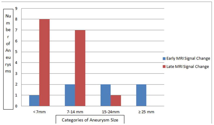
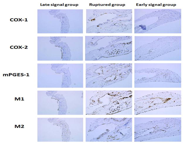
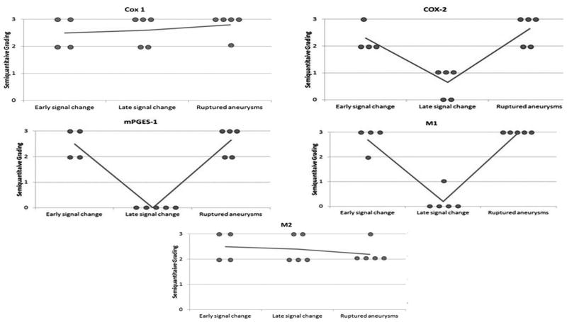
Methods: Thirty unruptured aneurysms in 22 patients were imaged with magnetic resonance imaging 24 hours after infusion of ferumoxytol. Eighteen aneurysms were also imaged 72 hours after infusion of ferumoxytol. Aneurysm dome tissue was collected from 4 patients with early magnetic resonance imaging signal changes, 5 patients with late signal changes, and 5 other patients with ruptured aneurysms. The tissue was immunostained for expression of cyclooxygenase-1, cyclooxygenase-2, microsomal prostaglandin E2 synthase-1, and macrophages.

Results: In 23% (7/30) of aneurysms, there was pronounced early uptake of ferumoxytol. Four aneurysms were clipped. The remaining 3 aneurysms were managed conservatively; all 3 ruptured within 6 months. In 53% (16 of 30) of aneurysms, there was pronounced uptake of ferumoxytol at 72 hours. Eight aneurysms were surgically clipped, and 8 were managed conservatively; none ruptured or increased in size after 6 months. Expression of cyclooxygenase-2, microsomal prostaglandin E2 synthase-1, and macrophages was similar in unruptured aneurysms with early uptake of ferumoxytol and ruptured aneurysms. Expression of these inflammatory molecules was significantly higher in aneurysms with early uptake of ferumoxytol versus aneurysms with late uptake.

Conclusions: Uptake of ferumoxytol in aneurysm walls within the first 24 hours strongly suggests aneurysm instability and probability of rupture within 6 months, and may warrant urgent intervention.


Conflict of interest statement

Conflict of interest/Disclosure

None

Figures

Figure 1
Figure 1
Schematic presentation of the study.
Figure 2
Figure 2
Figure 2(A-B-C): A:T2* GE MRI sequenceat baseline and 24 hours post-infusion showing early signal changes in the walls of three cerebral aneurysms (Panel A1–4 corresponds to patient # 1 with a right vertebral artery aneurysm, panel B1–4 corresponds to patient # 2 with a left supraclinoid ICA aneurysm, and panel C1–4 corresponds to patient # 3 with a fusiform vertebrobasilar artery aneurysm). Difference images (last two panels) demonstrate the relative signal loss after ferumoxytol infusion. All three aneurysms ruptured within six months. B: T2* GE MRI sequence at baseline and 24 hours post-infusion, and subtraction images showing no early signal changes in three aneurysms from patient # 3 (Panel A 1–3, right ICA terminus aneurysm; panel B 1–3, anterior communicating artery aneurysm; and panel C 1–3, right cavernous ICA aneurysm). None of these aneurysms ruptured during the follow-up period. C: T2* GE MRI sequenceat five different times and magnified subtraction image at 72- hours post – infusion, showing late MRI signal changes in three aneurysms. Panel A 1–6 corresponds to patient # 8 with a right middle cerebral artery aneurysm, panel B 1–6 corresponds to patient # 19 with a left middle cerebral artery aneurysm, and panel C 1–6 corresponds to patient # 9 with anterior communicating artery and basilar tip aneurysms (the left middle cerebral artery aneurysm was on a different cut). MRI signal changes are best detected in the wall of these aneurysms at 72 hours post-infusion of ferumoxytol. None of these aneurysms ruptured during the follow-up period.
Figure 2
Figure 2
Figure 2(A-B-C): A:T2* GE MRI sequenceat baseline and 24 hours post-infusion showing early signal changes in the walls of three cerebral aneurysms (Panel A1–4 corresponds to patient # 1 with a right vertebral artery aneurysm, panel B1–4 corresponds to patient # 2 with a left supraclinoid ICA aneurysm, and panel C1–4 corresponds to patient # 3 with a fusiform vertebrobasilar artery aneurysm). Difference images (last two panels) demonstrate the relative signal loss after ferumoxytol infusion. All three aneurysms ruptured within six months. B: T2* GE MRI sequence at baseline and 24 hours post-infusion, and subtraction images showing no early signal changes in three aneurysms from patient # 3 (Panel A 1–3, right ICA terminus aneurysm; panel B 1–3, anterior communicating artery aneurysm; and panel C 1–3, right cavernous ICA aneurysm). None of these aneurysms ruptured during the follow-up period. C: T2* GE MRI sequenceat five different times and magnified subtraction image at 72- hours post – infusion, showing late MRI signal changes in three aneurysms. Panel A 1–6 corresponds to patient # 8 with a right middle cerebral artery aneurysm, panel B 1–6 corresponds to patient # 19 with a left middle cerebral artery aneurysm, and panel C 1–6 corresponds to patient # 9 with anterior communicating artery and basilar tip aneurysms (the left middle cerebral artery aneurysm was on a different cut). MRI signal changes are best detected in the wall of these aneurysms at 72 hours post-infusion of ferumoxytol. None of these aneurysms ruptured during the follow-up period.
Figure 3
Figure 3
Distribution of aneurysms with early MRI signal change vs. late MRI signal change in relation to aneurysm size. Early MRI signal change was independent of aneurysm size. Late MRI signal change was noted in 50% of aneurysms < 7mm, and 44% in aneurysms 7–14mm.
Figure 4
Figure 4
Immunostaining for three different aneurysm categories: Aneurysms with late MRI signal changes, ruptured aneurysms, and aneurysms with early MRI signal changes. Immunostaining for COX-1 and M2 cells is similar in the three categories of aneurysms. Immunostaining for COX-2, mPGES-1, and M1 cells indicates upregulation in aneurysms with early MRI signal changes in a similar manner to ruptured aneurysms.
Figure 5
Figure 5
Semiquantitative grading for tissues collected from aneurysms with early MRI signal changes (n=4), aneurysms with late MRI signal changes (n=5), and ruptured aneurysms (n=5). Immunostaining for COX-1 and M2 cells is similar in the three categories of aneurysms. Immunostaining for COX-2, mPGES-1, and M1 cells indicates upregulation in aneurysms with early MRI signal changes in a similar manner to ruptured aneurysms.

References

    1. Lu M, Cohen MH, Rieves D, Pazdur R. Fda report: Ferumoxytol for intravenous iron therapy in adult patients with chronic kidney disease. Am J Hematol. 2010;85:315–319. - PubMed
    1. Spinowitz BS, Kausz AT, Baptista J, Noble SD, Sothinathan R, Bernardo MV, et al. Ferumoxytol for treating iron deficiency anemia in ckd. J Am Soc Nephrol. 2008;19:1599–1605. - PMC - PubMed
    1. Dosa E, Tuladhar S, Muldoon LL, Hamilton BE, Rooney WD, Neuwelt EA. Mri using ferumoxytol improves the visualization of central nervous system vascular malformations. Stroke. 2011;42:1581–1588. - PMC - PubMed
    1. Neuwelt EA, Varallyay CG, Manninger S, Solymosi D, Haluska M, Hunt MA, et al. The potential of ferumoxytol nanoparticle magnetic resonance imaging, perfusion, and angiography in central nervous system malignancy: A pilot study. Neurosurgery. 2007;60:601–611. discussion 611-602. - PubMed
    1. Herborn CU, Vogt FM, Lauenstein TC, Dirsch O, Corot C, Robert P, et al. Magnetic resonance imaging of experimental atherosclerotic plaque: Comparison of two ultrasmall superparamagnetic particles of iron oxide. J Magn Reson Imaging. 2006;24:388–393. - PubMed

Publication types

MeSH terms