Review
. 2013 Jan;23(1):43-50.
doi: 10.1097/MOU.0b013e32835ad3ee.

MRI-ultrasound fusion for guidance of targeted prostate biopsy

Affiliations
Review

MRI-ultrasound fusion for guidance of targeted prostate biopsy

Leonard Marks et al. Curr Opin Urol. 2013 Jan.

Abstract

Purpose of review: Prostate cancer (CaP) may be detected on MRI. Fusion of MRI with ultrasound allows urologists to progress from blind, systematic biopsies to biopsies, which are mapped, targeted and tracked. We herein review the current status of prostate biopsy via MRI/ultrasound fusion.

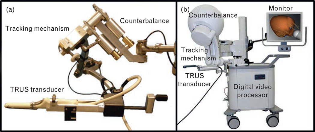
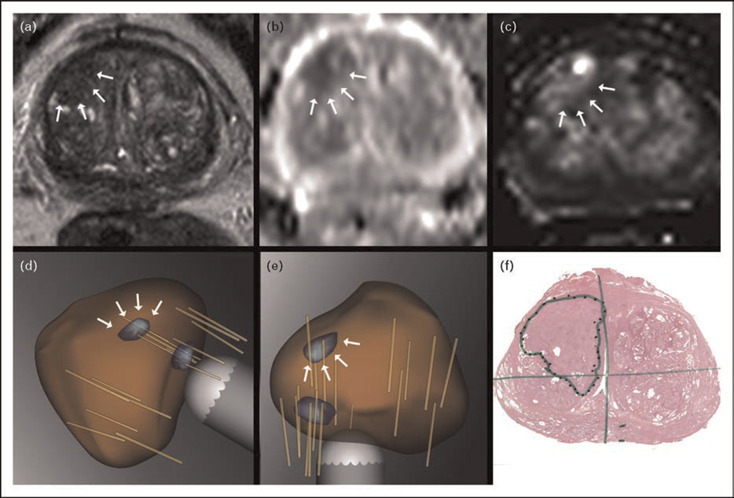
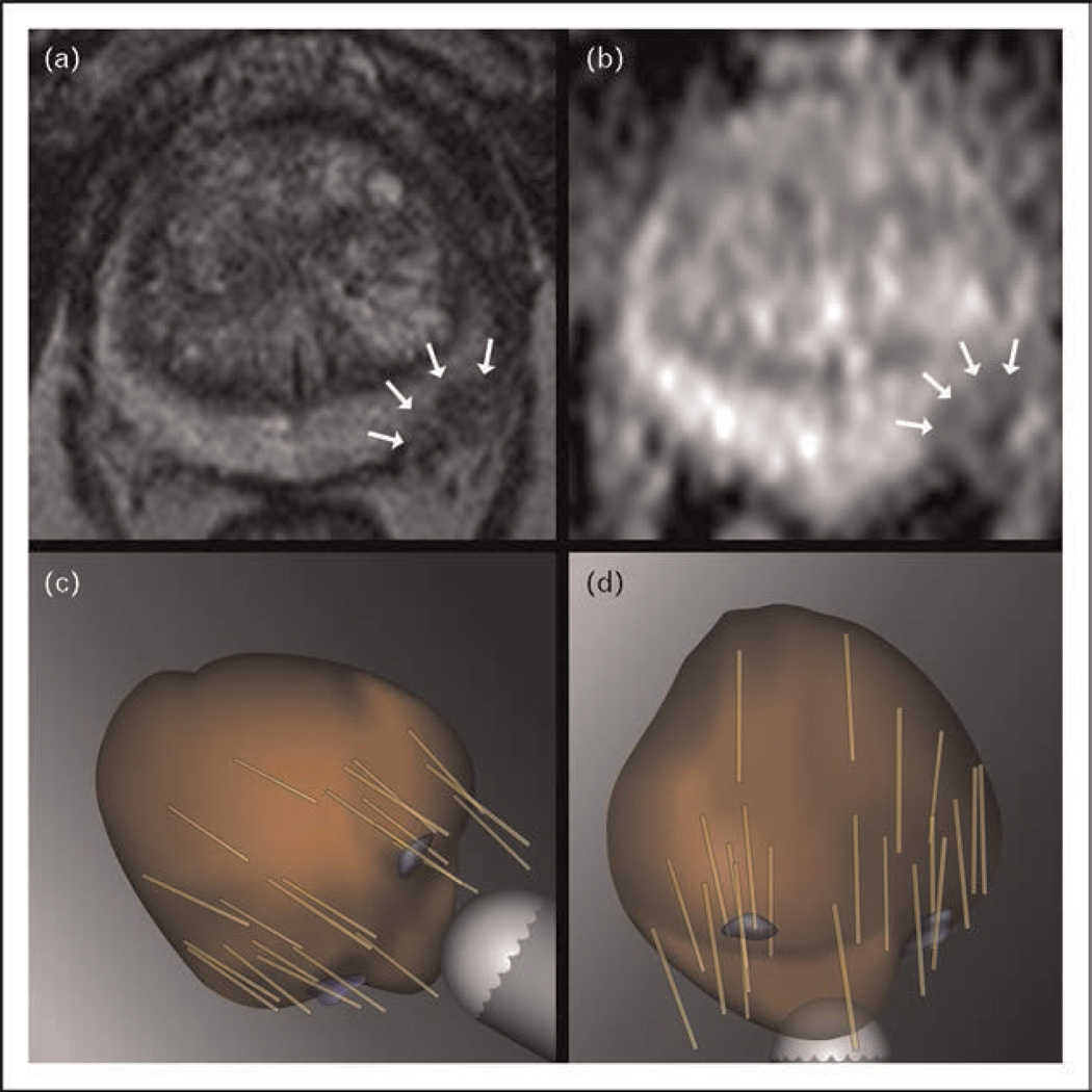
Recent findings: Three methods of fusing MRI for targeted biopsy have been recently described: MRI-ultrasound fusion, MRI-MRI fusion ('in-bore' biopsy) and cognitive fusion. Supportive data are emerging for the fusion devices, two of which received US Food and Drug Administration approval in the past 5 years: Artemis (Eigen, USA) and Urostation (Koelis, France). Working with the Artemis device in more than 600 individuals, we found that targeted biopsies are two to three times more sensitive for detection of CaP than nontargeted systematic biopsies; nearly 40% of men with Gleason score of at least 7 CaP are diagnosed only by targeted biopsy; nearly 100% of men with highly suspicious MRI lesions are diagnosed with CaP; ability to return to a prior biopsy site is highly accurate (within 1.2 ± 1.1 mm); and targeted and systematic biopsies are twice as accurate as systematic biopsies alone in predicting whole-organ disease.

Summary: In the future, MRI-ultrasound fusion for lesion targeting is likely to result in fewer and more accurate prostate biopsies than the present use of systematic biopsies with ultrasound guidance alone.


Conflict of interest statement

Conflicts of interest

There are no conflicts of interest.

Figures

FIGURE 1
FIGURE 1. Artemis fusion device
(a) Robarts prototype and (b) FDA-approved commercial model (Eigen). When the TRUS probe is rotated, encoders in the tracking mechanism transmit orientation and position of the transducer tip to software that displays and records location on the monitor. The tracking arm is stabilized and held stationary during the rotation, preventing change in pitch, yaw and depth of penetration. During the scan, 2D images are digitized with a frame grabber and reconstructed into a 3D image. A model of the prostate is then generated from the 3D image; biopsy, tracking of biopsy site and MRI fusion are then performed on the reconstructed model [23]. 2D, two-dimensional; 3D, three-dimensional; FDA, US Food and Drug Administration.
FIGURE 2
FIGURE 2. A 60-year-old man with eight prior negative biopsies over a 10-year period
During that time, a PSA increase from 4 to 50 ng/ml was observed. Arrows show regions of interest (a) T2-weighted axial MR image demonstrating a dominant lesion in the right anterior prostate with a focal low signal. (b) Diffusion-weighted axial MR image with an ADC value of 0.865 × 10⁻³m2/s in the corresponding area. (c) Dynamic contrast enhancement image of the corresponding area showing increased local perfusion. The lesion was classified as an image grade 4 on the basis of multiparametric features [22▪▪]. The radiologist outlined the lesion in T2 axial images. Open-source imaging software [25] was then used to produce a 3D model of the prostate including the target. A second 3D model was then generated on the basis of an outline of the prostate on ultrasound. (d) and (e) The two models were then dynamically fused, generating the composite virtual 3D model seen in a sagittal (d) and axial (e) views. The prostate is mapped in brown and the target identified in blue. A second, smaller target located peripherally was also identified. Systematic and targeted biopsies were obtained, generating the final 3D model demonstrating the location of all biopsy cores (light brown cylinders). Targeted biopsies in this patient revealed Gleason 3+4=7 CaP in the primary target. (f) Radical prostatectomy specimen demonstrated the presence of a 4.5-cm Gleason 3+4 cancer in the anterior prostate, confirming biopsy localization of the lesion. 2D, two-dimensional; 3D, three-dimensional; MR, magnetic resonance; PSA, prostate-specific antigen.
FIGURE 3
FIGURE 3. A 70-year-old man enrolled in active surveillance with a PSA of 4.1
(a) T2-weighted axial MR image demonstrating a dominant lesion (arrows) in the left posterior prostate with a focal low signal. (b) Diffusion-weighted axial MR image with an ADC value of 1.071 × 10⁻³m2/s in the corresponding area. The lesion was classified as image grade 3 on the basis of multiparametric features [22▪▪]. 3D image processing was performed as in Figure 2. Systematic and targeted biopsies were obtained, generating the final 3D model demonstrating the location of all biopsy cores (light brown cylinders). Targeted biopsies in this patient revealed Gleason 3+4=7 CaP in the primary target. 2D, two-dimensional; 3D, three-dimensional; MR, magnetic resonance.

Similar articles

Cited by

References

    1. Welch HG, Fisher ES, Gottlieb DJ, Barry MJ. Detection of prostate cancer via biopsy in the Medicare-SEER population during the PSA era. J Natl Cancer Inst. 2007;99:1395–1400. - PubMed
    1. Hodge KK, McNeal JE, Stamey TA. Ultrasound guided transrectal core biopsies of the palpably abnormal prostate. J Urol. 1989;142:66–70. - PubMed
    1. Hodge KK, McNeal JE, Terris MK, Stamey TA. Random systematic versus directed ultrasound guided transrectal core biopsies of the prostate. J Urol. 1989;142:71–74. discussion 4–5. - PubMed
    1. Cooperberg MR, Broering JM, Kantoff PW, Carroll PR. Contemporary trends in low risk prostate cancer: risk assessment and treatment. J Urol. 2007;178:S140–S149. - PMC - PubMed
    1. Taira AV, Merrick GS, Galbreath RW, et al. Performance of transperineal template-guided mapping biopsy in detecting prostate cancer in the initial and repeat biopsy setting. Prostate Cancer Prostatic Dis. 2010;13:71–77. - PMC - PubMed

Publication types

MeSH terms