. 2013 Jan;22(1):83-92.
doi: 10.1002/pro.2190. Epub 2012 Nov 29.

Structural insight into the separate roles of inositol tetraphosphate and deacetylase-activating domain in activation of histone deacetylase 3

Affiliations

Structural insight into the separate roles of inositol tetraphosphate and deacetylase-activating domain in activation of histone deacetylase 3

Mehrnoosh Arrar et al. Protein Sci. 2013 Jan.

Abstract

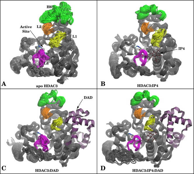
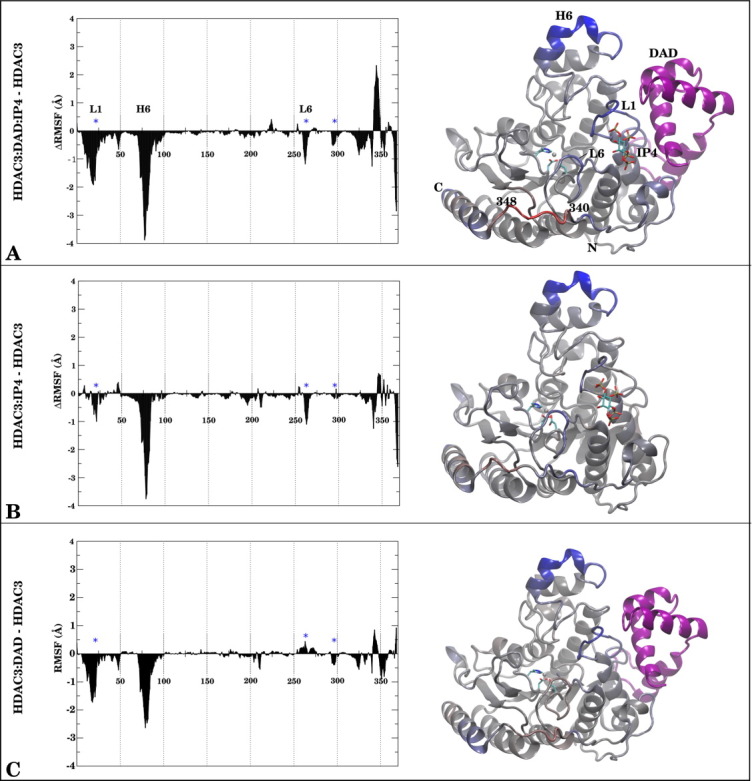
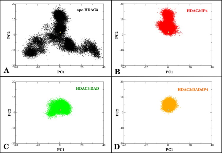
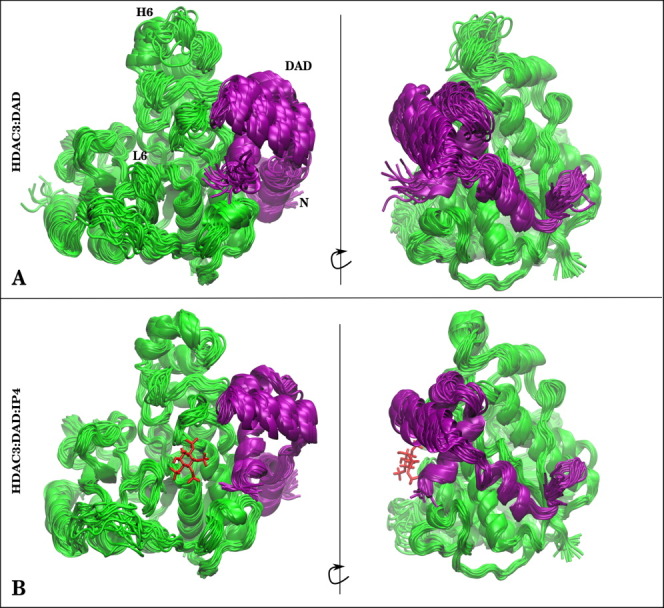
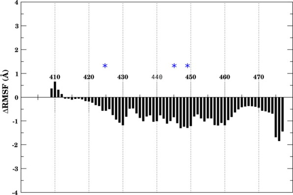
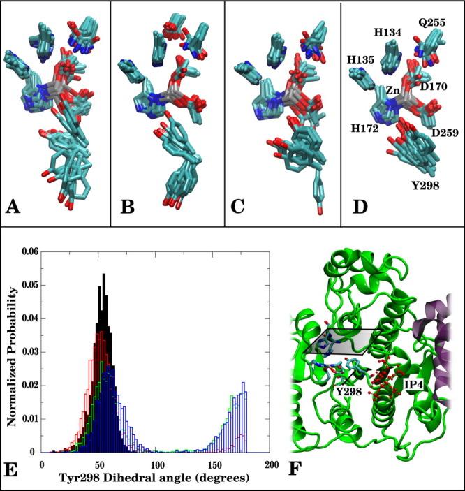
Histone deacetylases (HDACs) repress transcription by deacetylating acetyllysines on specific histone tails. HDAC3 is implicated in neurodegenerative diseases, certain leukemias, and even in disrupting HIV-1 latency. A recent crystal structure of HDAC3 in complex with the deacetylase-activating domain (DAD) of its corepressor complex revealed an inositol tetraphosphate (IP4) molecule at the protein-protein interface. IP4 was shown to play an important, yet mechanistically ambiguous, role in the activity of HDAC3. The goal of this article is to explore the conformational ensemble of HDAC3 in its inactive apo state and in the presence of each or both of DAD and IP4. Using triplicate, 100 ns molecular dynamic simulations, we study the apo, ternary, and intermediate DAD-bound or IP4-bound HDAC3 states. We find that a population-shift effect is induced by the presence of each corepressor, and is most notable in the presence of both. Our results offer new insights into the change in dynamics necessary for the activation of HDAC3 and highlight the roles of IP4 and DAD in this process.


Figures

Figure 1
Figure 1
Superimposed snapshots from MD simulations for (A) apo HDAC3, (B) HDAC3:IP4, (C) HDAC3:DAD, and (D) HDAC3:IP4:DAD. Helix 6 (green), Loop1 (yellow), Loop2 (orange), and Loop 6 (magenta) are highlighted to show differences in dynamics. If present in the simulation, DAD and IP4 are shown as purple ribbons or red ball-and-stick representations, for reference. [Color figure can be viewed in the online issue, which is available at wileyonlinelibrary.com.]
Figure 2
Figure 2
The ΔRMSF (Å) per residue in HDAC3 between (A) HDAC3:DAD:IP4, (B) HDAC3:IP4, (C) HDAC3:DAD and the apo state is shown on the left side of each panel. The same values are projected onto a structure of the protein for reference, using a Blue (Negative)→Gray (Zero)→Red (Positive) scale. A range from −3.00 to +3.00 Å was used to make the figure. DAD and IP4, when present in the simulations are shown for reference in purple ribbons and stick representations, respectively.
Figure 3
Figure 3
PCA of HDAC3. First two principal components were calculated from apo HDAC3 Cα dynamics. Structural ensembles of HDAC3 Cα atoms from (A) apo HDAC3, (B) HDAC3:IP4, (C), HDAC3:DAD, and (D) HDAC3:DAD:IP4 simulations were projected onto these two principal components. A yellow diamond in each graph marks the crystal structure conformation of HDAC3 for reference. [Color figure can be viewed in the online issue, which is available at wileyonlinelibrary.com.]
Figure 4
Figure 4
Structures from (A) HDAC3:DAD and (B) HDAC3:DAD:IP4 simulations are presented to show difference in stabilities of the protein–protein complex owing to the presence of IP4. HDAC3 (green) is shown with the DAD domain (purple). If IP4 was present in the simulation, a single snapshot of IP4 is shown as red sticks for reference. [Color figure can be viewed in the online issue, which is available at wileyonlinelibrary.com.]
Figure 5
Figure 5
ΔRMSF of DAD backbone (HDAC3:DAD:IP4 minus HDAC3:DAD). Negative values indicate stabilization of backbone owing to the presence of IP4. Blue asterisks indicate residues near IP4-binding site. [Color figure can be viewed in the online issue, which is available at wileyonlinelibrary.com.]
Figure 6
Figure 6
Surface representation of stable tunnel observed in ternary complex simulation. Zinc metal center is shown as a silver sphere. Tyr298 side-chain lining the tunnel is shown as sticks. [Color figure can be viewed in the online issue, which is available at wileyonlinelibrary.com.]
Figure 7
Figure 7
Superimposed snapshots from MD simulations show displacements of active site residues for (A) HDAC3, (B) HDAC3:IP4, (C) HDAC3:DAD, (D) HDAC3:DAD:IP4. (E) Tyr298 (N[sbond]Cα[sbond]Cβ[sbond]Cγ) dihedral angle probability distributions shown for HDAC3:DAD:IP4 (solid, black), HDAC3:DAD (red), HDAC3:IP4(green), and HDAC3 (blue). (F) Cartoon representation of HDAC3:IP4:DAD complex, showing active site resides as sticks; the horizontal plane above the active site marks the top-down perspective of (A–D).

References

    1. Bhaskara S, Knutson SK, Jiang G, Chandrasekharan MB, Wilson AJ, Zheng S, Yenamandra A, Locke K, Yuan J, Bonine-Summers AR, Wells CE, Kaiser JF, Washington MK, Zhao Z, Wagner FF, Sun ZW, Xa F, Holson EB, Khabele D, Hiebert SW. Hdac3 is essential for the maintenance of chromatin structure and genome stability. Cancer Cell. 2010;18:436–447. - PMC - PubMed
    1. Pogo BG, Allfrey VG, Mirsky AE. RNA synthesis and histone acetylation during the course of gene activation in lymphocytes. Proc Natl Acad Sci USA. 1966;55:805. - PMC - PubMed
    1. Witt O, Deubzer HE, Milde T, Oehme I. HDAC family. what are the cancer relevant targets? Cancer Lett. 2009;277:8–21. - PubMed
    1. Jia H, Pallos J, Jacques V, Lau A, Tang B, Cooper A, Syed A, Purcell J, Chen Y, Sharma S, Sangrey GR, Darnell SB, Plasterer H, Sadri-Vakili G, Gottesfeld JM, Thompson LM, Rusche JR, Marsh JL, Thomas EA. Histone deacetylase (HDAC) inhibitors targeting HDAC3 and HDAC1 ameliorate polyglutamine-elicited phenotypes in model systems of Huntington's disease. Neurobiol Dis. 2012;46:351–361. - PMC - PubMed
    1. Debacker K, Frizzell A, Gleeson O, Kirkham-McCarthy L, Mertz T, Lahue RS. Histone deacetylase complexes promote trinucleotide repeat expansions. PLoS Biol. 2012;10:e1001257. - PMC - PubMed

Publication types

MeSH terms

Substances

LinkOut - more resources