. 2013 Jan 15;304(2):G142-56.
doi: 10.1152/ajpgi.00483.2011. Epub 2012 Nov 8.

Capsaicin induces NKCC1 internalization and inhibits chloride secretion in colonic epithelial cells independently of TRPV1

Affiliations

Capsaicin induces NKCC1 internalization and inhibits chloride secretion in colonic epithelial cells independently of TRPV1

Patrice G Bouyer et al. Am J Physiol Gastrointest Liver Physiol. .

Abstract

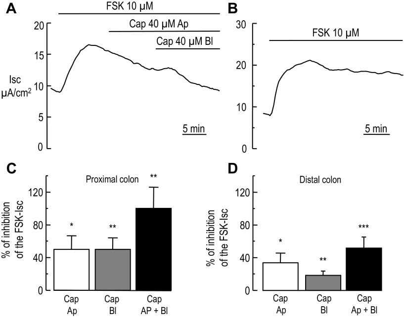
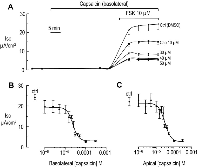
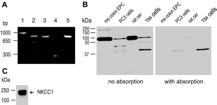
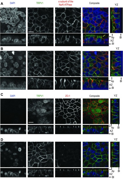
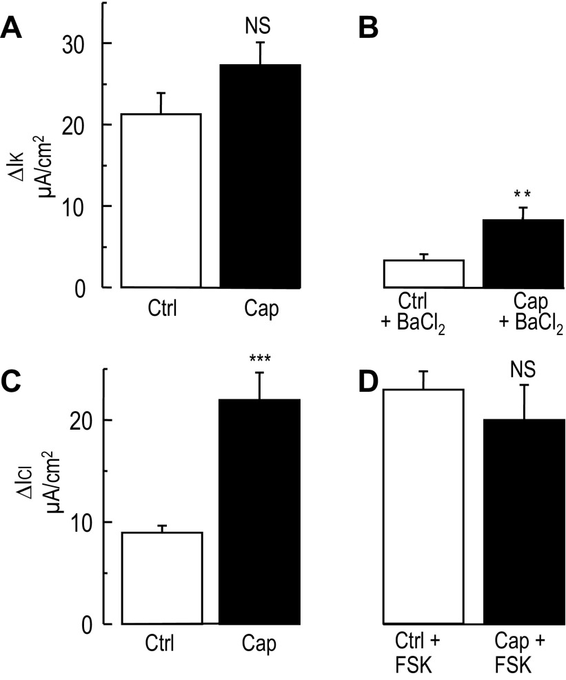
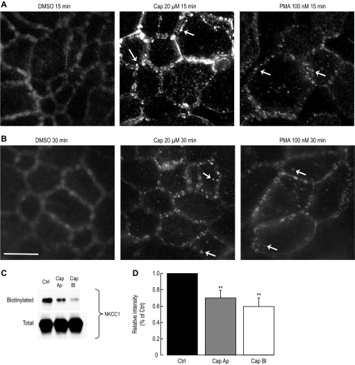
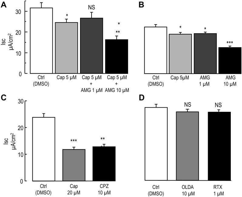
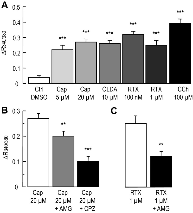
Colonic chloride secretion is regulated via the neurohormonal and immune systems. Exogenous chemicals (e.g., butyrate, propionate) can affect chloride secretion. Capsaicin, the pungent ingredient of the chili peppers, exerts various effects on gastrointestinal function. Capsaicin is known to activate the transient receptor potential vanilloid type 1 (TRPV1), expressed in the mesenteric nervous system. Recent studies have also demonstrated its presence in epithelial cells but its role remains uncertain. Because capsaicin has been reported to inhibit colonic chloride secretion, we tested whether this effect of capsaicin could occur by direct action on epithelial cells. In mouse colon and model T84 human colonic epithelial cells, we found that capsaicin inhibited forskolin-dependent short-circuit current (FSK-I(sc)). Using PCR and Western blot, we demonstrated the presence of TRPV1 in colonic epithelial cells. In T84 cells, TRPV1 localized at the basolateral membrane and in vesicular compartments. In permeabilized monolayers, capsaicin activated apical chloride conductance, had no effect on basolateral potassium conductance, but induced NKCC1 internalization demonstrated by immunocytochemistry and basolateral surface biotinylation. AMG-9810, a potent inhibitor of TRPV1, did not prevent the inhibition of the FSK-I(sc) by capsaicin. Neither resiniferatoxin nor N-oleoyldopamine, two selective agonists of TRPV1, blocked the FSK-I(sc). Conversely capsaicin, resiniferatoxin, and N-oleoyldopamine raised intracellular calcium ([Ca(2+)](i)) in T84 cells and AMG-9810 blocked the rise in [Ca(2+)](i) induced by capsaicin and resiniferatoxin suggesting the presence of a functional TRPV1 channel. We conclude that capsaicin inhibits chloride secretion in part by causing NKCC1 internalization, but by a mechanism that appears to be independent of TRPV1.


Figures

Fig. 1.
Fig. 1.
Inhibition of the forskolin-stimulated short-circuit current by luminal and basolateral capsaicin in mouse colon. A: representative Ussing chamber recording of the short-circuit (Isc) in mouse proximal colon. When the baseline Isc was stable, addition of forskolin (FSK) activated chloride secretion depicted by an increase of the Isc, which subsequently reaches a steady state. At the indicated time (horizontal bar) the tissue was exposed first to apical (Ap) capsaicin (Cap). When the Isc reached a plateau basolateral (Bl) capsaicin was added causing further inhibition of the forskolin-stimulated short-circuit current (FSK-Isc). B: representative recording of Isc in a distal colon exposed only to FSK. At the indicated time FSK was added causing an increase of the Isc, which remained constant during the time of application of FSK. C and D: summary data (n = 8 and 9 animals for proximal and distal colon, respectively) of the inhibition of the FSK-Isc by apical and basolateral capsaicin in the proximal colon (C) and distal colon (D), respectively. Paired t-test, *P < 0.05, **P < 0.01, ***P < 0.001.
Fig. 2.
Fig. 2.
Inhibition of the forskolin-stimulated short-circuit current by basolateral and apical capsaicin in T84 cells. A: short-circuit current recording in T84 cells cultured on Millicell inserts (n = 3 inserts for each condition for this experiment). T84 cells were incubated in HPBSS (see materials and methods for the salt composition) and then exposed to basolateral capsaicin (Cap) or vehicle (DMSO) for 30 min. At the indicated time (horizontal bar) 10 μM FSK was added to stimulate chloride secretion. B: each point represents the mean Isc measured at the end of the Isc plateau in A during FSK exposure in presence of increasing basolateral capsaicin concentration (n ≥ 7 for each concentration). Ctrl represents the Isc in absence of capsaicin. From the adjustment of the data to a single-site inhibition curve an IC50 of 23.5 ± 3.1 (SD) was computed. C: same as B except capsaicin was applied to the apical side (n ≥ 8 for each concentration). From the adjustment of the data to a single-site inhibition curve an IC50 of 30.7 ± 5.0 (SD) was computed.
Fig. 3.
Fig. 3.
Expression of TRPV1 in colonic epithelial cell. A: RT-PCR products of TRPV1, GAPDH, and NKCC1. The agarose gel shows the products of the PCR designed to amplify a 962 bp (lane 1), 805 bp (lane 2), and 769 bp (lane 3) product for TRPV1, 226 bp (lane 4) for GAPDH, and 800 bp (lane 5) NKCC1 from T84 cells cDNA (representative gel of a triplicate experiment). B: TRPV1 expression in mouse colon epithelial cells, PC3 cells (human prostate epithelial line), rat cerebellum (rat cer), and T84 cells. Whole cell lysate was prepared using a RIPA lysis buffer. Proteins were separated by SDS-PAGE and immunoblotted for TRPV1 (left blot) using a rabbit anti-TRPV1 antibody (1:800 dilution), or after antibody preabsorption by the antigen peptide (right blot). Representative image of a triplicate experiment. C: from the mouse colonic epithelial cells harvested by calcium-EDTA chelation method and used in B, NKCC1 expression was tested using T4 mouse anti-NKCC1 (1:7,500 dilution). Representative image of n = 4 experiments.
Fig. 4.
Fig. 4.
Immunolocalization of TRPV1 in polarized T84 cells. A: TRPV1 localization with the Na-K-ATPase. T84 cells grown on Millicell inserts to develop a polarized epithelium were fixed in cold methanol and mounted for confocal microscopy. Cells were dual-stained for TRPV1 (primary antibody 1:200 dilution) using a goat anti-rabbit DyLight 488 (1:200 dilution) and the α-subunit of the Na-K-ATPase (primary antibody 1:200 dilution) using a donkey anti-mouse DyLight 594 (1:200 dilution). Top four planes represent en face views (focal plane adjusted on TRPV1 staining) of the nuclear staining (DAPI), TRPV1, the α-subunit of the Na-K-ATPase, and composite of the three colors for colocalization. Below each en face view is the XZ optical section, with a supplemental YZ section for the composite. The black arrows indicate apical (Ap) and basolateral (Bl) sides. The yellow arrows point to colocalization of TRPV1 and Na-K-ATPase, whereas the yellow stars point to the subapical TRPV1 staining. B: same as A except the focal plane was adjusted to the staining of the Na-K-ATPase. Representative images of n = 6 experiments, 3 inserts for each condition. C: TRPV1 localization with ZO-1 in T84 cells. Cells were dual-stained for TRPV1 using a goat anti-rabbit DyLight 488 and ZO-1 (primary antibody 1:200 dilution) using a donkey anti-mouse DyLight 594 (1:200 dilution). The top four planes represent en face views (focal plane adjusted on ZO-1 staining) of the nuclear staining (DAPI), TRPV1, ZO-1 and composite of the three colors for localization. Below each en face view is the XZ optical section, with a supplemental YZ section for the composite. D: same as C except the focal plane for the en face views was focused on TRPV1 staining. The yellow stars point to the subapical TRPV1 staining. Representative images of n = 6 experiments, 3 inserts for each condition. Scale bar, 10 μm.
Fig. 5.
Fig. 5.
Effect of capsaicin on the basolateral potassium and apical chloride currents in T84 cells. After equilibrium in a bilateral low K+ solution, T84 cells were exposed to high K+ (140 mM K+) apical to low basolateral K+ (5 mM) gradient. The apical membrane was permeabilized using 500 U/ml of nystatin, causing an increase of basolateral potassium current (IK). A: histograms represent the change in IK induced in control (vehicle) and capsaicin (Cap) treated T84 monolayers during the imposition of the K+ gradient. No significant difference was found between Ctrl and Cap-treated cells (n = 48 and 47 for Ctrl and Cap, respectively). B: the histograms represent the inhibition of IK by 3 mM barium in control (n = 24) and capsaicin-treated cells (n = 23). NS, nonsignificant, **P < 0.01 (unpaired t-test). C: T84 cells were exposed to a high basolateral KCl (140 mM) to high K+, Cl-free (140 mM K+-gluconate) apical gradient. The basolateral membrane was permeabilized with 500 U/ml nystatin, which caused an increase of apical chloride current (ICl). Histograms represent the change in ICl in control (n = 48) and capsaicin-treated cells (n = 48). In this condition capsaicin induced a significant stimulation of the apical ICl compared with control. ***P < 0.001 (unpaired t-test). D: histograms represent the change in ICl at the peak of the forskolin stimulation in control (n = 48) and capsaicin-treated cells (n = 48). No difference between the two conditions was found.
Fig. 6.
Fig. 6.
Loss of NKCC1 membrane expression during capsaicin treatment in T84 cells. A: NKCC1 internalization during capsaicin and PMA treatment. T84 cells grown on Millicell inserts to develop a polarized epithelium were exposed to vehicle (DMSO), capsaicin (Cap), or phorbol 12-myristate 13-acetate (PMA), subsequently fixed with 3% paraformaldehyde, and mounted for fluorescent microscopy. Images were captured using a brightfield microscope equipped for fluorescence. Cells were stained for NKCC1 (primary antibody 1:200 dilution) using a donkey anti-mouse DyLight 594 (1:200 dilution). En face view of NKCC1 staining during vehicle, 20 μM capsaicin and 100 nM PMA exposure (15 min). White arrows point to NKCC1 staining in endocytic vesicles. B: same as A except cells were treated for 30 min with vehicle, capsaicin, or PMA (n = 5 experiments, 2 to 3 inserts per condition). Scale bar, 10 μm. C: NKCC1 surface expression during apical and basolateral capsaicin exposure. T84 cells were treated for 1 h with 20 μM capsaicin applied either to the apical (Ap) or basolateral (Bl) side or with DMSO (Ctrl). Basolateral membrane was exposed to sulfo-NHS-SS-biotin and biotinylated proteins were captured by streptavidin and subjected to Western blotting. NKCC1 proteins in the biotinylated and total fraction were detected using a mouse anti-NKCC1 (T4 1:7,500 dilution). D: data summary of the experiment in C. Films were scanned and relative intensity was measured using ImageJ and normalized to control (n = 7 for each condition). **P < 0.01 (unpaired t-test).
Fig. 7.
Fig. 7.
Pharmacological characterization of the inhibition of the forskolin-stimulated short-circuit current by capsaicin. A: effect of AMG-9810 on capsaicin-induced inhibition of the FSK-Isc. The experimental design was similar to the one shown in Fig. 2A. T84 cells grown on Millicell inserts were exposed to DMSO (Ctrl), 5 μM capsaicin, or AMG-9810 (AMG) (1 μM or 10 μM) plus capsaicin (AMG-9810 was added 10 min before capsaicin) for 30 min, before stimulation of chloride secretion by FSK (10 μM). Histograms represent the mean Isc values measured at the end of the FSK-Isc plateau (n = 12 for each condition). B: same as A, except that the cells were incubated with either 5 μM capsaicin, AMG-9810 at 1 μM or 10 μM alone for 30 min. Histograms represent the mean Isc values measured at the end of the FSK-Isc plateau (n = 6 for each condition). C: same as A except that cells were incubated in presence of 20 μM capsaicin or 10 μM capsazepine (CPZ). Histograms represent the mean Isc values measured at the end of the FSK-Isc plateau (n = 19 for Ctrl, n = 16 for Cap and n = 19 for CPZ). D: same as A except that cells were incubated in presence of 10 μM N-oleoyldopamine (OLDA) or 1 μM resiniferatoxin (RTX). Histograms represent the mean Isc values measured at the end of the FSK-Isc plateau (n = 24 for each condition). Unpaired t-test between Ctrl and experimental condition are *P < 0.05, **P < 0.01, and ***P < 0.001.
Fig. 8.
Fig. 8.
Calcium measurement in T84 cells during exposure to capsaicin, OLDA, and RTX. T84 cells plated on 96 well glass bottom and black wall plates were loaded with the calcium fluorescent dye fura-2. The fluorescent ratio (R340/380) of the emitted light at 510 nm after excitation at 340 and 380 nm was computed and used as an index of change in [Ca2+]i. A: histograms represent the change in R340/380 in control (DMSO), in presence of DMSO (n = 81 wells from five plates), capsaicin 5 μM (n = 62 wells obtained from four plates), capsaicin 20 μM (n = 83 wells obtained from five plates) OLDA (n = 62 wells obtained from four plates), 100 nM RTX (n = 16 wells obtained from two plates) 1 μM RTX (n = 38 wells obtained from two plates) and carbachol (CCh, n = 48 wells obtained from three plates). Unpaired t-test between Ctrl and experimental condition are ***P < 0.001. B: histograms represent the change in R340/380 induced by 20 μM capsaicin alone or in presence of 100 nM AMG-9810 (n = 84 wells obtained from five plates) or 10 μM CPZ (n = 57 wells obtained from four plates). **P < 0.01 and ***P < 0.001 unpaired t-test between 20 μM capsaicin and in presence of AMG-9810 and CPZ respectively. C: Histograms represent the change in R340/380 induced by 1μM RTX alone and in presence of 100 nM AMG-9810 (n = 34 wells obtained from two plates). **P < 0.01, unpaired t-test between RTX alone and in presence of 100 nM AMG-9810.

Similar articles

Cited by

References

    1. Ai T, Bompadre SG, Wang X, Hu S, Li M, Hwang TC. Capsaicin potentiates wild-type and mutant cystic fibrosis transmembrane conductance regulator chloride-channel currents. Mol Pharmacol 65: 1415–1426, 2004. - PubMed
    1. Auzanneau C, Norez C, Antigny F, Thoreau V, Jougla C, Cantereau A, Becq F, Vandebrouck C. Transient receptor potential vanilloid 1 (TRPV1) channels in cultured rat Sertoli cells regulate an acid sensing chloride channel. Biochem Pharmacol 75: 476–483, 2008. - PubMed
    1. Baird AW, Skelly MM, O′Donoghue DP, Barrett KE, Keely SJ. Bradykinin regulates human colonic ion transport in vitro. Br J Pharmacol 155: 558–566, 2008. - PMC - PubMed
    1. Barrett KE, Keely SJ. Chloride secretion by the intestinal epithelium: molecular basis and regulatory aspects. Annu Rev Physiol 62: 535–572, 2000. - PubMed
    1. Bevan S, Hothi S, Hughes G, James IF, Rang HP, Shah K, Walpole CS, Yeats JC. Capsazepine: a competitive antagonist of the sensory neurone excitant capsaicin. Br J Pharmacol 107: 544–552, 1992. - PMC - PubMed

Publication types

MeSH terms

LinkOut - more resources