. 2012 Dec 19;53(13):8350-66.
doi: 10.1167/iovs.12-10495.

Age-related susceptibility to apoptosis in human retinal pigment epithelial cells is triggered by disruption of p53-Mdm2 association

Affiliations

Age-related susceptibility to apoptosis in human retinal pigment epithelial cells is triggered by disruption of p53-Mdm2 association

Sujoy Bhattacharya et al. Invest Ophthalmol Vis Sci. .

Abstract

Purpose: Relatively little is known about the contribution of p53/Mdm2 pathway in apoptosis of retinal pigment epithelial (RPE) cells or its possible link to dysfunction of aging RPE or to related blinding disorders such as age-related macular degeneration (AMD).

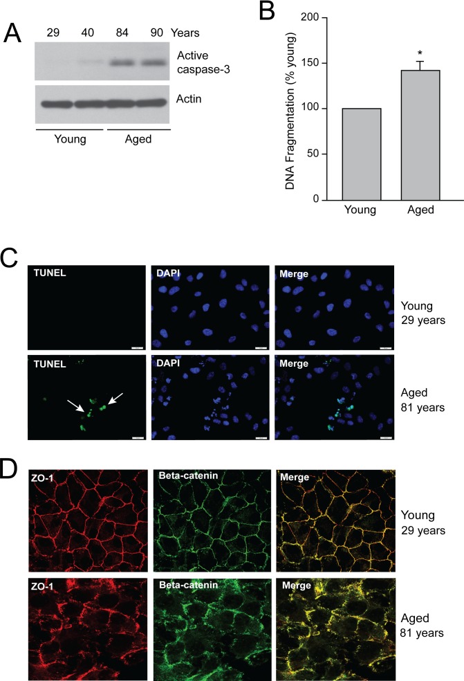
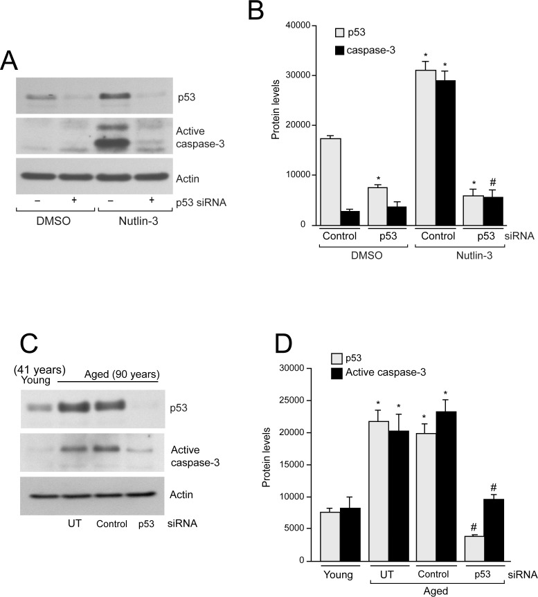
Methods: Age-associated changes in p53 activation were evaluated in primary RPE cultures from human donor eyes of various ages. Apoptosis was evaluated by activation of caspases and DNA fragmentation. Gene-specific small interfering RNA was used to knock down expression of p53.

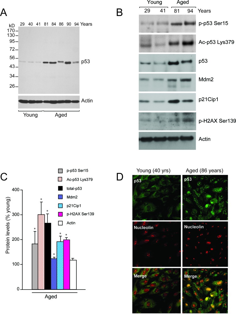
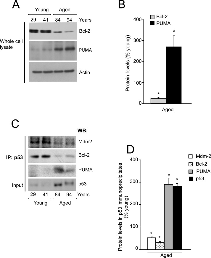
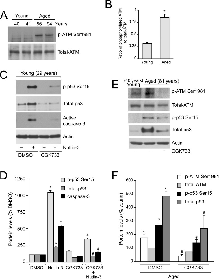
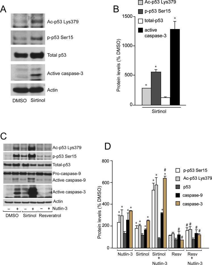
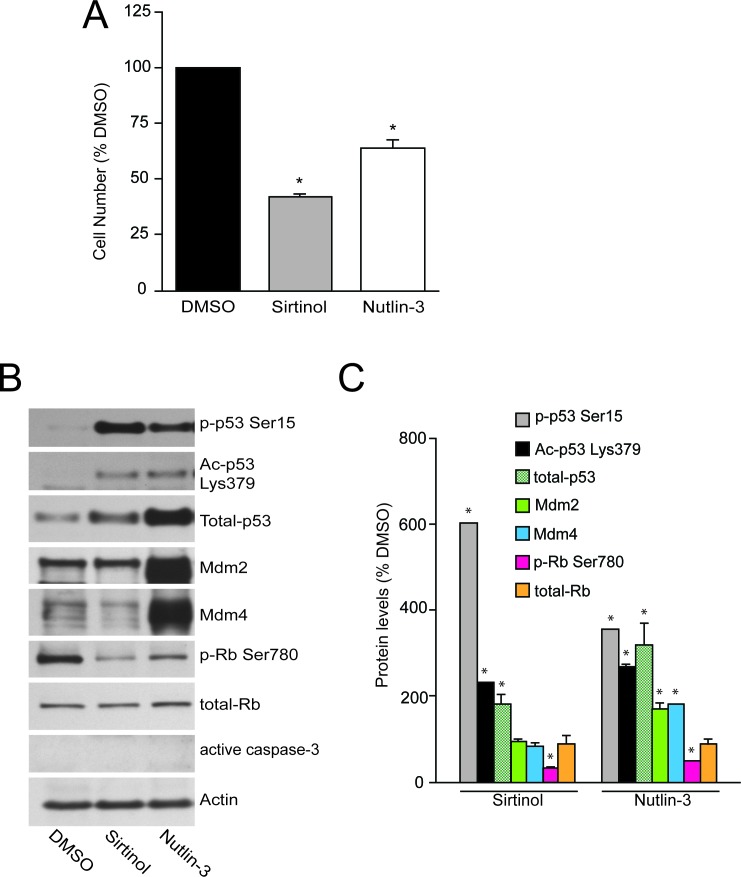
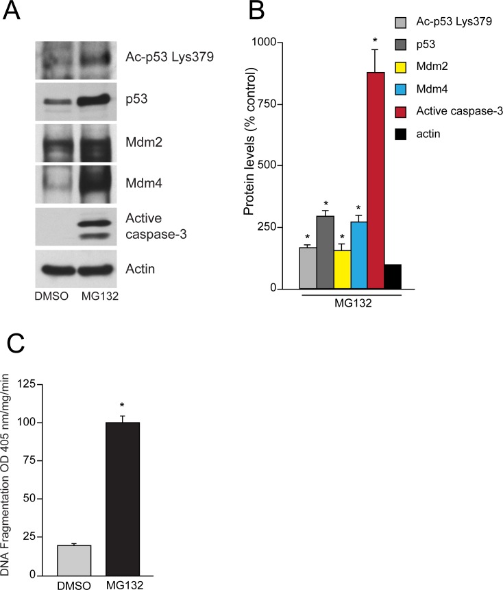
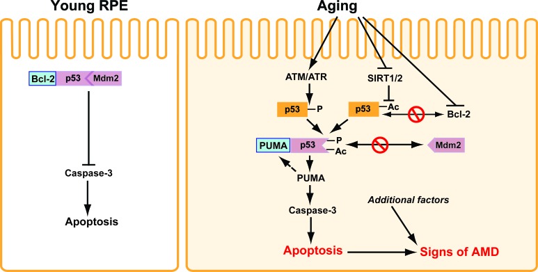
Results: We observed that the basal rate of p53-dependent apoptosis increased in an age-dependent manner in human RPE. The age-dependent increase in apoptosis was linked to alterations in several aspects of the p53 pathway. p53 phosphorylation Ser15 was increased through the stimulation of ATM-Ser1981. p53 acetylation Lys379 was increased through the inhibition of SIRT1/2. These two posttranslational modifications of p53 blocked the sequestration of p53 by Mdm2, thus resulting in an increase in free p53 and of p53 stimulation of apoptosis through increased expression of PUMA (p53 upregulated modulator of apoptosis) and activation of caspase-3. Aged RPE also had reduced expression of antiapoptotic Bcl-2, which contributed to the increase in apoptosis. Of particular interest in these studies was that pharmacologic treatments to block p53 phosphorylation, acetylation, or expression were able to protect RPE cells from apoptosis.

Conclusions: Our studies suggest that aging in the RPE leads to alterations of specific checkpoints in the apoptotic pathway, which may represent important molecular targets for the treatment of RPE-related aging disorders such as AMD.


Conflict of interest statement

Disclosure: S. Bhattacharya, None; E. Chaum, None; D.A. Johnson, None; L.R. Johnson, None

Figures

Figure 1.
Figure 1.
Aging induces apoptosis of RPE cells. RPE cells were isolated from young and aged donor eyes, and primary cultures were grown to confluence. (A) Cell lysates were separated on SDS-PAGE, and Western blot analysis was carried out using an antibody specific for active caspase-3. (B) DNA fragmentation was measured by ELISA (mean ± SEM, n = 3 from young [donor age 29, 40, and 41 years] and aged groups [donor age 81, 86, and 94 years]). *, significantly different compared with young RPE cells (P < 0.05). (C) Cell monolayers were analyzed by TUNEL staining using a kit from Roche and following the manufacturer's instructions. DAPI-containing mounting medium was used to stain nucleus. Arrowheads represent apoptotic cells. Scale bar, 20 μm. (D) Age-related disruption of tight and adherens junctions in RPE cells. RPE cells were obtained from young and aged human eye donors. Primary cultures were grown to confluence and monolayers were fixed and stained for ZO-1 (red) and beta-catenin (green) by the immunofluorescence. Proteins were localized using confocal microscopy. Scale bar, 20 μm.
Figure 2.
Figure 2.
Age-induced posttranslational modification of p53. Primary cultures of RPE cells obtained from cohorts of young and aged donor eyes were grown to confluence. (A) RPE cell lysates were analyzed for p53 expression using specific antibody. Blot was stripped and probed for actin as an internal loading control. (B) Cell lysates were analyzed by Western blot for phospho Ser15, Acetyl-Lys389, and total-p53, Mdm2, p21Cip1, and phospho-H2AX Ser139 using specific antibodies. Actin was used as an internal loading control. (C) Densitometric analysis of results shown in (A). Values from young RPE cells were set at 100%. *, significantly different compared with young RPE cells (P < 0.05). (D) Nuclear translocation of p53 in aged RPE cells. Confluent RPE monolayers were fixed and stained using antibodies specific for p53 (green) and nucleolin (red). Localization of proteins was carried out using confocal microscopy. Scale bar, 20 μm.
Figure 3.
Figure 3.
Age-dependent disruption of p53–Mdm2 binding. RPE cells isolated from young and aged donors were grown to confluence. (A) Cell lysates were analyzed by Western blot for the expression of Bcl-2 and PUMA. Actin was used as a loading control. (B) Densitometric analysis of results shown in (A). Values from young RPE cells were set at 100%. *, significantly different compared with young RPE cells (P < 0.05). (C) Cell lysates were immunoprecipitated using p53 antibody and immunoprecipitates were analyzed by Western blot using Mdm2, Bcl-2, and PUMA-specific antibodies. Blots were stripped and probed using p53 antibody (total input). (D) Densitometric analysis of results shown in (C). Values from young RPE cells were set at 100%. *, significantly different compared with young RPE cells (P < 0.05).
Figure 4.
Figure 4.
ATM/ATR kinase activates p53 in RPE cells. (A) Confluent RPE monolayers obtained from young and aged human donors were analyzed by Western blot for the levels of phospho-ATM Ser1981 and total-ATM using specific antibodies. (B) Densitometric analysis of phosphorylated and total ATM levels. *, significantly different compared with young RPE cells (P < 0.05). (C) RPE cells from young donors were pretreated with ATM/ATR kinase inhibitor (CGK733, 10 μM) for 1 hour followed by treatment with 60 μM Nutlin-3 for 2 hours. Equal volume of DMSO was used as control. Samples were analyzed for phospho-p53 Ser15, total-p53, and active caspase-3. (D) Densitometric analysis of results shown in (C). *, significantly different compared with young RPE cells treated with DMSO; #, significantly different from young RPE cells treated with Nutlin-3 (P < 0.05). (E) Confluent monolayers of aged RPE cells were treated overnight with CGK733 (10 μM). Levels of phospho-ATM Ser1981, total-ATM, phospho-p53 Ser15, and total-p53 were compared with young RPE cells using specific antibodies. Actin was used as an internal loading control. (F) Densitometric analysis of results shown in (E). Values from young RPE cells were set at 100%. *, significantly different compared with young RPE cells; #, significantly different compared with aged RPE cells treated with DMSO (P < 0.05).
Figure 5.
Figure 5.
SIRT1/2 and RPE apoptosis. (A) Confluent serum starved RPE cells from young donor eyes (29 years of age) were treated with 50 μM Sirtinol for 48 hours and cell lysates were prepared. Western blot for changes in protein levels for phospho Ser15, acetyl-Lys379, total p53, active caspase-3, and actin are shown. (B) Densitometric analysis of results shown in (A). Values from DMSO-treated cells were set at 100%. *, significantly different compared with cells treated with DMSO (P < 0.05). (C) Confluent serum-starved RPE cells from young donor eyes (40 years of age) were pretreated with or without 50 μM Sirtinol and 25 μM Resveratrol for 18 to 20 hours followed by treatment with 60 μM Nutlin-3 for 2 hours. Cell lysates were analyzed for the levels of phospho-53 Ser15, acetyl-p53 Lys379, total p53, active caspase-9, and active caspase-3 using specific antibodies. Actin was used as a loading control. (D) Densitometric analysis of results shown in (C). Values from DMSO-treated cells were set at 100%. *, significantly different compared with untreated cells; #, significantly different compared with Nutlin-3–treated cells (P < 0.05). Resv, Resveratrol.
Figure 6.
Figure 6.
Sirtinol and Nutlin-3 inhibit proliferation of RPE cells. RPE cells from young donor eyes (40 years of age) were trypsinized and equal numbers of cells were seeded in serum-containing medium. Eighteen hours later the attached cells were treated with 25 μM Sirtinol and 5 μM Nutlin-3 and grown for 48 hours in serum-containing medium. (A) The cells were trypsinized and counted. *, significantly different compared with DMSO-treated cells (P < 0.05). (B) Cell lysates were analyzed by Western blot for the levels of phospho-p53 Ser15, acetyl-p53 Lys379, total p53, Mdm2, Mdm4, phospho-Rb Ser780, total-Rb, and active caspase-3. Actin was used an internal loading control. (C) Densitometric analysis of results shown in (B). Values from DMSO treated cells were set at 100%. *, significantly different compared with cells grown in the presence of DMSO (P < 0.05).
Figure 7.
Figure 7.
Inhibition of ubiquitination by MG132 sensitizes young RPE cells to p53-dependent apoptosis. (A) RPE cells from young donor eyes (41 years of age) were treated with 10 μM MG132 for 18 hours, and lysates were subjected to Western blot analysis for the levels of acetylated p53 Lys379, total p53, Mdm2, Mdm4, and active caspase-3 using specific antibodies. Actin was used as a loading control. (B) Densitometric analysis of results shown in (A). Values from DMSO-treated young cells were set at 100%. *, significantly different compared with cells treated with DMSO (P < 0.05). (C) Confluent monolayers of RPE cells from young donor eyes (40 years of age) were treated with DMSO or 10 μM MG132 overnight. DNA fragmentation was measured by ELISA (mean ± SEM, n = 3). *, significantly different compared with cells treated with DMSO (P < 0.05).
Figure 8.
Figure 8.
Knockdown of p53 by gene-specific siRNA inhibits RPE apoptosis. RPE cells obtained from young donor eyes (29 years of age) were transfected with control or p53-specific siRNA as described in experimental procedures and treated with 60 μM Nutlin-3 for 2 hours. (A) Cell lysates were analyzed by Western blots for the levels of total p53, and active caspase-3 using specific antibodies. Membranes were stripped and probed for actin as an internal loading control. (B) Densitometric analysis of results shown in (A). *, significantly different compared with untreated cells; #, significantly different compared with Nutlin-3 treated cells (P < 0.05). (C). RPE cells obtained from aged donors were either left untreated (UT) or transfected with control or p53-specific siRNA. Total-p53 and active caspase-3 levels were compared with RPE cells from young donors using specific antibodies. (D) Densitometric analysis of results shown in (C). *, significantly different compared with young RPE; #, significantly different compared with aged RPE cells transfected with control siRNA (P < 0.05).
Figure 9.
Figure 9.
Schematic representation of age-dependent p53–Mdm2 signaling in human RPE. In RPE cells from young donors, the p53 protein is kept at low activity levels via its association with Mdm2 (E3 ubiquitin ligase). As a result, p53 cannot upregulate synthesis of proapoptotic PUMA. Furthermore, antiapoptotic Bcl-2 binds and sequesters p53 and consequently cells are resistant to apoptosis. In RPE cells from aged donors, decreased expression of antiapoptotic Bcl-2 and its reduced association with age-modified p53 increase susceptibility to apoptosis. Aging of RPE results in the phosphorylation (-P) of p53 by ATM/ATR kinases. Simultaneously, age-related inhibition of histone deacetylase (SIRT1/2) favors acetylation (-Ac) of p53. Both phosphorylation and acetylation of p53 inhibit its binding with Mdm2 and lead to its stabilization and activation. Overactive p53 in RPE from aged human donors allows nascent synthesis of PUMA. In addition, PUMA complexes with p53 and, consequently, triggers caspase-3–dependent apoptosis. Increased RPE apoptosis coupled with environmental and genetic factors set the stage for the pathogenesis of AMD.

References

    1. Friedman DS, O'Colmain BJ, Muñoz B, et al. Eye Diseases Prevalence Research Group. Prevalence of age-related macular degeneration in the United States. Arch Ophthalmol. 2004;122:564–572 - PubMed
    1. Green WR. Histopathology of age-related macular degeneration. Mol Vis. 1999;3:5–27 - PubMed
    1. Bonilha VL. Age and disease-related structural changes in the retinal pigment epithelium. Clin Ophthalmol. 2008;2:413–424 - PMC - PubMed
    1. Nowak JZ. Age-related macular degeneration (AMD): pathogenesis and therapy. Pharmacol Rep. 2006;58:353–363 - PubMed
    1. Marmor MF, Wolfensberger TJ. The Retinal Pigment Epithelium: Function and Disease. New York: Oxford University Press; 1998

Publication types

MeSH terms