

Temporal development of retinal arteriolar endothelial dysfunction in porcine type 1 diabetes
- PMID: 23139282
- PMCID: PMC3513275
- DOI: 10.1167/iovs.12-11005
Temporal development of retinal arteriolar endothelial dysfunction in porcine type 1 diabetes
Abstract
Purpose: Although hyperglycemia is implicated in retinal vascular dysfunction associated with the development of diabetic retinopathy, the temporal influence of hyperglycemia on retinal arteriolar reactivity remains unclear. Development of a large animal model of diabetes relevant to the human retina for evaluation of vascular function is also lacking. Herein, we examined nitric oxide (NO)-mediated dilation and endothelin-1 (ET-1)-induced constriction in retinal arterioles at various time periods in a porcine model of type 1 diabetes.
Methods: Retinal arterioles were isolated from streptozocin-induced diabetic pigs (2, 6, and 12 weeks of hyperglycemia, 427 ± 23 mg/dL) and age-matched control pigs (73 ± 4 mg/dL), and then cannulated and pressurized for vasoreactivity study using videomicroscopic techniques.
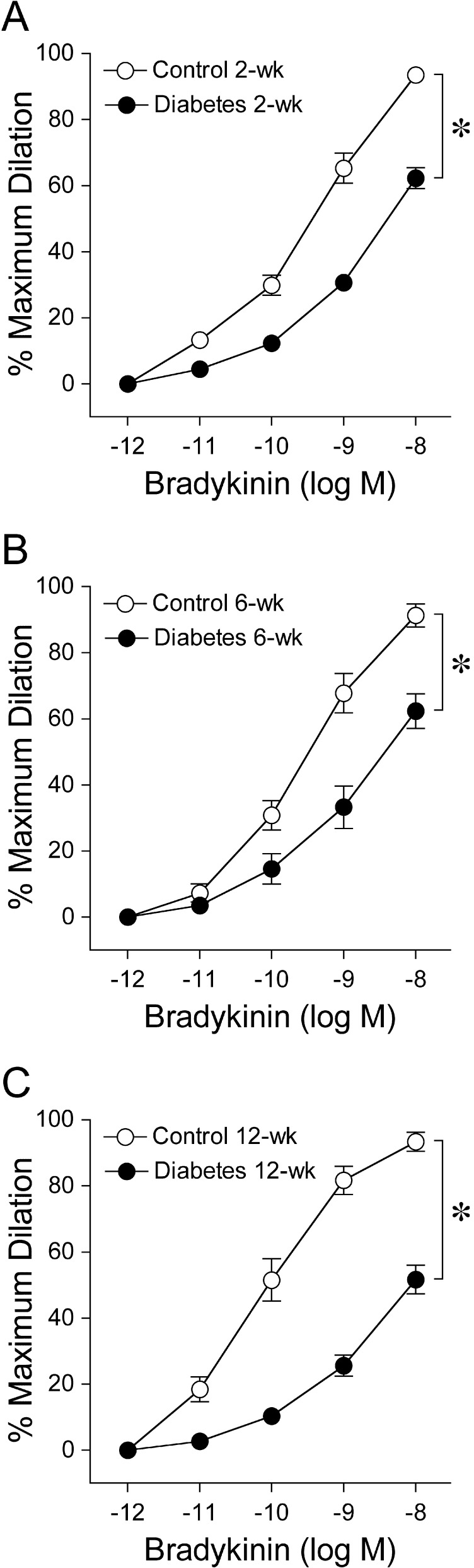
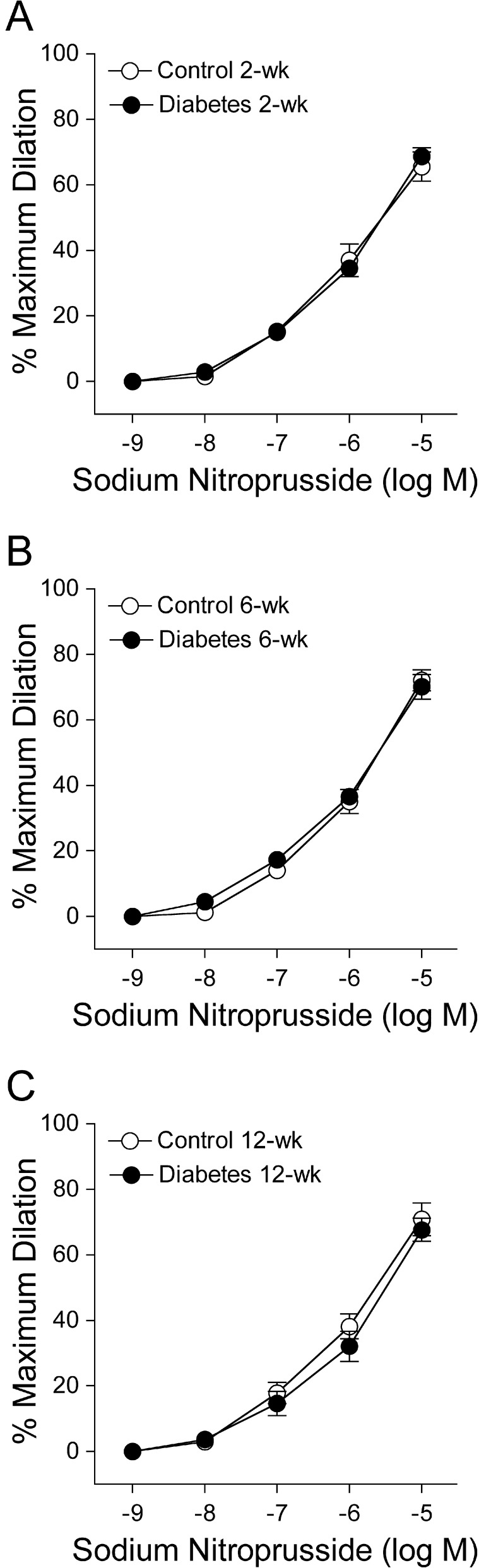
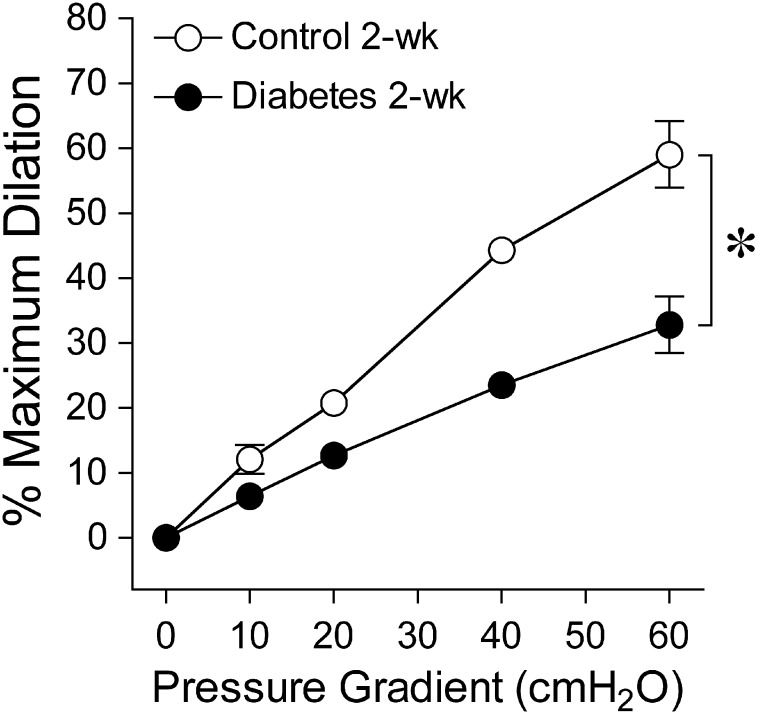
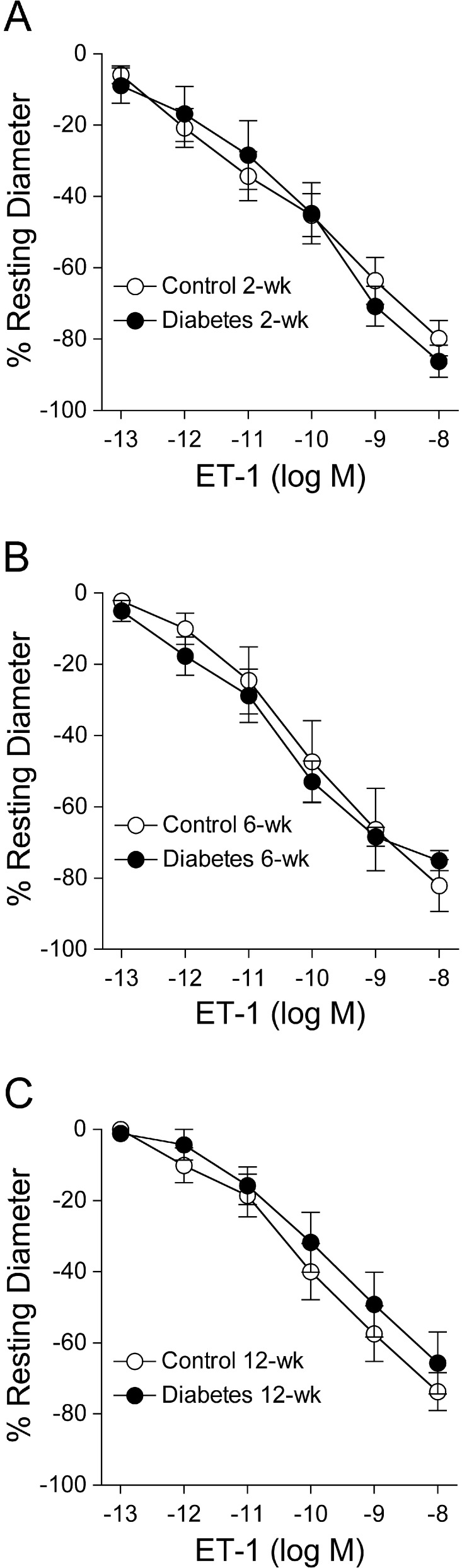
Results: Retinal arterioles isolated from control and diabetic pigs developed comparable levels of myogenic tone. The endothelium-dependent NO-mediated vasodilations to bradykinin and stepwise increases in luminal flow were significantly reduced within 2 weeks of hyperglycemia. The inhibitory effect was comparable following 6 and 12 weeks of hyperglycemia. However, the endothelium-independent vasodilation to sodium nitroprusside was unaffected. Constriction of retinal arterioles to ET-1 was unaltered at all time periods of hyperglycemia.
Conclusions: Our findings provide the first direct evidence for selective impairment of endothelium-dependent NO-mediated dilation of retinal arterioles within 2 weeks of hyperglycemia in a pig model of diabetes. By contrast, the ability of arteriolar smooth muscle to dilate to NO donor or contract to ET-1 was unaffected throughout the study period. This endothelial vasodilator dysfunction during early diabetes may contribute to development of retinopathy with chronic hyperglycemia.
Conflict of interest statement
Disclosure:
Figures
References
-
- Sivaprasad S, Gupta B, Crosby-Nwaobi R, Evans J. Prevalence of diabetic retinopathy in various ethnic groups: a worldwide perspective. Surv Ophthalmol. 2012;57:347–370 - PubMed
-
- Durham JT, Herman IM. Microvascular modifications in diabetic retinopathy. Curr Diab Rep. 2011;11:253–264 - PubMed
-
- Clermont AC, Brittis M, Shiba T, McGovern T, King GL, Bursell SE. Normalization of retinal blood flow in diabetic rats with primary intervention using insulin pumps. Invest Ophthalmol Vis Sci. 1994;35:981–990 - PubMed
Publication types
MeSH terms
Substances
Grants and funding
LinkOut - more resources
Full Text Sources
Medical
