

TRAF family member-associated NF-κB activator (TANK) induced by RANKL negatively regulates osteoclasts survival and function
- PMID: 23139637
- PMCID: PMC3492797
- DOI: 10.7150/ijbs.5079
TRAF family member-associated NF-κB activator (TANK) induced by RANKL negatively regulates osteoclasts survival and function
Abstract
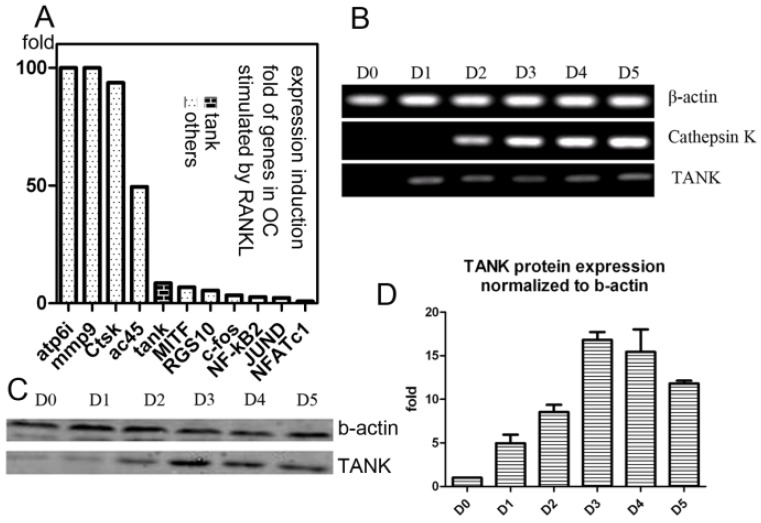
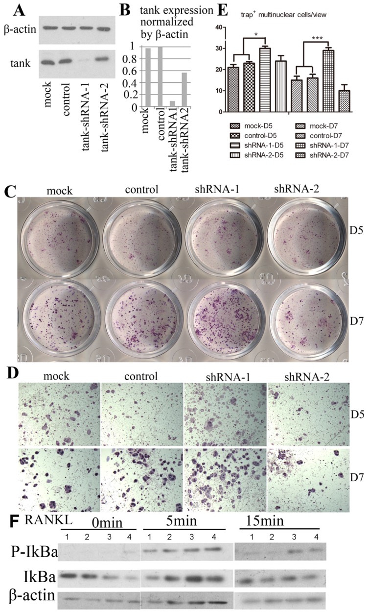
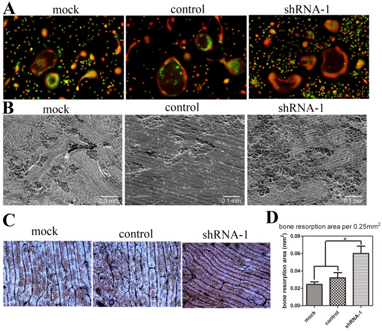
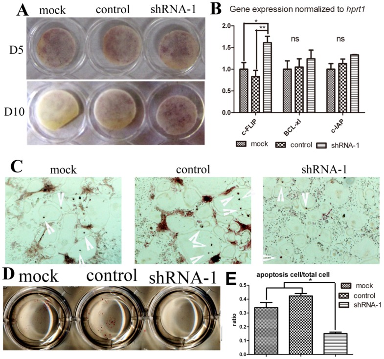
Osteoclasts are the principle bone-resorbing cells. Precise control of balanced osteoclast activity is indispensable for bone homeostasis. Osteoclast activation mediated by RANK-TRAF6 axis has been clearly identified. However, a negative regulation-machinery in osteoclast remains unclear. TRAF family member-associated NF-κB activator (TANK) is induced by about 10 folds during osteoclastogenesis, according to a genome-wide analysis of gene expression before and after osteoclast maturation, and confirmed by western blot and quantitative RT-PCR. Bone marrow macrophages (BMMs) transduced with lentivirus carrying tank-shRNA were induced to form osteoclast in the presence of RANKL and M-CSF. Tank expression was downregulated by 90% by Tank-shRNA, which is confirmed by western blot. Compared with wild-type (WT) cells, osteoclastogenesis of Tank-silenced BMMs was increased, according to tartrate-resistant acid phosphatase (TRAP) stain on day 5 and day 7. Number of bone resorption pits by Tank-silenced osteoclasts was increased by 176% compared with WT cells, as shown by wheat germ agglutinin (WGA) stain and scanning electronic microscope (SEM) analysis. Survival rate of Tank-silenced mature osteoclast is also increased. However, acid production of Tank-knockdown cells was not changed compared with control cells. IκBα phosphorylation is increased in tank-silenced cells, indicating that TANK may negatively regulate NF-κB activity in osteoclast. In conclusion, Tank, whose expression is increased during osteoclastogenesis, inhibits osteoclast formation, activity and survival, by regulating NF-κB activity and c-FLIP expression. Tank enrolls itself in a negative feedback loop in bone resorption. These results may provide means for therapeutic intervention in diseases of excessive bone resorption.
Keywords: NF-κB; Osteoclast.; RANKL; TANK.
Conflict of interest statement
Competing Interests: The authors declare no competing financial interests.
Figures

Similar articles
-
TRAF family member-associated NF-κB activator (TANK) is a negative regulator of osteoclastogenesis and bone formation.J Biol Chem. 2012 Aug 17;287(34):29114-24. doi: 10.1074/jbc.M112.347799. Epub 2012 Jul 6. J Biol Chem. 2012. PMID: 22773835 Free PMC article.
-
A medium-chain fatty acid, capric acid, inhibits RANKL-induced osteoclast differentiation via the suppression of NF-κB signaling and blocks cytoskeletal organization and survival in mature osteoclasts.Mol Cells. 2014 Aug;37(8):598-604. doi: 10.14348/molcells.2014.0153. Epub 2014 Aug 18. Mol Cells. 2014. PMID: 25134536 Free PMC article.
-
[Effect of lipopolysaccharide on osteoclasts formation and bone resorption function and its mechanism].Zhongguo Xiu Fu Chong Jian Wai Ke Za Zhi. 2018 May 15;32(5):568-574. doi: 10.7507/1002-1892.201712044. Zhongguo Xiu Fu Chong Jian Wai Ke Za Zhi. 2018. PMID: 29806344 Free PMC article. Chinese.
-
Osteoclasts: New Insights.Bone Res. 2013 Mar 29;1(1):11-26. doi: 10.4248/BR201301003. eCollection 2013 Mar. Bone Res. 2013. PMID: 26273491 Free PMC article. Review.
-
Osteoclast motility: putting the brakes on bone resorption.Ageing Res Rev. 2011 Jan;10(1):54-61. doi: 10.1016/j.arr.2009.09.005. Epub 2009 Sep 27. Ageing Res Rev. 2011. PMID: 19788940 Free PMC article. Review.
Cited by
-
A sustained-release Trametinib bio-multifunction hydrogel inhibits orthodontically induced inflammatory root resorption.RSC Adv. 2022 Jun 6;12(26):16444-16453. doi: 10.1039/d2ra00763k. eCollection 2022 Jun 1. RSC Adv. 2022. PMID: 35754868 Free PMC article.
-
Muscone Ameliorates Ovariectomy-Induced Bone Loss and Receptor Activator of Nuclear Factor-κb Ligand-Induced Osteoclastogenesis by Suppressing TNF Receptor-Associated Factor 6-Mediated Signaling Pathways.Front Pharmacol. 2020 Mar 20;11:348. doi: 10.3389/fphar.2020.00348. eCollection 2020. Front Pharmacol. 2020. PMID: 32265718 Free PMC article.
-
GPR125 positively regulates osteoclastogenesis potentially through AKT-NF-κB and MAPK signaling pathways.Int J Biol Sci. 2022 Mar 6;18(6):2392-2405. doi: 10.7150/ijbs.70620. eCollection 2022. Int J Biol Sci. 2022. PMID: 35414778 Free PMC article.
-
F2r negatively regulates osteoclastogenesis through inhibiting the Akt and NFκB signaling pathways.Int J Biol Sci. 2020 Mar 12;16(9):1629-1639. doi: 10.7150/ijbs.41867. eCollection 2020. Int J Biol Sci. 2020. PMID: 32226307 Free PMC article.
-
Guizhi-Shaoyao-Zhimu decoction attenuates bone erosion in rats that have collagen-induced arthritis via modulating NF-κB signalling to suppress osteoclastogenesis.Pharm Biol. 2021 Dec;59(1):262-274. doi: 10.1080/13880209.2021.1876100. Pharm Biol. 2021. PMID: 33626293 Free PMC article.
References
-
- Karsenty G, Wagner EF. Reaching a genetic and molecular understanding of skeletal development. Dev Cell. 2002;2:389–406. - PubMed
-
- Boyle WJ, Simonet WS, Lacey DL. Osteoclast differentiation and activation. Nature. 2003;423:337–42. - PubMed
-
- Grigoriadis AE, Wang ZQ, Cecchini MG, Hofstetter W, Felix R, Fleisch HA. et al. c-Fos: a key regulator of osteoclast-macrophage lineage determination and bone remodeling. Science. 1994;266:443–8. - PubMed
-
- Takayanagi H, Kim S, Koga T, Nishina H, Isshiki M, Yoshida H. et al. Induction and activation of the transcription factor NFATc1 (NFAT2) integrate RANKL signaling in terminal differentiation of osteoclasts. Dev Cell. 2002;3:889–901. - PubMed
Publication types
MeSH terms
Substances
Grants and funding
LinkOut - more resources
Full Text Sources
Research Materials
