. 2012;7(11):e47452.
doi: 10.1371/journal.pone.0047452. Epub 2012 Nov 5.

Large-scale screen for modifiers of ataxin-3-derived polyglutamine-induced toxicity in Drosophila

Affiliations

Large-scale screen for modifiers of ataxin-3-derived polyglutamine-induced toxicity in Drosophila

Hannes VoSSfeldt et al. PLoS One. 2012.

Abstract

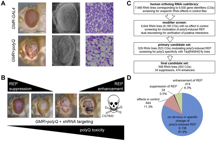
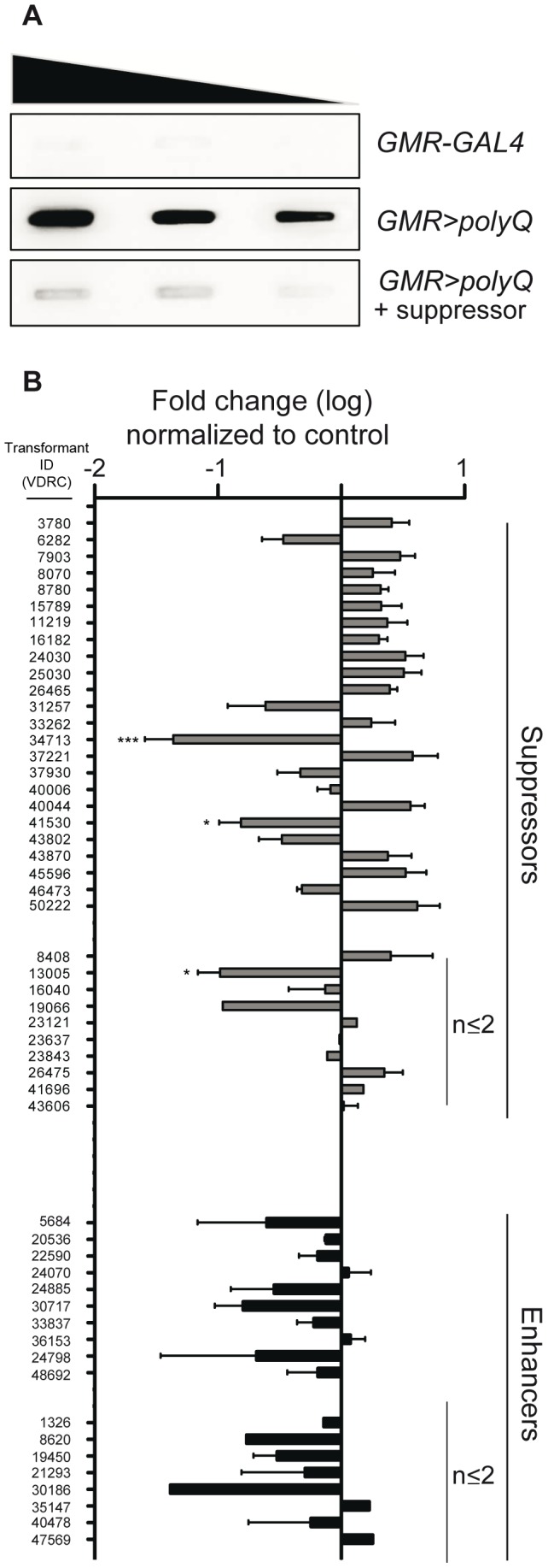
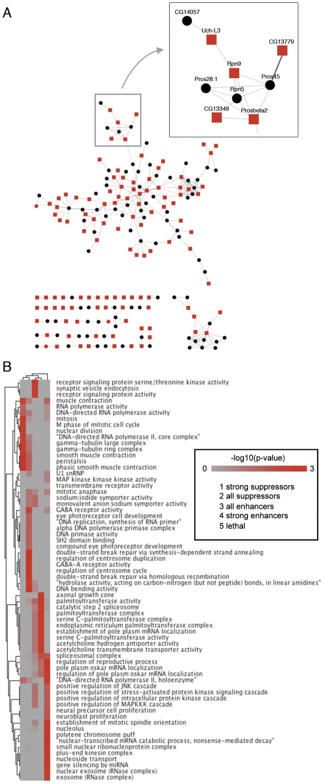
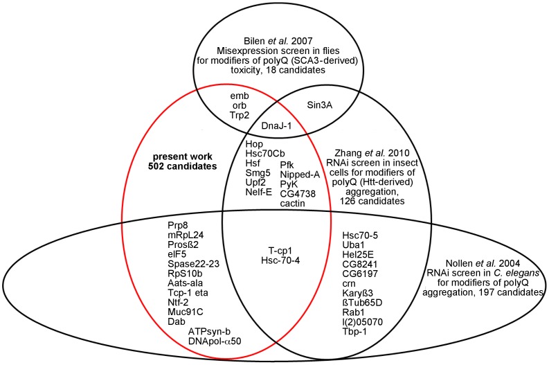
Polyglutamine (polyQ) diseases represent a neuropathologically heterogeneous group of disorders. The common theme of these disorders is an elongated polyQ tract in otherwise unrelated proteins. So far, only symptomatic treatment can be applied to patients suffering from polyQ diseases. Despite extensive research, the molecular mechanisms underlying polyQ-induced toxicity are largely unknown. To gain insight into polyQ pathology, we performed a large-scale RNAi screen in Drosophila to identify modifiers of toxicity induced by expression of truncated Ataxin-3 containing a disease-causing polyQ expansion. We identified various unknown modifiers of polyQ toxicity. Large-scale analysis indicated a dissociation of polyQ aggregation and toxicity.


Conflict of interest statement

Competing Interests: The authors have declared that no competing interests exist.

Figures

Figure 1
Figure 1. Screening for modifiers of polyQ-induced toxicity.
(A) Rough eye phenotype (REP) used as a primary readout for screening. Compared to control (upper panels), eye-specific (GMR-GAL4) expression of polyQ (lower panels) induces disturbances of the external eye texture, e. g. depigmentation of the compound eye observed by light microscopy (left) and as depicted in scanning electron micrographs (middle). Toluidine blue-stained semi-thin eye sections reveal that the disturbance of external eye structures is accompanied by degeneration of retinal cells (right). (B) Modification of the polyQ-induced REP by enhancers and suppressors. VDRC transformants used to silence respective genes: CG3284 (11219), CG16807 (23843), CG15399 (19450) and CG7843 (22574). (C) Flow chart of the screening procedures to identify modifiers of polyQ-induced toxicity. (D) Brief summary of screen results. Scale bars represent either 200 µm in eye pictures or 50 µm in semi-thin eye sections.
Figure 2
Figure 2. Analysis of polyQ aggregate load.
(A) Exemplified filter retardation analysis to visualize polyQ aggregates. Decreasing amounts of loaded protein derived from fly heads of control (GMR-GAL4, top), GMR>polyQ (middle) or GMR>polyQ in combination with a candidate suppressor (bottom). (B) Densitometric measures of filter retardation analysis. Data depicted as fold change compared to control (GMR>polyQ) for suppressors and enhancers of polyQ-induced toxicity. Independent homogenates (if available) were used for repetitions. In case of none or only one independent repetition n≤2 is indicated. In all other cases, number of independent repetitions is n≥3. Significant changes are indicated * p<0.05; *** p<0.001.
Figure 3
Figure 3. Computational analysis of modifiers of polyQ-induced toxicity.
(A) Meta-interaction network displaying modifiers of polyQ toxicity. Only candidates causing a robust modification of the REP (red) as well as directly interacting subtle modifiers (black) were retained from an initial network of more than 5 k genes with 20 k interactions . One local cluster of functionally interacting modifiers is highlighted. (B) Gene Ontology analysis of these candidate gene groups. Shown are -log10(p-value) scores for GO term enrichment for candidate gene groups (horizontal axis, see inset for group identities) and GO term (vertical). The matrix incorporates the structure of the GO hierarchy and is based on the Topology Weighted Term-algorithm as implemented in Ontologizer (terms with a p-value<0.005 are shown).
Figure 4
Figure 4. Overlap between screens for genetic modifiers of polyQ-induced toxicity or aggregation.
The Venn-like diagram displays only candidate genes shared by the different screens. Mode of modification (enhancement/suppression) is not addressed, due to the different readouts (aggregation/toxicity), model systems (Drosophila, insect cells, C. elegans) and elongated polyQ-containing proteins used in the diverse screening approaches.

Similar articles

Cited by

References

    1. Durr A Autosomal dominant cerebellar ataxias: polyglutamine expansions and beyond. Lancet Neurol 9: 885–894. - PubMed
    1. Kawaguchi Y, Okamoto T, Taniwaki M, Aizawa M, Inoue M, et al. (1994) CAG expansions in a novel gene for Machado-Joseph disease at chromosome 14q32.1. Nat Genet 8: 221–228. - PubMed
    1. La Spada AR, Taylor JP (2010) Repeat expansion disease: progress and puzzles in disease pathogenesis. Nat Rev Genet 11: 247–258. - PMC - PubMed
    1. Mangiarini L, Sathasivam K, Seller M, Cozens B, Harper A, et al. (1996) Exon 1 of the HD gene with an expanded CAG repeat is sufficient to cause a progressive neurological phenotype in transgenic mice. Cell 87: 493–506. - PubMed
    1. Orr HT, Zoghbi HY (2007) Trinucleotide repeat disorders. Annu Rev Neurosci 30: 575–621. - PubMed

Publication types

MeSH terms

LinkOut - more resources