

ROCK/Cdc42-mediated microglial motility and gliapse formation lead to phagocytosis of degenerating dopaminergic neurons in vivo
- PMID: 23139861
- PMCID: PMC3492875
- DOI: 10.1038/srep00809
ROCK/Cdc42-mediated microglial motility and gliapse formation lead to phagocytosis of degenerating dopaminergic neurons in vivo
Abstract
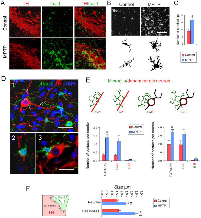
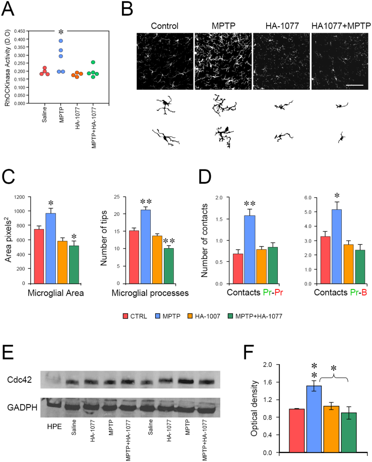
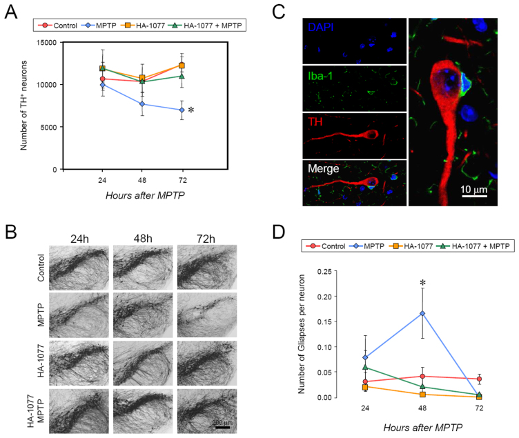
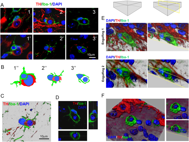
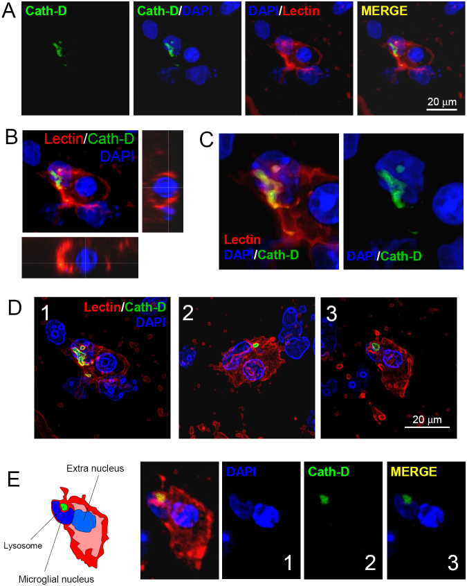
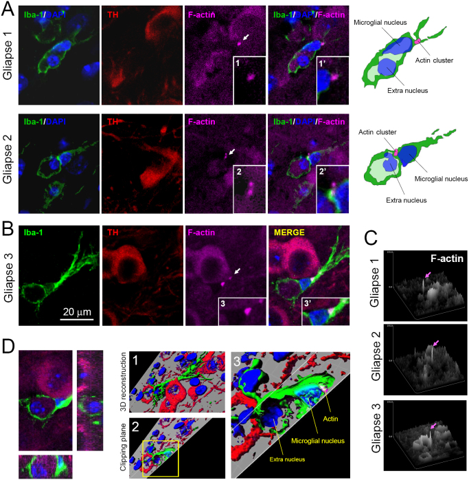
The role of microglial motility in the context of adult neurodegeneration is poorly understood. In the present work, we investigated the microanatomical details of microglia-neuron interactions in an experimental mouse model of Parkinson's disease following the intraperitoneal injection of MPTP. The specific intoxication of dopaminergic neurons induces the cellular polarization of microglia, leading to the formation of body-to-body neuron-glia contacts, called gliapses, which precede neuron elimination. Inhibiting ROCK/Cdc42-mediated microglial motility in vivo blocks the activating features of microglia, such as increased cell size and number of filopodia and diminishes their phagocyting/secreting domains, as the reduction of the Golgi apparatus and the number of microglia-neuron contacts has shown. High-resolution confocal images and three-dimensional rendering demonstrate that microglia engulf entire neurons at one-to-one ratio, and the microglial cell body participates in the formation of the phagocytic cup, engulfing and eliminating neurons in areas of dopaminergic degeneration in adult mammals.
Figures


References
-
- Nimmerjahn A., Kirchhoff F. & Helmchen F. Resting microglial cells are highly dynamic surveillants of brain parenchyma in vivo. Science (New York, N.Y) 308, 1314–1318 (2005). - PubMed
-
- Davalos D. et al. ATP mediates rapid microglial response to local brain injury in vivo. Nature neuroscience 8, 752–758 (2005). - PubMed
-
- Inoue K., Koizumi S., Kataoka A., Tozaki-Saitoh H. & Tsuda M. P2Y(6)-Evoked Microglial Phagocytosis. International review of neurobiology 85, 159–163 (2009). - PubMed
Publication types
MeSH terms
Substances
LinkOut - more resources
Full Text Sources
Other Literature Sources
Molecular Biology Databases
Miscellaneous
