. 2012 Sep;2(5):595-605.
doi: 10.1002/brb3.86. Epub 2012 Aug 10.

Role of C-terminal negative charges and tyrosine residues in fibril formation of α-synuclein

Affiliations

Role of C-terminal negative charges and tyrosine residues in fibril formation of α-synuclein

Yasutaka Izawa et al. Brain Behav. 2012 Sep.

Abstract

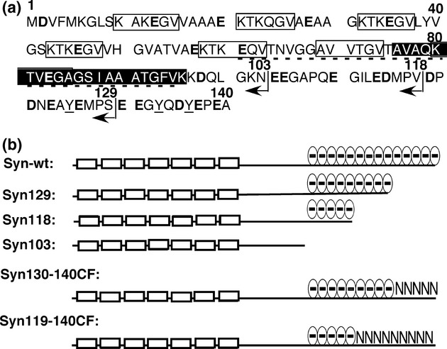
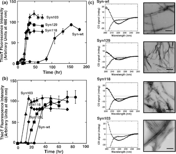
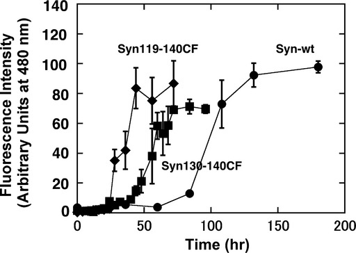
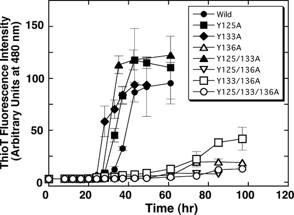
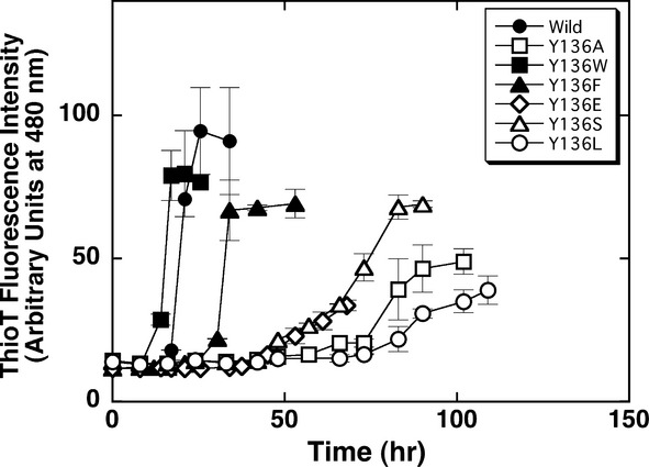
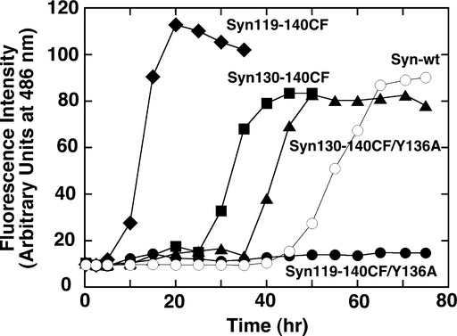
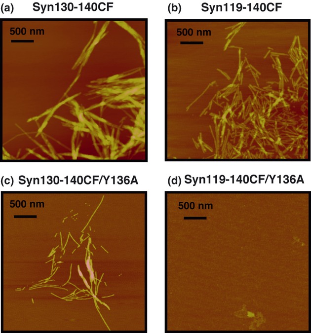
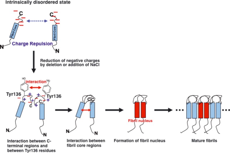
α-Synuclein (140 amino acids), one of the causative proteins of Parkinson's disease, forms amyloid fibrils in brain neuronal cells. In order to further explore the contributions of the C-terminal region of α-synuclein in fibril formation and also to understand the overall mechanism of fibril formation, we reduced the number of negatively charged residues in the C-terminal region using mutagenesis. Mutants with negative charges deleted displayed accelerated fibril formation compared with wild-type α-synuclein, demonstrating that negative charges located in the C-terminal region of α-synuclein modulate fibril formation. Additionally, when tyrosine residues located at position 125, 133, and 136 in the C-terminal region were changed to alanine residue(s), we found that all mutants containing the Tyr136Ala mutation showed delays in fibril formation compared with wild type. Mutation of Tyr136 to various amino acids revealed that aromatic residues located at this position act favorably toward fibril formation. In mutants where charge neutralization and tyrosine substitution were combined, we found that these two factors influence fibril formation in complex fashion. These findings highlight the importance of negative charges and aromatic side chains in the C-terminal region of α-synuclein in fibril formation.

Keywords: Amyloid; Parkinson's disease; amyloid formation mechanism; protein aggregation; site-directed mutagenesis; α-Synuclein.


Figures

Figure 1
Figure 1
Amino acid sequence of α-syn and the schematic representation of mutants used in this study. (a) Amino acid sequence of α-syn. Open squares indicate the imperfect KTKEGV repeats. The closed square indicates the fibril core region determined previously. The NAC region is indicated by a dotted line under the sequence. Acidic amino acid residues are represented in bold and the tyrosine residues mutated in this study are underlined. The position of the C-terminal amino acid in the truncated mutants are denoted by arrows. (b) Schematic drawing of the relative polypeptide length and negative charge distributions of the C-terminal truncated or mutated proteins. The open boxes represent the imperfect KTKEGV region.
Figure 2
Figure 2
Fibril formation characteristics of the C-terminal truncated mutants. (a) and (b) Amyloid fibril formation monitored by ThioT binding assay. Conditions were 1 mg/mL protein in 25 mmol/L Tris–HCl buffer, pH 7.5, containing 0 mol/L (a) and 150 mmol/L (b) NaCl, at 37°C. Closed circles indicate Syn-wt; closed squares, Syn129; closed diamonds, Syn118; closed triangles, Syn103. Standard error bars derived from at least three independent measurements are also shown. (c) CD spectra and TEM images of Syn-wt, Syn129, Syn118, and Syn103. In the CD spectra, open and closed symbols represent for samples before and after incubation for fibril formation, respectively. The scale bar in each panel of TEM image represents 200 nm.
Figure 3
Figure 3
Fibril formation characteristics of the charge-free full-length α-syn mutants. Conditions were 1 mg/mL protein in 25 mmol/L Tris–HCl buffer, pH 7.5 at 37°C. Closed circles indicate Syn-wt; closed squares, Syn130-140CF; closed diamonds, Syn119-140CF. Standard error bars derived from at least three independent measurements are also shown.
Figure 4
Figure 4
Fibril formation characteristics of various tyrosine substitution α-syn mutants. Conditions were 1 mg/ml protein in 25 mmol/L Tris–HCl buffer, containing 1 mol/L NaCl, pH 7.5 at 37°C. Representations of symbols are explained in the figure. Standard error bars derived from at least three independent measurements are also shown.
Figure 5
Figure 5
Fibril formation characteristics of various Tyr136 substitution α-syn mutants. Conditions were the same as described in the legend of Figure 4. Representations of symbols are explained in the figure. Standard error bars derived from at least three independent measurements are also shown.
Figure 6
Figure 6
Fibril formation characteristics of Tyr136Ala mutants of Syn130-140CF and Syn119-140CF. Conditions were 1 mg/mL protein in 25 mmol/L Tris–HCl buffer, containing 150 mmol/L NaCl, pH 7.5 at 37°C. Plate readers of ARVO X4 (Perkin Elmer) was used for measurements by shaking. Representations of symbols: Syn-wt (open circles), Syn130-140CF (closed squares), Syn119-140CF (closed diamonds), Syn130-140CF/Y136A (closed triangles), Syn119-140CF/Y136A (closed circles).
Figure 7
Figure 7
AFM images of α-syn mutants prepared in Figure 6. (A) Syn130-140CF, (B) Syn119-140CF, (C) Syn130-140CF/Y136A, and (D) Syn119-140C/Y136A. The scale bars represent 500 nm.
Figure 8
Figure 8
A schematic model of α-syn fibril formation mechanism. Roles of the C-terminal negative charges and Tyr136 on the fibril formation, especially on the fibril nucleus formation step, are shown. The long blue squares represent the fibril core region site of the α-syn (Yagi et al. 2010). See text for details.

Similar articles

Cited by

References

    1. Baba M, Nakajo S, Tu PH, Tomita T, Nakaya K, Lee VM, et al. Aggregation of alpha-synuclein in Lewy bodies of sporadic Parkinson's disease and dementia with Lewy bodies. Am. J. Pathol. 1998;152:879–884. - PMC - PubMed
    1. Bisaglia M, Mammi S, Bubacco L. Structural insights on physiological functions and pathological effects of alpha-synuclein. FASEB J. 2009;23:329–340. - PubMed
    1. Carrell RW, Lomas DA. Conformational disease. Lancet. 1997;350:134–138. - PubMed
    1. Cho MK, Nodet G, Kim HY, Jensen MR, Bernado P, Fernandez CO, et al. Structural characterization of alpha-synuclein in an aggregation prone state. Protein Sci. 2009;18:1840–1846. - PMC - PubMed
    1. Conway KA, Harper JD, Lansbury PT., Jr Fibrils formed in vitro from alpha-synuclein and two mutant forms linked to Parkinson's disease are typical amyloid. Biochemistry. 2000;39:2552–2563. - PubMed

LinkOut - more resources