. 2012 Sep;2(5):628-39.
doi: 10.1002/brb3.84. Epub 2012 Aug 23.

The expression of nicotinic receptor alpha7 during cochlear development

Affiliations

The expression of nicotinic receptor alpha7 during cochlear development

Scott W Rogers et al. Brain Behav. 2012 Sep.

Abstract

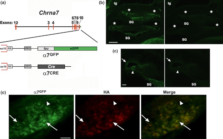
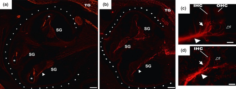
Nicotinic acetylcholine receptor alpha7 expression was examined in the developing and adult auditory system using mice that were modified through homologous recombination to coexpress either GFP (alpha7GFP) or Cre (alpha7Cre), respectively. The expression of alpha7GFP is first detected at embryonic (E) day E13.5 in cells of the spiral prominence. By E14.5, sensory regions including the putative outer hair cells and Deiters' cells express alpha7GFP as do solitary efferent fibers. This pattern diminishes after E16.5 in a basal to apex progression, as Hensen's cells and cells of the spiral ligament acquire alpha7GFP expression. At birth and thereafter alpha7GFP also identifies a subset of spiral ganglion cells whose processes terminate on inner hair cells. Efferent fibers identified by peripherin or calcitonin gene-related protein do not coexpress alpha7GFP. In addition to cochlear structures, there is strong expression of alpha7GFP by cells of the central auditory pathways including the ventral posterior cochlear nucleus, lateral lemniscus, central inferior colliculus, and the medial geniculate nucleus. Our findings suggest that alpha7 expression by both neuronal and non-neuronal cells has the potential to impact multiple auditory functions through mechanisms that are not traditionally attributed to this receptor.

Keywords: Alpha7; auditory system; cochlear; development; mouse; nicotinic acetylcholine receptor.


Figures

Figure 1
Figure 1
Mouse models used to examine nicotinic receptor α7 expression. (A) A diagram showing how the α7 gene (Chrna7) was modified using homologous recombination to add a C-terminus epitope tag (hemagglutinin [HA]) and inserted into the 3′ terminus of Chrna7 a reporter bicistronic internal ribosome entry sequence (IRES)-tau fusion to enhanced green fluorescent protein (eGFP) fusion protein cassette (α7GFP; see Methods and Rogers and Gahring 2012; Rogers et al. 2012). This construct was subsequently altered by replacing the tau:GFP cassette with the Cre-recombinase gene (α7Cre). (B, C) The visualization of the Chrna7 transcription using immunological detection of GFP compared with background. Shown are sagittal sections of the cochlear sensory structures of an E16.5 α7GFP embryo in (B) and at greater magnification in (C). The panels on the left are stained for GFP expression (see Methods), whereas the image on the right shows an adjacent serial section that received the same staining treatment, only primary antibody was omitted. Photographs were collected at the same gain and exposure. The asterisk identifies cochlear ducts and the arrow points to the spiral prominence and the arrow head points to cell giving rise to the outer hair cells and Deiters' cells. Abbreviations are SG, spiral ganglion; and tg, trigeminal ganglion. In (B), the bar = 100 μm and in (C), the bar = 400 μm. (D) Examples of colabeling for α7GFP (green) and anti-HA (HA) in cells associated with the spiral ganglion at E16.5. Examples of double-labeled cells are identified by with arrows. Some processes are also colabeled (arrow head). Bar = 50 μm.
Figure 2
Figure 2
The expression of α7GFP varies with cochlear development. The expression of α7GFP identified by immunohistochemical detection of GFP (Methods) is shown for the embryonic cochlear structures in the sagittal section plane. (A) E12.5 shows one early cochlear structure and associated background fluorescence. (B) E13.5, the first identified expression of α7GFP in cells of the spiral prominence (sp). (C) An E14.5 cochlear structure shows the expression of α7GFP in the presumptive sensory region (sr) and pioneering efferents are also identified (arrows). (D) The E14.5 sr is shown at greater magnification. (E) E15.5 increased expression of α7GFP by cells of the sr is present as ar sp cells. Labeled processes associated with the spiral ganglion (SG) are identified by (arrows). (F) E16.5 α7GFP is seen in outer hair cells (OHC) and Deiters' cells (D). The expression of α7GFP in afferent processes is noted (arrow). The insert box shows a similar section colabeled with α7GFP (green) and S100beta (red; Buckiova and Syka 2009) identifies the inner hair cell (open arrow). (G) Increased magnification of the E16.5 sensory region showing the OHC and Deiters' (D) cell labeling (open arrow). (H) The E18.5 cochlear structure exhibits decreased α7GFP expression in OHCs and acquisition of label by Hensen's cells (H). Also labeled are SG afferents (arrow) and their terminals on the base of IHCs (arrow head). The asterisk identifies autofluorescence in the tectorial membrane that is inconsistently present. (I) The E18.5 sensory region is shown at greater magnification. (J) The postnatal day 6 (P6) α7GFP expression pattern reveals the SG afferents (arrow) that terminate as part of the extensive synaptic terminals at the base of the IHCs (arrow head). A Hensen's cell (H) is identified. (K) The P6 α7GFP expression image in Panel I is superimposed over a phase contrast image of the cochlear structure. Single afferents (arrow) and their terminals (arrow head) are evident. Tectorial membrane (TM). (L) Diagrams depicting the adult cochlear structures (as labeled) and the major expression patterns of α7GFP (green) at the corresponding developmental stage. Bars = 100 μm (A, B, C, E, F, H) or 20 μm (D, F-insert,G, I).
Figure 3
Figure 3
Remodeling of α7GFP Expression is from basal to apical. (A) A sagittal section showing the E16.5 cochlear structure and α7GFP expression (green). At this stage, all cochlear structures exhibit a similar pattern of α7GFP expression by cells of the lesser epithelial ridge and presumptive sensory region (arrow heads). Also labeled are cells of the SG, the cochlear nerve (cn), trigeminal ganglion (tg), and the trigeminal nucleus (V) and spiral prominence (sp). (B) A similar view of the E18.5 cochlear structure shows four cochlear ducts (arrow heads) that are numbered in a basal-to-apex direction. (C) Greater magnification of the numbered cochlear ducts in B shows the corresponding α7GFP expression. The arrow indicates the site of shift in α7GFP expression. The asterisks identify autofluorescence in the tectorial membrane. (D) Diagrams illustrating the approximate site of each numbered duct in the cochlear structure and identification of the α7GFP labeling of the individual cochlear sensory cells regions. Bars = 100 μm.
Figure 4
Figure 4
Postnatal expression of α7GFP in the cochlear structure. (A) An image of a sagittal section showing the P6 cochlear structure and the expression of α7GFP in afferents originating from spiral ganglion (SG) cells that terminate (arrow head) at the base of the inner cell. Hensen's cells (H) and cells in the spiral ligament are also labeled (SL). (B) A low-magnification image of an adjacent section shows the expression by cells of the spiral prominence (SP). Other labels are as in (A). (C) Another serial section reveals expression of α7GFP in cells of the spiral ligament (SL). (D) Increased magnification shows the α7GFP signal to be in SL cell bodies and the elaborated processes of these cells. (E) SL cell bodies in the P12 mouse. The labeled cell bodies (arrow heads) and their associated processes extend throughout this region.
Figure 5
Figure 5
The α7GFP expression during cochlear innervation. Innervation of the developing cochlear structure is revealed by α7GFP labeling. (A) An E13.5 sagittal section shows a group of efferent processes (arrow) that distribute to solitary fibers that are strongly labeled for α7GFP expression (arrow heads). Cells of the putative sensory region (sr) and the spiral prominence (SP) are identified. (B) At greater magnification, these fibers (arrow heads) have a beaded appearance and project towards the base of sr. (C) The possible origin of the pioneering efferent fibers is suggested by the intense expression of α7GFP in the E13.5 cell groups (arrow) located caudal to the trigeminal sensory nucleus (V) consistent with the early olive in this horizontal section through the posterior brain stem. At increased magnification (Insert), the cell clustering (arrow) and their projections (arrowhead) are identified. Serial sections (not shown) reveal continuity between these cells and those entering the cochlear structures (arrow heads). (D) E15.5 α7GFP expression and colabeling with other neuronal process markers (red). The processes that express peripherin (arrow) end mostly in the vicinity of the inner hair cells (IHC). Occasional solitary fibers (arrow) extend towards the base of the outer hair cells (OHC) at the dorsal boarder of the Deiters' cells (D). (E) The E16.5 cochlear innervation pattern looks much the same as E15.5, although the peripherin-labeled fibers (arrow) are more distinct. These processes lack detectable α7GFP expression. (F) Olivocochlear efferents identified by calcitonin gene-related proteins (CGRP; arrows). (G) The E18.5 embryo exhibits afferents detected by α7GFP expression (arrows). These extend from SG cells that are not colabeled with peripherin (not shown). (H) At birth (P0), there are distinctly labeled α7GFP afferents (arrowhead) and peripherin-labeled efferents that extend to the Deiters' cells (D) and then turn (arrows) to contact the base of the OHCs. Hensen's cells are noted (H). (I) The P12 innervation pattern is similar to the P0. In this merged image of α7GFP expression (green) and peripherin (red), many spiral ganglion (SG) cells and processes are labeled, but the labels only rarely overlap in the same processes (see insert). The α7GFP identify mostly processes reaching the IHCs (arrow). Peripherin-labeled processes mostly terminate at the base of the outer hair cells (OHC) or onto the α7GFP-labeled afferent fiber near the base of the IHC. Hensen's cells expressing α7GFP is identified (H). The inset shows the sensory cell region at increased magnification. The arrows identify the α7GFP-expressing afferent ending at the base of nonlabeled IHC, whereas the double arrow heads point to the peripherin-labeled terminal. Other peripherin processes extend to the base of the OHCs (individual arrow heads). (J) Diagrams as in Fig. 2 depicting the basic innervation patterns observed in this study. Green is α7GFP and red is peripherin. Afferent (af) and efferents (ef). Bars = 50 μm
Figure 6
Figure 6
The ablation of α7 cell lineages is consistent with α7GFP expression. Comparison of a cochlear structure labeled for expression of the filament marker peripherin from an E16.5 α7GFP mouse (A) and similarly timed α7Cre:DTA ablated embryo (B). The basic patterning of the cochlear structures is outlined by white dots and the spiral ganglia (SG) and trigeminal nerve (TG) are identified, Arrow heads point to cochlear nerves and the asterisk identifies in A the cochlear ducts (asterisk in A). (C) Greater magnification of the cochlear sensory cell region of an α7GFP or (D) α7Cre:DTA embryo. The arrow heads identify prominent cochlear nerve bundles and filled arrows point to fibers in the inner cell (IHC) region. Open arrows identify fibers extending to the sensory cell region, giving rise to outer hair cells and Deiters' cells. Bars: 100 μm (A, B); 20 μm (C, D).
Figure 7
Figure 7
Central auditory systems express α7GFP. Central auditory nuclei identified by α7GFP expression. (A) At E18.5 in this sagittal image of the entire otic complex and the adjacent basal brainstem is included. The cochlear nucleus (C) and the eighth cranial nerve (8n) are visible as is the fifth cranial nerve (5n), the trigeminal nucleus (TGN), and trigeminal ganglion (TGG). Also noted are cochlear ducts (asterisk) and a spiral ganglion (SG). (B) At P12, α7GFP expression of cochlear complex reveals the strongly labeled cells of the ventral-posterior cochlear nucleus (VCP). The dorsal cochlear nucleus (DC) and ventral-anterior cochlear nucleus (VCA) are identified and is the eighth nerve (8n) and a cochlear duct (asterisk). The inset shows the VCP at increased magnification. Cells clusters expressing α7GFP (arrowhead) and individual cells that resemble the morphology of octopus cells described previously (Morley and Happe 2000; Morley 2005) to express a7 (arrow) are noted. (C) Another P12 sagittal section reveals α7GFP expression in the ascending central auditory pathways. (D) The expression of α7GFP in the inferior colliculus (CIC) of this horizontal section reveals staining of the commissural fibers (arrow). Structures identified are the brachium of the inferior colliculus (BIC); dentate gyrus (DG), inferior colliculus, central nucleus (CIC); lateral lemniscus, dorsal nucleus (DLL); lateral lemniscus, ventral nucleus (VLL); medial geniculate nucleus (MGN), and the substantia nigra (SN). Bars = 100 μm (A, B0; 20 μm (B-insert), and 1 mm (C, D).

Similar articles

Cited by

References

    1. Albuquerque EX, Pereira EF, Alkondon M, Rogers SW. Mammalian nicotinic acetylcholine receptors: from structure to function. Physiol. Rev. 2009;89:73–120. - PMC - PubMed
    1. Araki H, Suemaru K, Gomita Y. Neuronal nicotinic receptor and psychiatric disorders: functional and behavioral effects of nicotine. Jpn. J. Pharmacol. 2002;88:133–138. - PubMed
    1. Brown AS. The environment and susceptibility to schizophrenia. Prog. Neurobiol. 2011;93:23–58. - PMC - PubMed
    1. Buckiova D, Syka J. Calbindin and S100 protein expression in the developing inner ear in mice. J. Comp. Neurol. 2009;513:469–482. - PubMed
    1. Elgoyhen AB, Johnson DS, Boulter J, Vetter DE, Heinemann S. Alpha 9: an acetylcholine receptor with novel pharmacological properties expressed in rat cochlear hair cells. Cell. 1994;79:705–715. - PubMed

LinkOut - more resources