Review
. 2013:53:531-56.
doi: 10.1146/annurev-pharmtox-032112-135923. Epub 2012 Nov 8.

Structure-function of the G protein-coupled receptor superfamily

Affiliations
Review

Structure-function of the G protein-coupled receptor superfamily

Vsevolod Katritch et al. Annu Rev Pharmacol Toxicol. 2013.

Abstract

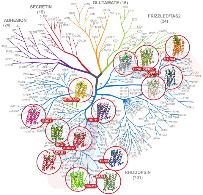
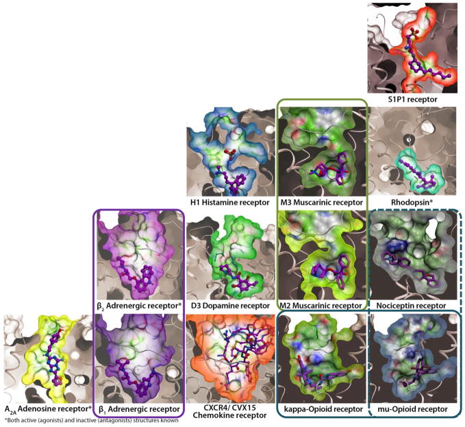
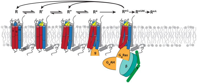
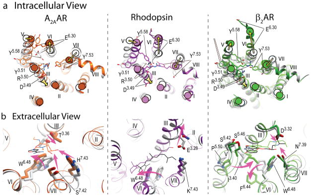
During the past few years, crystallography of G protein-coupled receptors (GPCRs) has experienced exponential growth, resulting in the determination of the structures of 16 distinct receptors-9 of them in 2012 alone. Including closely related subtype homology models, this coverage amounts to approximately 12% of the human GPCR superfamily. The adrenergic, rhodopsin, and adenosine receptor systems are also described by agonist-bound active-state structures, including a structure of the receptor-G protein complex for the β(2)-adrenergic receptor. Biochemical and biophysical techniques, such as nuclear magnetic resonance and hydrogen-deuterium exchange coupled with mass spectrometry, are providing complementary insights into ligand-dependent dynamic equilibrium between different functional states. Additional details revealed by high-resolution structures illustrate the receptors as allosteric machines that are controlled not only by ligands but also by ions, lipids, cholesterol, and water. This wealth of data is helping redefine our knowledge of how GPCRs recognize such a diverse array of ligands and how they transmit signals 30 angstroms across the cell membrane; it also is shedding light on a structural basis of GPCR allosteric modulation and biased signaling.


Figures

Figure 1
Figure 1
Dendrogram of the human GPCR superfamily with the crystal structures solved. The tree based on sequence similarity in the seven-transmembrane domain is redrawn from (2). According to this notation, human GPCRs include Class A (Rhodpsin family), Class B (Secreting and Adhesion families), Class C (Glutamate family) and Frizzled/TAS2 Family. The Rhodopsin family is divided into Groups (α-γ). GPCRs can be further provisionally divided into clusters (e.g., aminergic), subfamilies (e.g., adrenergic or opioid) and individual GPCR subtypes (e.g., dopamine receptor subtype D3). Olfactory receptors comprise the largest distinct cluster of 388 receptors (only 4 subtypes shown) in δ-group of the Class A (Rhodopsin family) of GPCRs.
Figure 2
Figure 2
Diversity of ligand binding pockets in GPCRs. Pockets are shown as molecular surfaces for available inactive-state GPCR structures in complex with corresponding antagonists. Receptor orientations and the surface clipping planes are the same for all receptors. Pairs of closely related GPCR subtypes with similar pockets are highlighted by colored frames.
Figure 3
Figure 3
Key intermediates in GPCR activation mechanism, characterized crystallographically (see Table 1 for PDB codes and references): R represents inactive (ground) states, which can be stabilized by binding of inverse agonists or antagonists. R’ represents inactive low affinity agonist-bound states, which differ from R by only small local changes in the receptor binding pocket. R’’ represents activated state(s), characterized by substantial global rearrangement of helices and side chain microswitches on the intracellular side that expose, at least partially, the G protein binding crevice. R* represents activated substates with initial insertion of G protein C-terminal α-helix (or its surrogate mimic g) into the intracellular (IC) crevice. Finally, R*G is a distinct G protein signaling conformation of a receptor, which can be achieved upon full engagement and activation of the GPCR-Gαβγ-complex. Other conformationally distinct active states (not depicted) also likely exist, for example for GPCR binding to G protein receptor kinases (R*GRK) and to β-arrestin (R*A). Note that transition from initial G protein binding (R*) to full signaling state R*G is accompanied by release of GDP and, therefore, proceeds unidirectionally; subsequent return to pre-signaling states requires dissociation of the protein complex and binding of a new Gαβγ-GDP unit to the receptor.
Figure 4
Figure 4
Major conformational rearrangements and ligand-dependent triggers in the three available structural models of GPCR activation. a) Intracellular view: common shifts of the intracellular tips of transmembrane helices (yellow arrows), include outward swinging of helix VI, accompanied by movement of helix V, as well as inward shift of helix VII and axial shift of helix III. The established conserved microswitches, shown by stick presentations and labeled, undergo rotamer changes upon activation. b) Extracellular view: the key ligand-dependent “triggers” of GPCR activation (highlighted by the shaped magenta arrows) found in the orthosteric site. Note that trigger residues and their Ballesteros-Weinstein positions are not conserved between these receptors, and the directions of the helical shifts are different. Ligands are shown by thin lines with black carbons for all antagonists (ZM241385, 11-cis retinal and carazolol, respectively) and colored carbons for agonists (NECA, all-trans retinal and BI-167107 colored orange, purple and green, respectively). In all panels, inactive conformations are shown in gray, and corresponding activated conformations are colored orange for A2AAR, PDB codes 3EML (45) and 3QAK (19), purple for Rhodopsin, PDB codes 1GZM (114) and 2X72 (17), and green for β2AR, PDB codes 2RH1 (27) and 3SN6 (21).
Figure 5
Figure 5
a) Structure of β2AR complex with agonist BI167107 and the G protein heterotrimer (PDB code 3SN6) (21). The receptor and G protein are shown by colored ribbons, while the agonist is illustrated by spheres with carbon atoms colored yellow. Stabilizing nanobody and T4 lysozyme used for crystallization are not shown for clarity. b) Conformational changes in helix VI upon β2AR activation. Structures of inactive R (PDB code 2RH1, gray) (27), nanobody-bound R* (PDB code 3P0G, yellow) (22) and G protein-bound R*G (PDB code 3SN6, orange) (21) states are superimposed at the extracellular part of the helix above Pro2886.50, framed by blue rectangle. While the bend angle of the Pro6.50-induced kink is maintained, the ligand-stabilized movement of Phe2826.44 unwinds the Pro6.50 kink, resulting in a swinging motion (combined tilt and rotation) of the intracellular portion of helix VI. Note also that the motion of the intracellular part cannot be described entirely in the rigid body terms, but shows substantial elastic behavior. This additional bend and displacement of the helix VI tip is apparently induced by insertion of G-protein or the nanobody mimic.
Figure 6
Figure 6
Allosteric sites in the high-resolution inactive A2AAR structure (blue ribbon and side chains) (57), as compared to the active state A2AAR complex (yellow ribbon) (19). (a) Highly conserved in Class A GPCR is an allosteric site in the middle of the transmembrane bundle. A Na+/water cluster is observed in the inactive state, but the collapsed pocket in the active state precludes Na+ binding. (b) Tight binding of a structured water in a non-proline kink in helix III of A2AAR, is abolished in the active state, where helix III is straightened (yellow ribbon). (c) Two cholesterol molecules (Clr2 and Clr3) sandwich the phenol ring of Phe2556.57 in close proximity of the binding pocket and stabilize the conformation of the extracellular part of helix VI.
Figure 7
Figure 7
Two major types of symmetric dimer interfaces observed in GPCR structures. A representative structure of dimer interface A with contacts via helices I, II, VIII is shown here for κ-opioid, PDB code 3DJH (36) (orange and magenta show receptor, the JDTic ligand is shown by spheres with white carbons). Interface A has been also observed within μ-opioid, rhodopsin and opsins structures. Another cluster of dimer interfaces B involves contacts via helices IV, V, VI (cyan and yellow) and is shown here for the CXCR4 complex with peptide antagonist PDB code 3OE0 (33). Similar orientation of subunits has also been observed in μ-opioid structure, PDB code 3DKL (34), with an extensive interface formed via helices V and VI.
Figure 8
Figure 8
NMR experiments shed light on major signaling pathways in β2AR (104). (a) 19F NMR spectra suggest existence of at least two distinct states in both Cys265 (Helix VI) and Cys327 (Helix VII). Equilibrium in each helix is differentially regulated by an agonist (isoproterenol), inverse agonist (carazolol), arrestin-biased agonist (isoetharine) and arrestin-biased antagonist (carvedilol). (b) Suggested differential effect of unbiased (top row) and biased (bottom row) ligands of G-protein-mediated and β-arrestin-mediated signaling. Helices V/VI and III/VII of β2AR are shown as two sets of bent boxes, of which the most highly occupied states are shown in dark gray, less occupied states in light gray, and minimally occupied states framed with a dotted line. The arrows at the bottom indicate the flow of signals through each helix to the corresponding downstream effector, with an increasing number of plus-signs (+) indicating higher levels of signaling, and a minus-sign (–) indicating reduced signaling, as compared to the basal level.

References

    1. Lagerstrom MC, Schioth HB. Structural diversity of G protein-coupled receptors and significance for drug discovery. Nat Rev Drug Discov. 2008;7:339–57. - PubMed
    1. Fredriksson R, Lagerstrom MC, Lundin LG, Schioth HB. The G-protein-coupled receptors in the human genome form five main families. Phylogenetic analysis, paralogon groups, and fingerprints. Mol Pharmacol. 2003;63:1256–72. - PubMed
    1. Overington JP, Al-Lazikani B, Hopkins AL. How many drug targets are there? Nat Rev Drug Discov. 2006;5:993–6. - PubMed
    1. Tyndall JD, Sandilya R. GPCR agonists and antagonists in the clinic. Med Chem. 2005;1:405–21. - PubMed
    1. Lappano R, Maggiolini M. G protein-coupled receptors: novel targets for drug discovery in cancer. Nat Rev Drug Discov. 2011;10:47–60. - PubMed

Publication types