. 2012 Nov 9;151(4):847-858.
doi: 10.1016/j.cell.2012.10.010.

The spinocerebellar ataxia-associated gene Tau tubulin kinase 2 controls the initiation of ciliogenesis

Affiliations

The spinocerebellar ataxia-associated gene Tau tubulin kinase 2 controls the initiation of ciliogenesis

Sarah C Goetz et al. Cell. .

Abstract

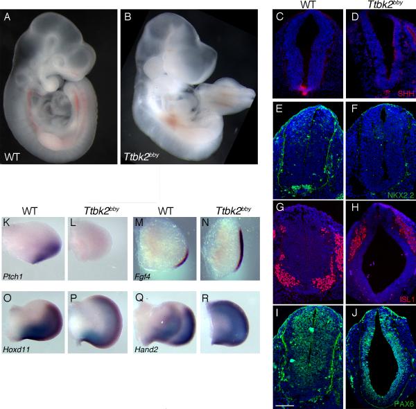
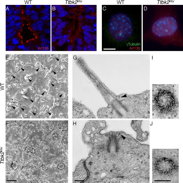
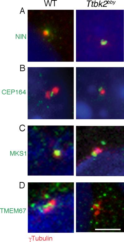
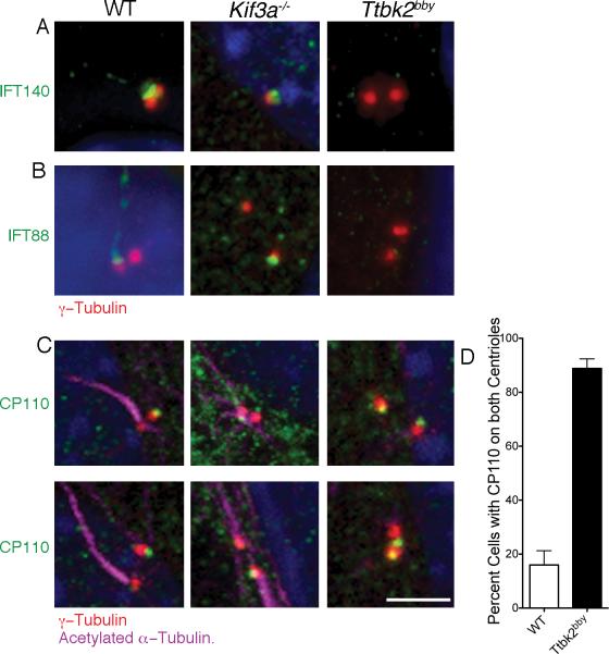
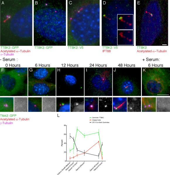
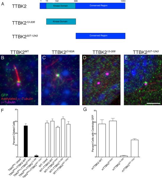
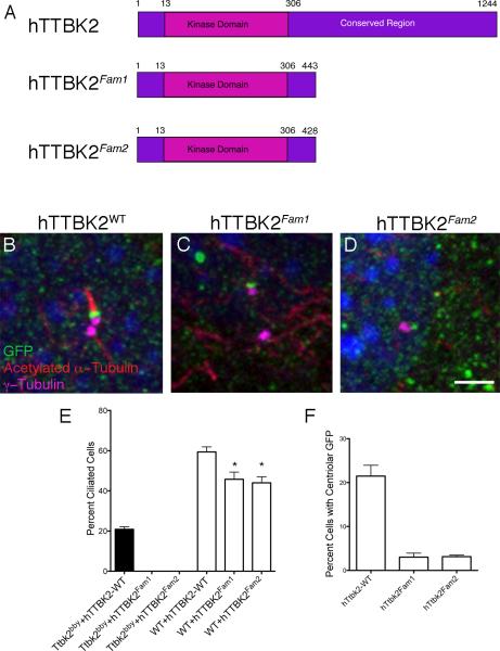
The primary cilium has critical roles in human development and disease, but the mechanisms that regulate ciliogenesis are not understood. Here, we show that Tau tubulin kinase 2 (TTBK2) is a dedicated regulator of the initiation of ciliogenesis in vivo. We identified a null allele of mouse Ttbk2 based on loss of Sonic hedgehog activity, a signaling pathway that requires the primary cilium. Despite a normal basal body template, Ttbk2 mutants lack cilia. TTBK2 acts at the distal end of the basal body, where it promotes the removal of CP110, which caps the mother centriole, and promotes recruitment of IFT proteins, which build the ciliary axoneme. Dominant truncating mutations in human TTBK2 cause spinocerebellar ataxia type 11 (SCA11); these mutant proteins do not promote ciliogenesis and inhibit ciliogenesis in wild-type cells. We propose that cell-cycle regulators target TTBK2 to the basal body, where it modifies specific targets to initiate ciliogenesis.


Figures

Figure 1
Figure 1. Altered Shh-dependent patterning in bby mutant embryos
(A and B) Whole wild-type (WT) (A) and bby (B) mutant embryos at E10.5. (C-J) Neural patterning in WT and bby embryos at E10.5. Sections were taken at the forelimb level; the same pattern of expression of neural markers was seen at lumbar levels. Shh (C, D), Nkx2.2 (E, F) and Isl1 (G, H) are not expressed in the neural tube of bby mutant embryos. Pax6 (I, J) is expanded ventrally in bby mutants. Dorsal is up. Nuclei are stained with DAPI (blue). Scale bar =100 μm. (K-R) Limb patterning in WT and bby mutant embryos. Ptch1 (K, L) is not expressed in limb buds of bby embryos. Fgf4 (M, N) is expanded anteriorly in E10.5 bby mutants; expression of Hoxd11 (O, P) and Hand2 (Q, R) are expanded anteriorly in E11.5 bby mutants. Anterior is up. Please see also Figure S1 for information on the bby mutation.
Figure 2
Figure 2. Ttbk2bby embryos lack neural and mesenchymal cilia
(A and B) Arl13b (red) staining shows cilia in the WT E10.5 ventral neural tube (A) and the absence of cilia in the Ttbk2bby neural tube (B). (C and D) Arl13b (red) and γ-tubulin (green) in embryonic fibroblasts derived from WT (C) and Ttbk2bby mutant embryos (D). DAPI is in blue. Scale bar = 10 μm. (E and F) Scanning electron micrographs of the apical surface of the neural tube (viewed en face) in E10.5 WT (E) and Ttbk2bby mutant (F) embryos. Scale bar = 1 μm. (G -J) Transmission electron micrographs of cilia in the E10.5 neural tube of WT (G) and Ttbk2bby mutant (H) embryos. Arrowheads denote the distal appendages. Cross sections through the basal body in WT (I) and Ttbk2bby mutant embryos (J) show the presence of the nine triplet microtubules in the centriole of the mutant. Scale bars = 200 nm. Please see Figure S2 for additional TEM images.
Figure 3
Figure 3. Normal localization of appendage and transition zone markers in Ttbk2bby basal bodies
(A) The subdistal appendage marker Ninein (NIN; green) is expressed normally in Ttbk2bby MEFs. γ-tubulin (red) labels the centrioles. Images shown are representative of 14 cells imaged for Ttbk2bby and 12 for wild-type. (B) Localization of the distal appendage-associated protein CEP164 (green) distal to the centrosome, marked by γ-tubulin (red) in MEFs derived from WT and Ttbk2bby mutant embryos. 23 cells were imaged for Ttbk2bby and 20 for wild-type. (C, D) Transition zone proteins MKS1 (C) and TMEM67 (D) (green) are expressed in the transition zone of WT and at the distal basal body in Ttbk2bby cells. Centrosomes are labeled by γ-tubulin (red). Scale bar = 2 μm. Total number of cells imaged for each marker was: MKS1- 17 for Ttbk2bby; 13 for wild-type; TMEM67- 19 for Ttbk2bby; 22 for wild-type. Similar images were obtained from a minimum of 2 independent experiments. Please see Figure S3 for additional images.
Figure 4
Figure 4. TTBK2 acts upstream of IFT and CP110
(A) IFT140 (green), an IFT-A complex protein, localizes primarily to the transition zone in WT and can be seen at lower levels in the axoneme. IFT140 is present at the mother centriole (red, γ-tubulin) in Kif3a-/- MEFs, but is not detectable at the Ttbk2bby centrosomes. 56 Ttbk2bby, 17 Kif3a-/- and 27 WT cells were examined. (B) The IFT-B complex component IFT88 (green) localizes to transition zone and ciliary axoneme in WT MEFS and to the mother centriole (red) in Kif3a-/- MEFs, but is not associated with the centrosomes of Ttbk2bby cells. Centrosomes are labeled with γ-tubulin (red). 52 Ttbk2bby cells, 21 Kif3a-/-- and 31 WT cells were examined. (C) CP110 (green) is present on the distal end of the WT daughter centriole (red), the centriole that does not template the ciliary axoneme (purple). CP110 is detected on one of the two centrosomes in Kif3a-/- MEFs. In Ttbk2bby cells, CP110 is present on both centrosomes in most cells. Scale bar is 2 μm. (D) The percentage of cells with CP110 seen on both centrosomes in WT (white bar, 16.0+/-5.2) and Ttbk2bby (black bar, 88.8+/-3.6) MEFs. Error bars represent the SEM. p < 0.0001, 5 fields of cells were counted for a total of 114 Ttbk2bby and 116 WT cells. Please see Figures S3 and S4 for additional images.
Figure 5
Figure 5. TTBK2 localizes to the basal body in response to serum withdrawal
(A-D) Localization of TTBK2 fusion protein constructs expressed in mouse cells. (A, B) WT MEFs were infected with a mouse N-terminal-eGFP-TTBK2 construct. TTBK2-eGFP (green) is present in the transition zone between the mother centriole, labeled with γ-tubulin (purple), and the axoneme, labeled by acetylated α-tubulin (red), in ciliated cells (A), and is localized to one of the two centrosomes in non-ciliated cells (B). (C, D) WT MEFs infected with mouse TTBK2 with a C-terminal V5 tag (green); TTBK2-V5 also localizes to the transition zone (C). TTBK2-V5 co-localizes with IFT88 (red) in the transition zone (D). Insets show high magnification images that highlight the overlap of IFT88 and TTBK2 at the base of the cilium. (E) An antibody to human TTBK2 (green) shows that the endogenous protein localizes to the transition zone of human RPE cells. Scale bar = 2 μm. (F-K) WT MEFs stably expressing mTTBK2::eGFP after shift to serum-free medium. 48 hours of serum withdrawal, cells were shifted back to growth media induce re-entry into the cell cycle (K). Localization of mTTBK2::eGFP was assessed using an antibody against GFP (green), and cells were counterstained with γ-tubulin (purple) to label the centrosome and acetylated α-tubulin (red) to label the ciliary axoneme. (L) Line graph shows the mean percentage of cells with centrosomal localization of mTTBK2::eGFP (green) the mean percentage of ciliated cells (red), and the mean percentage of cells with CP110 on both centrioles (black) at each time point from 4-6 randomly selected fields of cells from at least two different slides; see Supplemental Methods. Error bars indicate SEM. See also Figure S5.
Figure 6
Figure 6. TTBK2 kinase activity is required for ciliogenesis and the C-terminal domain is required for localization to the centriole
(A) Schematics of the mouse Ttbk2 truncations used in rescue experiments. (B-E) Cilia are marked by acetylated α-tubulin (red); centrosomes by γ-tubulin (magenta); GFP-tagged TTBK2 is in green. (B) WT mouse TTBK2::eGFP localizes to the transition zone and rescues the Ttbk2bby ciliogenesis defect. (C) The kinase-dead mTTBK2D163A::eGFP construct localizes to the centrosome but does not rescue ciliogenesis. (D) The kinase domain alone (mTTBK213-306::eGFP) localized inefficiently to the centrosome and rescued ciliogenesis weakly. (E) The C-terminal tail (mTTBK2307-1243::eGFP) localizes to the centrosome but does not rescue ciliogenesis. Scale bar = 2 μm. (F) Graph shows the percentage of ciliated cells observed in Ttbk2bby (black bars) or WT (white bars) MEFs infected with the indicated construct. (G) Graph showing the percentage of cells with centrosomal localization of GFP for the indicated construct. For F and G, bars represent the mean percentage of ciliated cells (F), or cells with centriolar GFP (G) from 4-8 randomly selected fields of cells per condition, and fields were selected from at least two different slides and had a minimum of 25 cells per field; see Supplemental Methods. Error bars indicate the SEM. Please see also Figure S6.
Figure 7
Figure 7. SCA11-associated variants of hTTBK2 do not rescue cilia formation in Ttbk2bby cells and interfere with ciliogenesis in WT cells
(A) Schematic of human TTBK2, which is 89% identical to mouse TTBK2; and two familial SCA11 mutations that truncate the protein at 443 or 428 AA. (B-D) Localization of WT hTTBK2::eGFP (A) and SCA11-associated Fam1 and Fam2 (C and D) variants expressed in Ttbk2bby MEFs. GFP is in green, γ-tubulin is in purple, and acetylated α-tubulin is in red. The Fam1 and Fam2 fusion proteins generally localized diffusely in the cytoplasm; C show a rare cell in which GFP is localized to centrosome. Scale bar = 2 μm. (E) Graph showing the percentage of ciliated cells observed in Ttbk2bby mutant (black bars) or WT (white bars) MEFs infected with the indicated construct. (F) Graph showing the percentage of cells with centrosomal localization of GFP for the indicated construct. For E and F, bars represent the mean percentage of ciliated cells (E) or cells with centriolar GFP (F) from 4-8 randomly selected fields of cells per condition. Fields were selected from at least two different slides and had a minimum of 25 cells per field; see Supplemental Methods. Error bars indicate the SEM. Please see also Figure S6.

Comment in

References

    1. Ahn S, Joyner AL. In vivo analysis of quiescent adult neural stem cells responding to Sonic hedgehog. Nature. 2005;437:894–897. - PubMed
    1. Aughsteen AA. The ultrastructure of primary cilia in the endocrine and excretory duct cells of the pancreas of mice and rats. Eur J Morphol. 2001;39:277–283. - PubMed
    1. Badano JL, Mitsuma N, Beales PL, Katsanis N. The ciliopathies: an emerging class of human genetic disorders. Annu Rev Genomics Hum Genet. 2006;7:125–148. - PubMed
    1. Bauer P, Stevanin G, Beetz C, Synofzik M, Schmitz-Hubsch T, Wullner U, Berthier E, Ollagnon-Roman E, Riess O, Forlani S, et al. Spinocerebellar ataxia type 11 (SCA11) is an uncommon cause of dominant ataxia among French and German kindreds. J Neurol Neurosurg Ps. 2010;81:1229–1232. - PubMed
    1. Berbari NF, Lewis JS, Bishop GA, Askwith CC, Mykytyn K. Bardet-Biedl syndrome proteins are required for the localization of G protein-coupled receptors to primary cilia. Proc Natl Acad Sci USA. 2008;105:4242–4246. - PMC - PubMed

Publication types