

Novel indices for left-ventricular dyssynchrony characterization based on highly automated segmentation from real-time 3-d echocardiography
- PMID: 23141901
- PMCID: PMC3513930
- DOI: 10.1016/j.ultrasmedbio.2012.08.019
Novel indices for left-ventricular dyssynchrony characterization based on highly automated segmentation from real-time 3-d echocardiography
Abstract
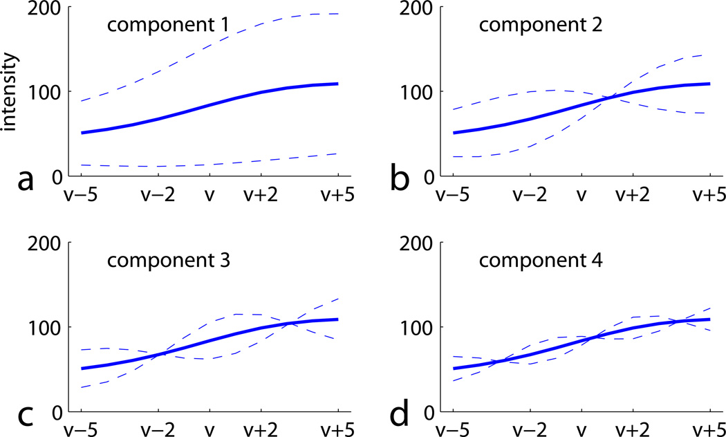
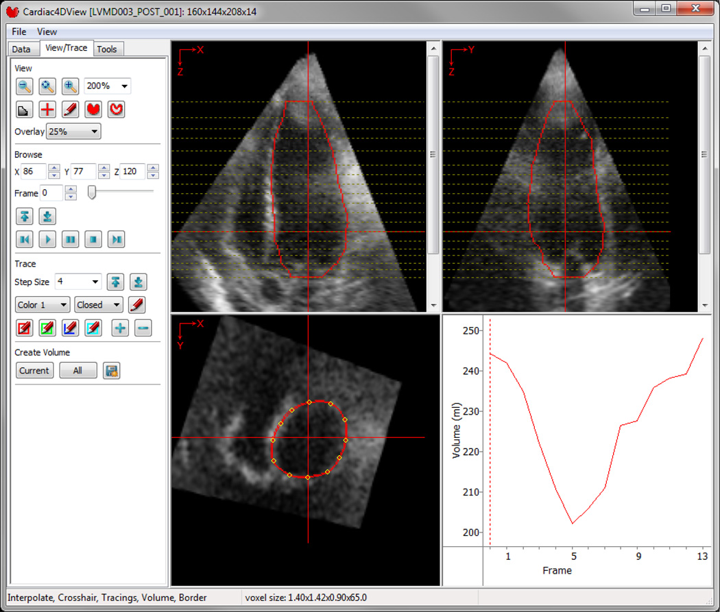
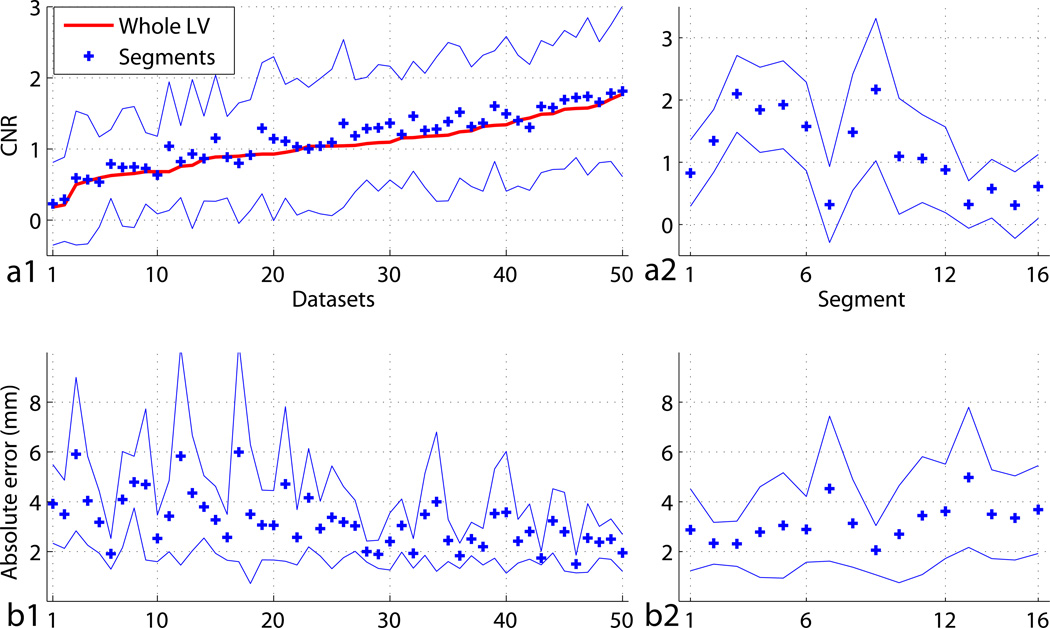
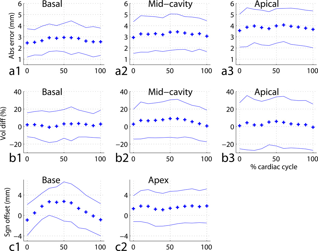
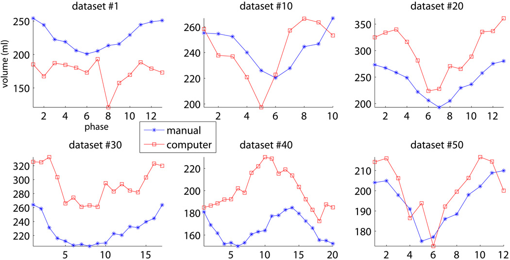
Cardiac resynchronization therapy (CRT) using a biventricular pacemaker is an invasive and expensive treatment option for left ventricular mechanical dyssynchrony (LVMD). The CRT candidate selection is a crucial issue due to the unreliability of the current standard CRT indicators. Real-time three-dimensional (3-D) echocardiography (RT3DE) provides four-dimensional (4-D) (3-D+time) information about the LV and is suitable for LVMD assessment. In this article, the complex left ventricle (LV) shape and motion of 50 RT3DE datasets are represented by novel 4-D descriptors - 4-D sphericity, volume and shape, from which novel indices were derived by principal component analysis (PCA) and subsequently analyzed by a support vector machine (SVM) classifier to assess their capability of LVMD characterization and CRT outcome prediction. These novel indices outperformed clinical indices and have promising capabilities in disease characterization and great potential in CRT outcome prediction. To enable efficient quantitative RT3DE analysis, a segmentation method was developed to combine the powers of active shape models and optimal graph search. Various aspects of the method were designed to handle varying RT3DE image quality among datasets and LV segments. An application with graphical user interface was developed to provide the user with simple and intuitive control. The developed method was robust to inter-observer variability and produced very good accuracy - 3.2±1.1 mm absolute surface positioning error, <1 mm mean signed error and <5% mean volume difference. The computer method's classification performance was compared with the independent standard, showing that the 4-D shape modal indices were not only the most capable of all tested options when employed for disease characterization but also the least sensitive to segmentation imperfections.
Copyright © 2013 World Federation for Ultrasound in Medicine & Biology. Published by Elsevier Inc. All rights reserved.
Figures


References
-
- Abraham WT, Fisher WG, Smith AL, Delurgio DB, Leon AR, Loh E, Kocovic DZ, Packer M, Clavell AL, Hayes DL, Ellestad M, Trupp RJ, Underwood J, Pickering F, Truex C, McAtee P, Messenger J MIRACLE Study Group. Cardiac resynchronization in chronic heart failure. N Engl J Med. 2002;34624:1845–1853. - PubMed
-
- Achilli A, Sassara M, Ficili S, Pontillo D, Achilli P, Alessi C, Spirito SD, Guerra R, Patruno N, Serra F. Long-term effectiveness of cardiac resynchronization therapy in patients with refractory heart failure and “narrow” QRS. J Am Coll Cardiol. 2003;4212:2117–2124. - PubMed
-
- Bax JJ, Bleeker GB, Marwick TH, Molhoek SG, Boersma E, Steendijk P, van der Wall EE, Schalij MJ. Left ventricular dyssynchrony predicts response and prognosis after cardiac resynchronization therapy. J Am Coll Cardiol. 2004;449:1834–1840. - PubMed
-
- Cerqueira MD, Weissman NJ, Dilsizian V, Jacobs AK, Kaul S, Laskey WK, Pennell DJ, Rumberger JA, Ryan T, Verani MS American Heart Association Writing Group on Myocardial Segmentation and Registration for Cardiac Imaging. Standardized myocardial segmentation and nomenclature for tomographic imaging of the heart. A statement for healthcare professionals from the cardiac imaging committee of the council on clinical cardiology of the American heart association. Int J Cardiovasc Imaging. 2002;181:539–542. - PubMed
-
- Chang CC, Lin CJ. LIBSVM: a library for support vector machines. 2001 Software available at http://www.csie.ntu.edu.tw/~cjlin/libsvm.
Publication types
MeSH terms
Grants and funding
LinkOut - more resources
Full Text Sources
Other Literature Sources
Research Materials
