. 2013 Jan 30;212(2):322-8.
doi: 10.1016/j.jneumeth.2012.10.017. Epub 2012 Nov 8.

Three variations in rabbit angiographic stroke models

Affiliations

Three variations in rabbit angiographic stroke models

William C Culp et al. J Neurosci Methods. .

Abstract

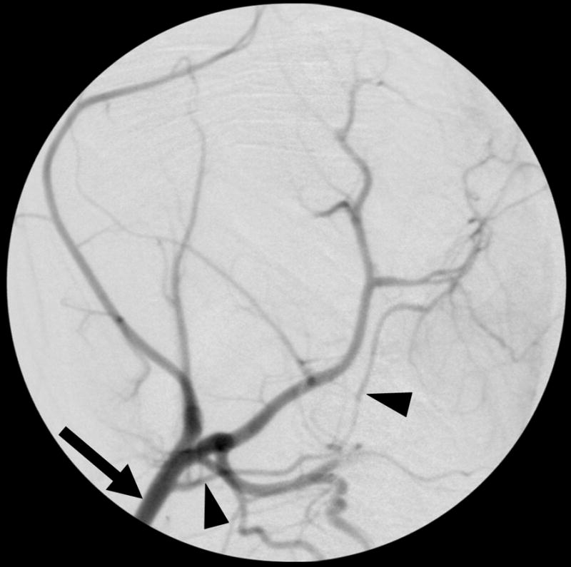
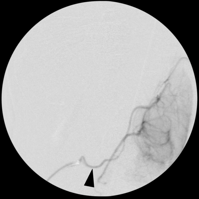
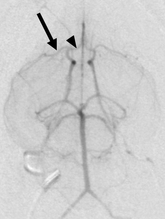
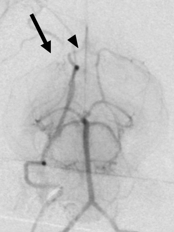
Purpose: To develop angiographic models of embolic stroke in the rabbit using pre-formed clot or microspheres to model clinical situations ranging from transient ischemic events to severe ischemic stroke.

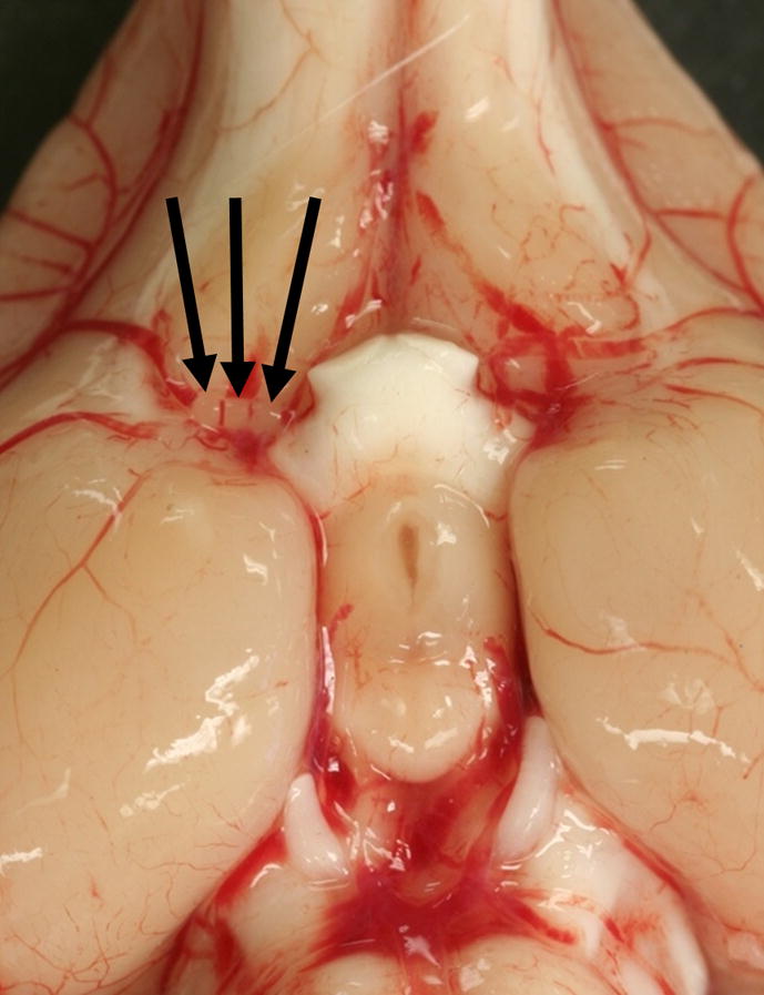
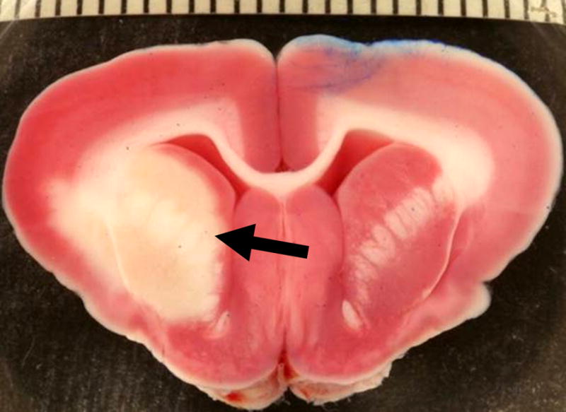
Materials and methods: New Zealand White rabbits (N=151) received angiographic access to the internal carotid artery (ICA) from a femoral approach. Variations of emboli type and quantity of emboli were tested by injection into the ICA. These included fresh clots (1.0-mm length, 3-6h), larger aged clots (4.0-mm length, 3 days), and 2 or 3 insoluble microspheres (700-900 μm). Neurological assessment scores (NAS) were based on motor, sensory, balance, and reflex measures. Rabbits were euthanized at 4, 7, or 24h after embolization, and infarct volume was measured as a percent of total brain volume using 2,3,5-triphenyltetrazolium chloride (TTC).

Results: Infarct volume percent at 24 h after stroke was lower for rabbits embolized with fresh clot (0.45±0.14%), compared with aged clot (3.52±1.31%) and insoluble microspheres (3.39±1.04%). Overall NAS (including posterior vessel occlusions) were positively correlated to infarct volume percent measurements in the fresh clot (r=0.50), aged clot (r=0.65) and microsphere (r=0.62) models (p<0.001).

Conclusion: The three basic angiographic stroke models may be similar to human transient ischemic attacks (TIA) (fresh clot), major strokes that can be thrombolysed (aged clot), or major strokes with insoluble emboli such as atheromata (microspheres). Model selection can be tailored to specific research needs.


Figures

Figure 1
Figure 1. Selective rabbit intracranial angiography
(a) Common carotid selection (arrow), lateral view. The internal carotid artery is seen coursing to the base of the brain (arrowheads). (b) Internal carotid sub-selection (arrowhead) in a lateral view demonstrates filling of the cerebral vasculature. (c) A frontal-view internal carotid angiogram clearly shows the Circle of Willis including the middle cerebral artery (MCA, arrow), and the anterior cerebral artery (ACA, arrowhead). (d) Repeat angiography after injection of three embolic microspheres (700–900 μm) shows the occlusion of the MCA (arrow) and persistent flow in the ACA (arrowhead).
Figure 2
Figure 2
Gross image of the ventral surface of the rabbit brain shows three 700–900 μm microsphere emboli (arrows) located in the middle cerebral artery origin at the connection with the internal carotid artery.
Figure 3
Figure 3. Infarct area in a rabbit brain section
2,3,5-triphenyltetrazolium chloride (TTC) stain at 24 hours after embolization with three 700–900 μm microspheres shows viable tissue as red or pink, and a pale infarct area (arrow) in the distribution of the right middle cerebral artery. The scale bar represents millimeters and the blue color is a laterality marker indicating the left hemisphere.

References

    1. Aronowski J, Samways E, Strong R, Rhoades HM, Grotta JC. An alternative method for the quantitation of neuronal damage after experimental middle cerebral artery occlusion in rats: analysis of behavioral deficit. J Cereb Blood Flow Metab. 1996;16:705–713. - PubMed
    1. Bederson JB, Pitts LH, Germano SM, Nishimura MC, Davis RL, Bartkowski HM. Evaluation of 2,3,5-triphenyltetrazolium chloride as a stain for detection and quantification of experimental cerebral infarction in rats. Stroke. 1986;17:1304–8. - PubMed
    1. Brown AT, Flores R, Hamilton E, Roberson PK, Borrelli MJ, Culp WC. Microbubbles improve sonothrombolysis in vitro and decrease hemorrhage in vivo in a rabbit stroke model. Invest Radiol. 2011;46:202–207. - PMC - PubMed
    1. Brown AT, Skinner RD, Flores R, Hennings L, Borrelli MJ, Lowery J, et al. Stroke location and brain function in an embolic rabbit stroke model. J Vasc Interv Radiol. 2010;21:903–909. - PMC - PubMed
    1. Caldwell B, Flores R, Lowery J, Brown AT, Culp WC. Variations in the circle of Willis in the New Zealand white rabbit. J Vasc Interv Radiol. 2011;22:1188–1192. - PMC - PubMed

Publication types