

Insights into the impact of phenolic residue incorporation at each position along secretin for receptor binding and biological activity
- PMID: 23142313
- PMCID: PMC3534899
- DOI: 10.1016/j.regpep.2012.10.001
Insights into the impact of phenolic residue incorporation at each position along secretin for receptor binding and biological activity
Abstract
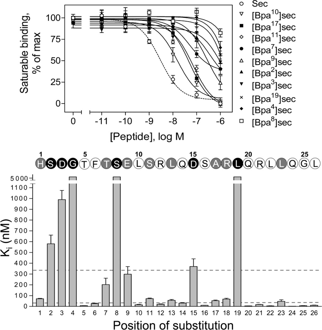
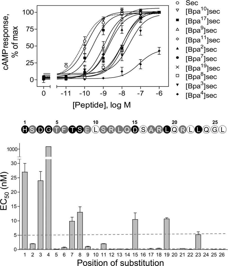
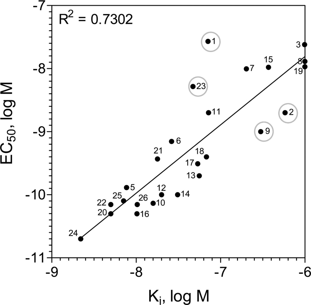
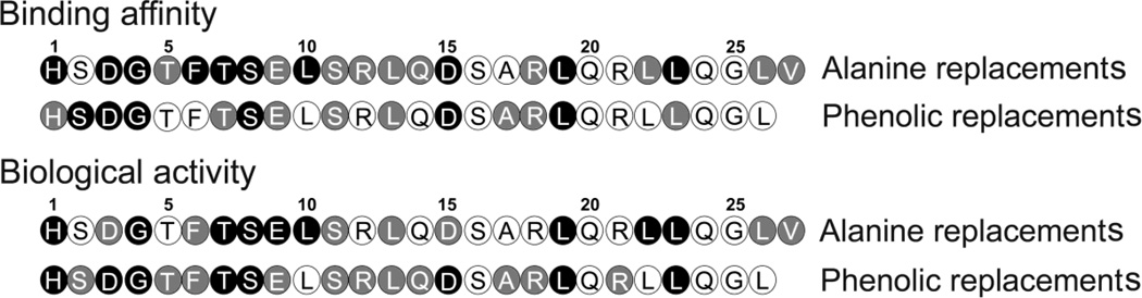
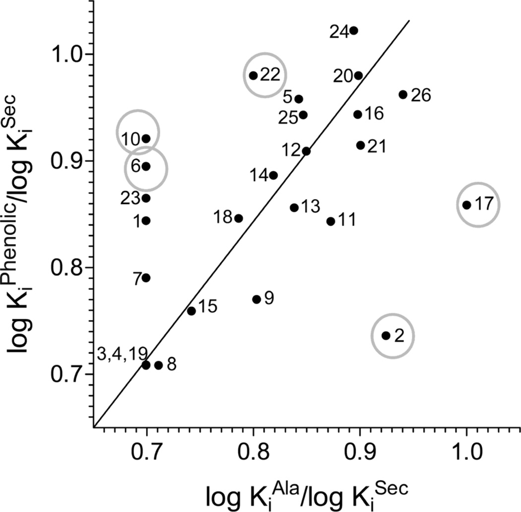
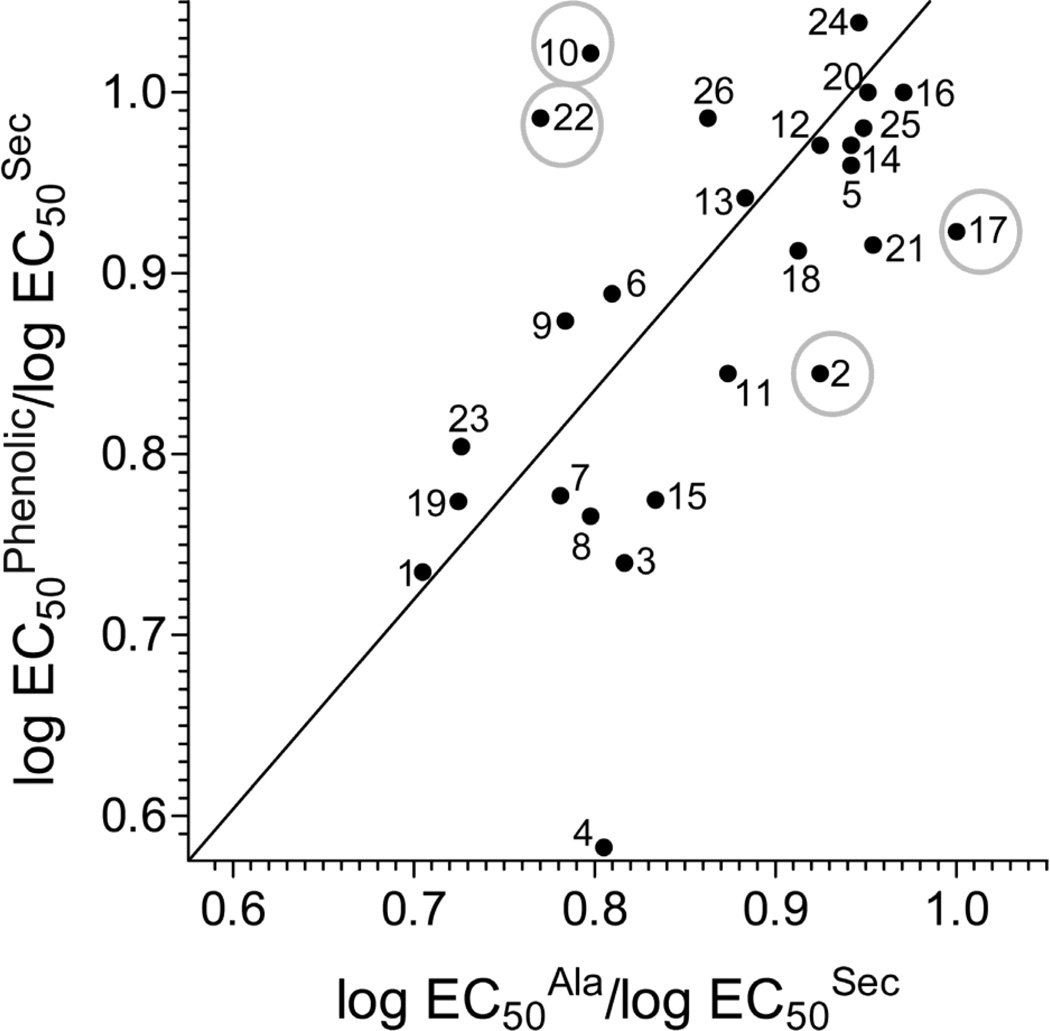
Understanding of the structural importance of each position along a peptide ligand can provide important insights into the molecular basis for its receptor binding and biological activity. This has typically been evaluated using serial replacement of each natural residue with an alanine. In the current report, we have further complemented alanine scanning data with the serial replacement of each residue within secretin-27, the natural ligand for the prototypic class B G protein-coupled secretin receptor, using a photolabile phenolic residue. This not only provided the opportunity to probe spatial approximations between positions along a docked ligand with its receptor, but also provided structure-activity insights when compared with tolerance for alanine replacement of the same residues. The pattern of sensitivity to phenolic residue replacement was periodic within the carboxyl-terminal region of this peptide ligand, corresponding with alanine replacements in that region. This was supportive of the alpha-helical conformation of the peptide in that region and its docking within a groove in the receptor amino-terminal domain. In contrast, the pattern of sensitivity to phenolic residue replacement was almost continuous in the amino-terminal region of this peptide ligand, again similar to alanine replacements, however, there were key positions in which either the phenolic residue or alanine was differentially preferred. This provided insights into the receptor environment of the portion of this ligand most critical for its biological activity. As the structure of the intact receptor is elucidated, these data will provide a guide for ligand docking to the core domain and to help clarify the molecular basis of receptor activation.
Copyright © 2012 Elsevier B.V. All rights reserved.
Figures
Similar articles
-
Spatial approximation between secretin residue five and the third extracellular loop of its receptor provides new insight into the molecular basis of natural agonist binding.Mol Pharmacol. 2008 Aug;74(2):413-22. doi: 10.1124/mol.108.047209. Epub 2008 May 8. Mol Pharmacol. 2008. PMID: 18467541 Free PMC article.
-
Molecular basis of secretin docking to its intact receptor using multiple photolabile probes distributed throughout the pharmacophore.J Biol Chem. 2011 Jul 8;286(27):23888-99. doi: 10.1074/jbc.M111.245969. Epub 2011 May 12. J Biol Chem. 2011. PMID: 21566140 Free PMC article.
-
Lactam constraints provide insights into the receptor-bound conformation of secretin and stabilize a receptor antagonist.Biochemistry. 2011 Sep 27;50(38):8181-92. doi: 10.1021/bi2008036. Epub 2011 Aug 30. Biochemistry. 2011. PMID: 21851058 Free PMC article.
-
Ligand binding and activation of the secretin receptor, a prototypic family B G protein-coupled receptor.Br J Pharmacol. 2012 May;166(1):18-26. doi: 10.1111/j.1476-5381.2011.01463.x. Br J Pharmacol. 2012. PMID: 21542831 Free PMC article. Review.
-
Structural basis of natural ligand binding and activation of the Class II G-protein-coupled secretin receptor.Biochem Soc Trans. 2007 Aug;35(Pt 4):709-12. doi: 10.1042/BST0350709. Biochem Soc Trans. 2007. PMID: 17635130 Review.
Cited by
-
Use of Cysteine Trapping to Map Spatial Approximations between Residues Contributing to the Helix N-capping Motif of Secretin and Distinct Residues within Each of the Extracellular Loops of Its Receptor.J Biol Chem. 2016 Mar 4;291(10):5172-84. doi: 10.1074/jbc.M115.706010. Epub 2016 Jan 6. J Biol Chem. 2016. PMID: 26740626 Free PMC article.
-
Secretin Receptor as a Target in Gastrointestinal Cancer: Expression Analysis and Ligand Development.Biomedicines. 2022 Feb 24;10(3):536. doi: 10.3390/biomedicines10030536. Biomedicines. 2022. PMID: 35327338 Free PMC article.
-
Transmembrane signal transduction by peptide hormones via family B G protein-coupled receptors.Front Pharmacol. 2015 Nov 5;6:264. doi: 10.3389/fphar.2015.00264. eCollection 2015. Front Pharmacol. 2015. PMID: 26594176 Free PMC article. Review.
References
-
- Adelhorst K, Hedegaard BB, Knudsen LB, Kirk O. Structure-activity studies of glucagon-like peptide-1. J. Biol. Chem. 1994;269:6275–6278. - PubMed
-
- Igarashi H, Ito T, Hou W, Mantey SA, Pradhan TK, Ulrich CD, 2nd, Hocart SJ, Coy DH, Jensen RT. Elucidation of vasoactive intestinal peptide pharmacophore for VPAC(1) receptors in human, rat guinea pig. J. Pharmacol. Exp. Ther. 2002;301:37–50. - PubMed
-
- Igarashi H, Ito T, Pradhan TK, Mantey SA, Hou W, Coy DH, Jensen RT. Elucidation of the vasoactive intestinal peptide pharmacophore for VPAC(2) receptors in human and rat and comparison to the pharmacophore for VPAC(1) receptors. J. Pharmacol. Exp. Ther. 2002;303:445–460. - PubMed
-
- Nicole P, Lins L, Rouyer-Fessard C, Drouot C, Fulcrand P, Thomas A, Couvineau A, Martinez J, Brasseur R, Laburthe M. Identification of key residues for interaction of vasoactive intestinal peptide with human VPAC1 and VPAC2 receptors and development of a highly selective VPAC1 receptor agonist. Alanine scanning and molecular modeling of the peptide. J. Biol. Chem. 2000;275:24003–24012. - PubMed
Publication types
MeSH terms
Substances
Grants and funding
LinkOut - more resources
Full Text Sources
Other Literature Sources
Research Materials
