

ALDH1B1 is a potential stem/progenitor marker for multiple pancreas progenitor pools
- PMID: 23142317
- PMCID: PMC3580293
- DOI: 10.1016/j.ydbio.2012.10.030
ALDH1B1 is a potential stem/progenitor marker for multiple pancreas progenitor pools
Abstract
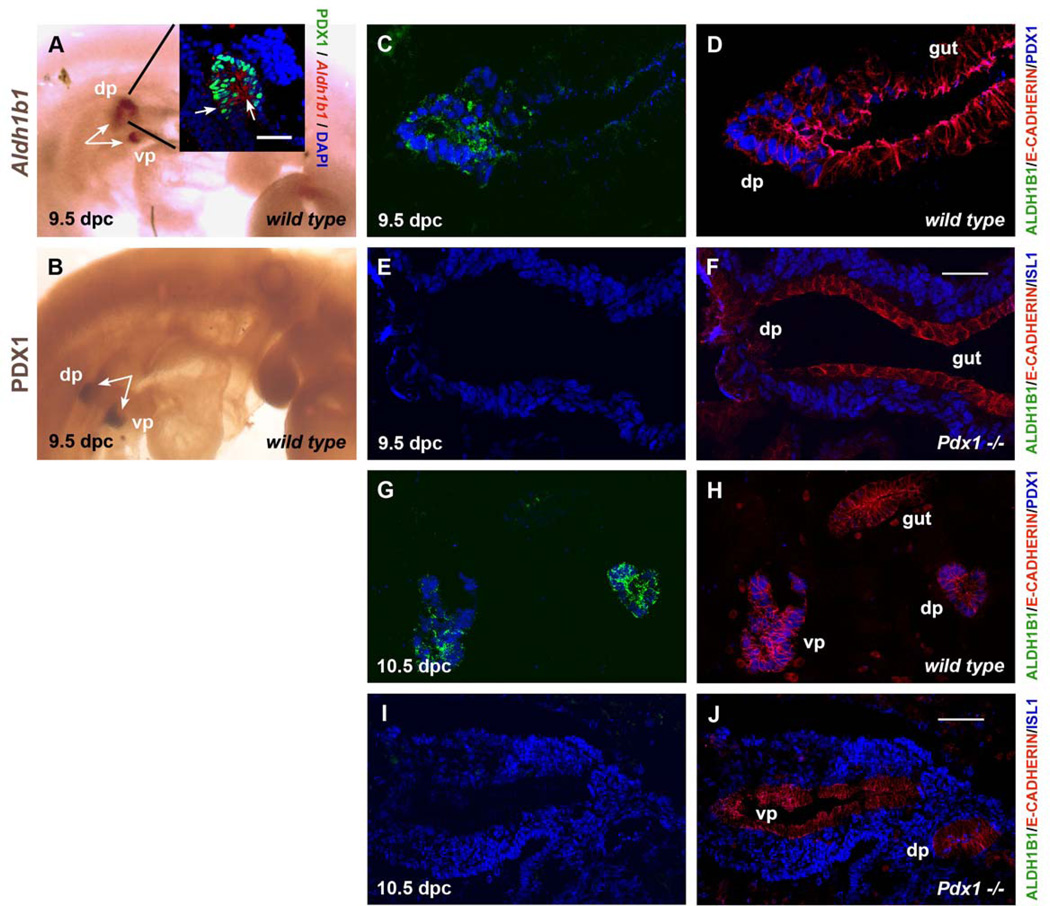
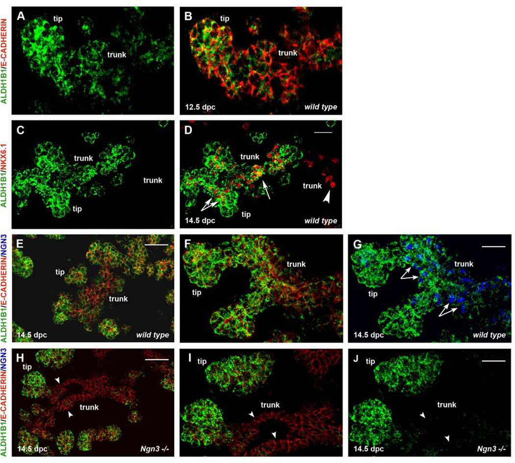
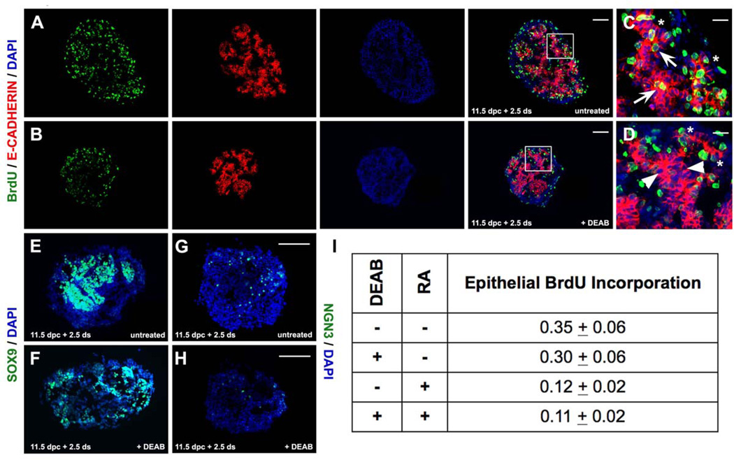
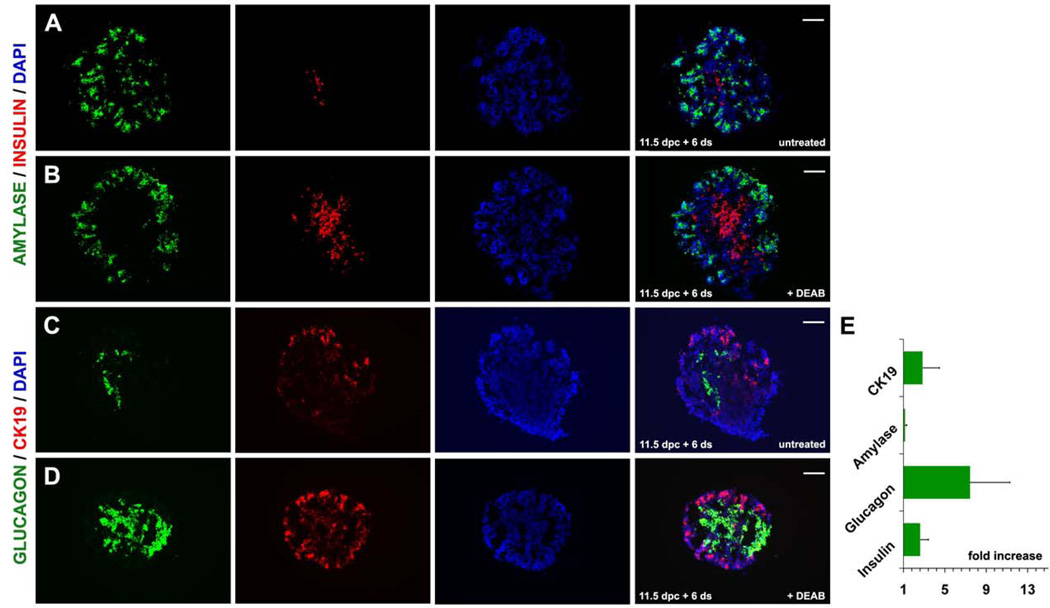
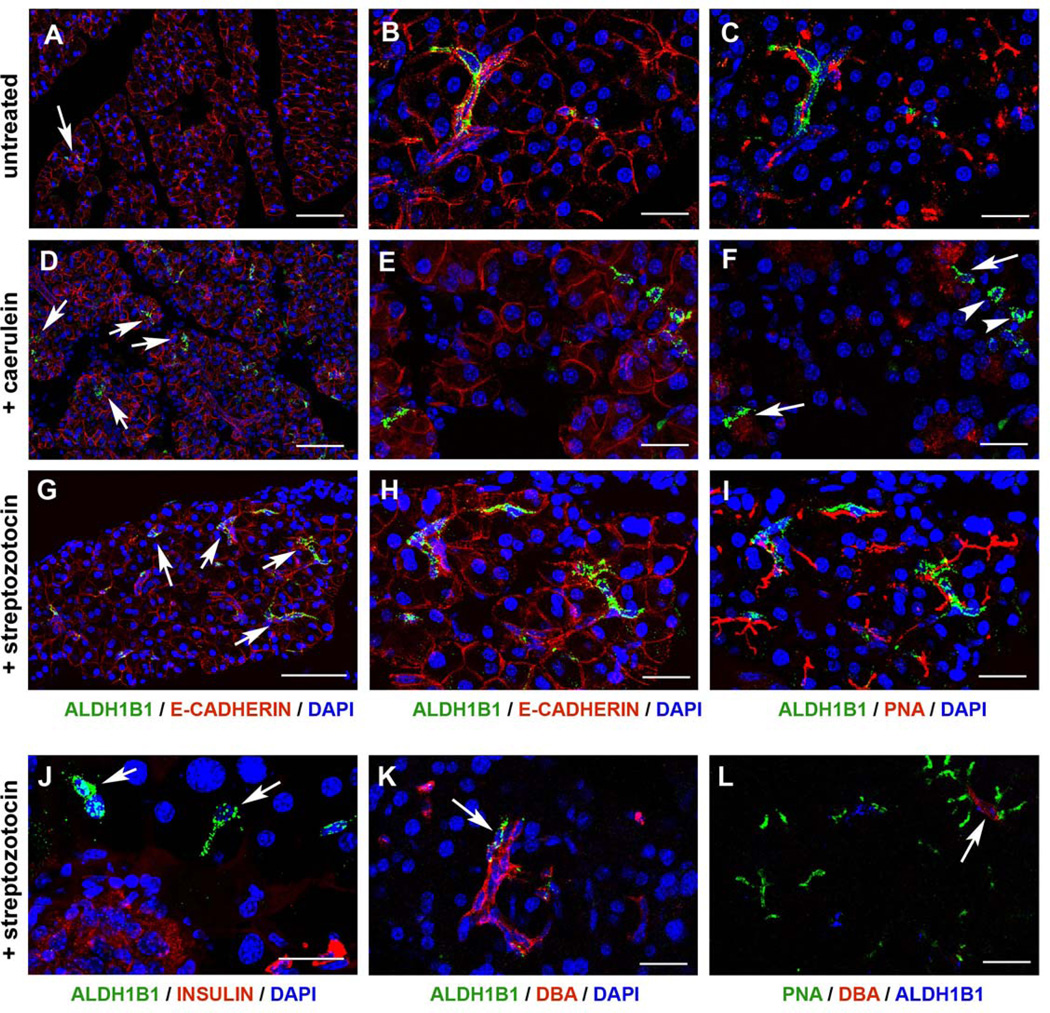
Aldehyde dehydrogenase (ALDH) genes are increasingly associated with stem/progenitor cell status but their role in the maintenance of pluripotency remains uncertain. In a screen conducted for downstream Ngn3 target genes using ES derived pancreas progenitors we identified Aldh1b1, encoding a mitochondrial enzyme, as one of the genes strongly up regulated in response to Ngn3 expression. We found both by in situ hybridization and immunofluorescence using a specific antibody that ALDH1B1 is exclusively expressed in the emerging pancreatic buds of the early embryo (9.5 dpc) in a Pdx1 dependent manner. Around the time of secondary transition, ALDH1B1 expression was restricted in the tip tripotent progenitors of the branching epithelium and in a subset of the trunk epithelium. Expression in the latter was Ngn3 dependent. Subsequently, ALDH1B1 expression persisted only in the tip cells that become restricted to the exocrine lineage and declined rapidly as these cells mature. In the adult pancreas we identified rare ALDH1B1(+) cells that become abundant following pancreas injury in either the caerulein or streptozotocin paradigms. Blocking ALDH catalytic activity in pancreas embryonic explants resulted in reduced size of the explants and accelerated differentiation suggesting for the first time that ALDH activity may be necessary in the developing pancreas for the maintenance and expansion of progenitor pools.
Copyright © 2012 Elsevier Inc. All rights reserved.
Figures
Similar articles
-
Protein Methyltransferase Inhibition Decreases Endocrine Specification Through the Upregulation of Aldh1b1 Expression.Stem Cells. 2019 May;37(5):640-651. doi: 10.1002/stem.2979. Epub 2019 Feb 13. Stem Cells. 2019. PMID: 30681750 Free PMC article.
-
Aldh1b1 expression defines progenitor cells in the adult pancreas and is required for Kras-induced pancreatic cancer.Proc Natl Acad Sci U S A. 2019 Oct 8;116(41):20679-20688. doi: 10.1073/pnas.1901075116. Epub 2019 Sep 23. Proc Natl Acad Sci U S A. 2019. PMID: 31548432 Free PMC article.
-
Aldehyde dehydrogenase activity is necessary for beta cell development and functionality in mice.Diabetologia. 2016 Jan;59(1):139-150. doi: 10.1007/s00125-015-3784-4. Epub 2015 Oct 31. Diabetologia. 2016. PMID: 26518685 Free PMC article.
-
Direct lineage tracing reveals the ontogeny of pancreatic cell fates during mouse embryogenesis.Mech Dev. 2003 Jan;120(1):35-43. doi: 10.1016/s0925-4773(02)00330-1. Mech Dev. 2003. PMID: 12490294 Review.
-
[Pancreatic development and stem cell-based regenerative medicine].Rinsho Byori. 2006 Apr;54(4):386-92. Rinsho Byori. 2006. PMID: 16722458 Review. Japanese.
Cited by
-
ALDH1B1 links alcohol consumption and diabetes.Biochem Biophys Res Commun. 2015 Aug 7;463(4):768-773. doi: 10.1016/j.bbrc.2015.06.011. Epub 2015 Jun 15. Biochem Biophys Res Commun. 2015. PMID: 26086111 Free PMC article.
-
Potential therapeutic effect of NK1R antagonist in diabetic non-healing wound and depression.Front Endocrinol (Lausanne). 2023 Jan 4;13:1077514. doi: 10.3389/fendo.2022.1077514. eCollection 2022. Front Endocrinol (Lausanne). 2023. PMID: 36686487 Free PMC article.
-
Aldehyde Dehydrogenase 1B1 Is Implicated in DNA Damage Response in Human Colorectal Adenocarcinoma.Cells. 2022 Jun 24;11(13):2017. doi: 10.3390/cells11132017. Cells. 2022. PMID: 35805102 Free PMC article.
-
Investigating Carvedilol's Repurposing for the Treatment of Non-Small Cell Lung Cancer via Aldehyde Dehydrogenase Activity Modulation in the Presence of β-Adrenergic Agonists.Curr Issues Mol Biol. 2023 Sep 29;45(10):7996-8012. doi: 10.3390/cimb45100505. Curr Issues Mol Biol. 2023. PMID: 37886948 Free PMC article.
-
Human ALDH1B1 polymorphisms may affect the metabolism of acetaldehyde and all-trans retinaldehyde--in vitro studies and computational modeling.Pharm Res. 2015 May;32(5):1648-62. doi: 10.1007/s11095-014-1564-3. Epub 2014 Nov 21. Pharm Res. 2015. PMID: 25413692 Free PMC article.
References
-
- Alison MR, Guppy NJ, Lim SM, Nicholson LJ. Finding cancer stem cells: are aldehyde dehydrogenases fit for purpose? J Pathol. 2010;222:335–344. - PubMed
-
- Balaban RS, Nemoto S, Finkel T. Mitochondria, oxidants, and aging. Cell. 2005;120:483–495. - PubMed
-
- Balber AE. Concise review: aldehyde dehydrogenase bright stem and progenitor cell populations from normal tissues: characteristics, activities, and emerging uses in regenerative medicine. Stem Cells. 2011;29:570–575. - PubMed
-
- Ballester M, Castello A, Ibanez E, Sanchez A, Folch JM. Real-time quantitative PCR-based system for determining transgene copy number in transgenic animals. Biotechniques. 2004;37:610–613. - PubMed
Publication types
MeSH terms
Substances
Grants and funding
LinkOut - more resources
Full Text Sources
Other Literature Sources
Molecular Biology Databases
