. 2012 Dec 21;19(12):1535-45.
doi: 10.1016/j.chembiol.2012.09.019. Epub 2012 Nov 8.

Validation of the proteasome as a therapeutic target in Plasmodium using an epoxyketone inhibitor with parasite-specific toxicity

Affiliations

Validation of the proteasome as a therapeutic target in Plasmodium using an epoxyketone inhibitor with parasite-specific toxicity

Hao Li et al. Chem Biol. .

Abstract

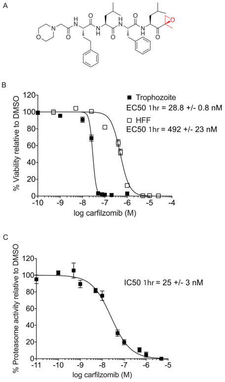
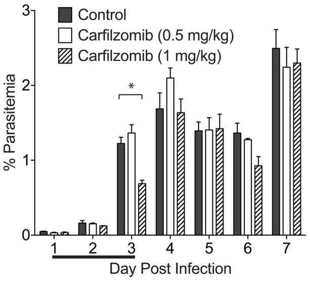
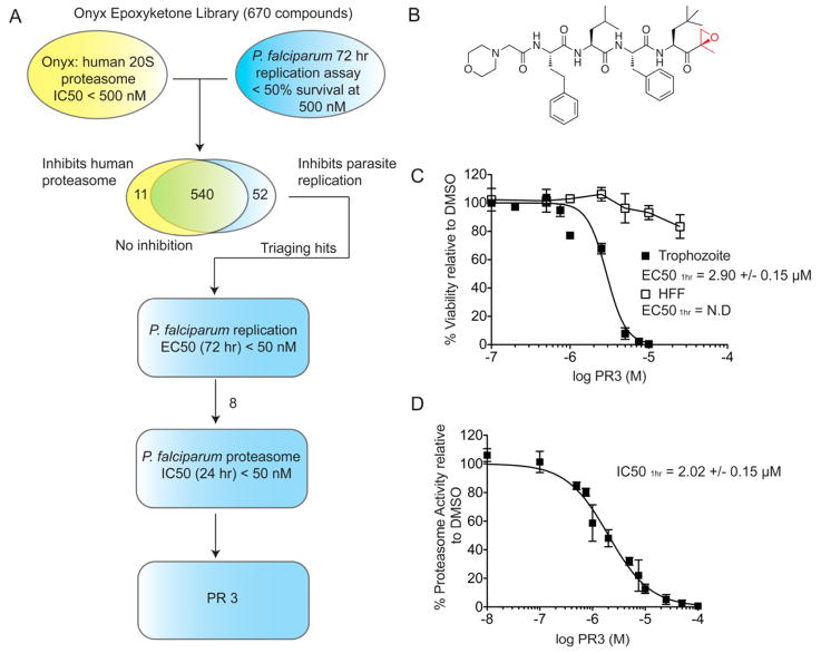
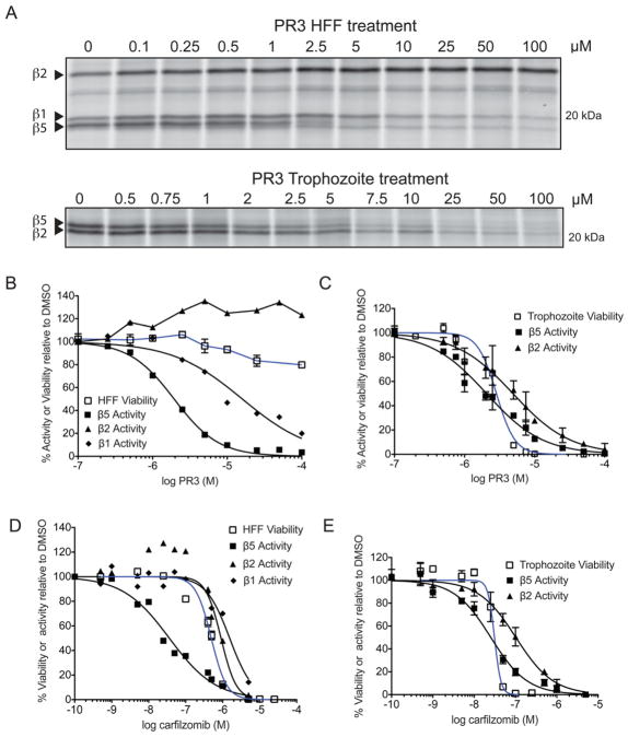
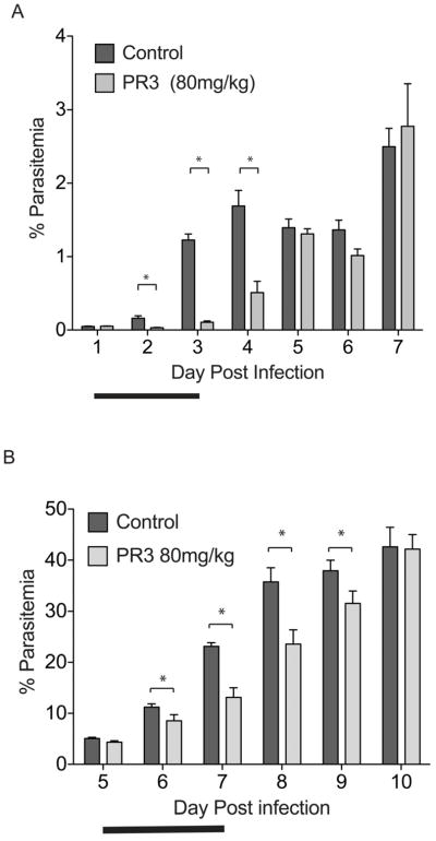
The Plasmodium proteasome has been suggested to be a potential antimalarial drug target; however, toxicity of inhibitors has prevented validation of this enzyme in vivo. We report a screen of a library of 670 analogs of the recent US Food and Drug Administration-approved inhibitor, carfilzomib, to identify compounds that selectively kill parasites. We identified one compound, PR3, that has significant parasite killing activity in vitro but dramatically reduced toxicity in host cells. We found that this parasite-specific toxicity is not due to selective targeting of the Plasmodium proteasome over the host proteasome, but instead is due to a lack of activity against one of the human proteasome subunits. Subsequently, we used PR3 to significantly reduce parasite load in Plasmodium berghei infected mice without host toxicity, thus validating the proteasome as a viable antimalarial drug target.


Figures

Figure 1
Figure 1
Anti-parasitic activity and therapeutic window of carfilzomib in vitro. A) Structure of carfilzomib. The epoxyketone reactive functional group is shown in red. B) Dose response of carfilzomib on P. falciparum culture and human foreskin fibroblasts (HFF). Synchronous P. falciparum trophozoites or non-confluent HFF cells were treated for 1 hr followed by washout of the inhibitor and incubation for 60 hr (trophozoite) or 72 hr (HFF). Each concentration was tested at least 3 times; error bars represent standard error of the mean (S.E.M) for each drug concentration from triplicates. EC50 value is shown as mean +/− standard deviation (SD). The dose response curve for 60 hr treatment is shown in Figure S2. C) Proteasome activity in intact trophozoites treated with carfilzomib. Proteasome activity was assessed by monitoring inhibition of the putative β5 subunit (assignment of labeled subunit is shown in Figure S1). Synchronous mid-trophozoite stage parasites were treated with the indicated concentrations of carfilzomib for 1 hr, followed by inhibitor washout and preparation of parasite lysates. Residual proteasome activity of parasite lysate under each drug concentration was determined by labeling with 2 μM MV151. The IC50 curve was obtained from 3 independent experiments; error bars represent S.E.M for each drug concentration from triplicates. A representative probe competition gel is shown in Figure S5. IC50 value is shown as mean +/− standard deviation (SD). (See also Figure S2).
Figure 2
Figure 2
Carfilzomib fails to effectively block parasite growth in vivo. Female Balb/c mice infected with 1×10⁶ P. berghei parasites via tail vein injection were dosed intravenously with vehicle (n=4), 0.5 mg/kg carfilzomib (n=3) or 1 mg/kg carfilzomib (n=3) on 3 consecutive days (as indicated by line below the x-axis). Parasitemia was quantified by microscopy counting of Giemsa-stained thin blood smears. The asterisk represents p < 0.05.
Figure 3
Figure 3
P. falciparum recovers proteasome activity after a short period of inhibition and prolonged proteasome inhibition prevents recovery. A) Recovery of P. falciparum proteasome activity after 1 hr treatment with carfilzomib. A synchronous culture of trophozoites was treated at the indicated concentrations of carfilzomib for 1 hr followed by extensive washout of the inhibitor. Parasites were kept in fresh media for a further 47 h, and proteasome activity in lysates obtained at different time points was determined by labeling with MV151 (see Figure S1). To adjust for variation of proteasome content due to parasite growth, proteasome activity was normalized to DMSO treated parasites obtained at each respective time point. Proteasome activity at each time point was assessed by intensity of probe labeling. Quantification of the putative β5 subunit labeling is shown in the graph below the gel image. B) Giemsa-stained thin blood smears of P. falciparum culture treated with carfilzomib for 1 hr followed by 47 hr washout. C) Synchronous trophozoites were treated with 100 nM or 1 μM carfilzomib for the indicated times followed by inhibitor wash out. Parasites were placed in fresh media, and proteasome activities of all samples were determined by MV151 labeling at 10 hr after inhibitor washout. Quantification of the putative β5 subunit labeling is shown in the graph below the gel image. (See also Figure S1)
Figure 4
Figure 4
Identification of PR3, an inhibitor of parasite growth with low host cell toxicity through screening of a focused library of carfilzomib analogs. A) Synchronous ring stage parasites were treated for 72 hr with 500 nM of each 670 carfilzomib analogs. Inhibition of parasite replication was quantified by flow cytometry and compared to the Onyx dataset for human 20S proteasome inhibition. The 52 preliminary hits were further tested in a dilution series in parasite replication assay. Eight compounds with EC50 < 50 nM were selected for a 24 hr proteasome inhibition assay with enriched P. falciparum 20S proteasome. PR3 was identified as the most potent anti-parasitic compound. B) Structure of PR3. C) Dose response of PR3 on P. falciparum culture and non-confluent HFF. Conditions were as described in Figure 1B. EC50 values are shown as mean +/− standard deviation (SD). Dose response for 60 hr treatment is shown in Figure S2, and effect of PR3 on P. falciparum gametocytes is shown in Figure S3. (D) Proteasome activity in intact trophozoites after PR3 treatment. Proteasome activity was determined by monitoring inhibition of the putative β5 subunit using MV151 (2 μM). The IC50 curve was obtained from 3 independent experiments (error bars are shown as S.E.M); IC50 value is shown as mean +/− SD. A representative probe competition gel is shown in Figure 5a (lower panel). (See also Figure S2 and S3)
Figure 5
Figure 5
PR3 is non-toxic to host cells due to partial inhibition of the human proteasome. A) MV151 labeling of intact HFF (top) and P. falciparum trophozoites (bottom) cells treated with PR3. Quantification of each labeled subunit (indicated by arrow) is shown below. B–E) Comparison of viability (blue line) versus inhibition of the catalytic proteasome subunit activities (black lines) after 1 hr incubation of HFF (B, D) or trophozoites (C, E) with PR3 (B–C) or carfilzomib (D–E). Proteasome activity was determined by competition of MV151 labeling from the gels shown in (A) and supplementary Figure S5. The effect of carfilzomib and PR3 in live trophozoites are obtained from 3 independent experiments; error bars represent S.E.M for each drug concentration from triplicates. (See also Figure S5).
Figure 6
Figure 6
PR3 significantly reduces parasite growth in an in vivo model of malaria. A) Treatment of mice with PR3 when parasitemia is low (one day after infection, initial parasitemia ~ 0.05% for all groups). Mice were inoculated (via i.v.) with 1×10⁶ of P. berghei parasites and treated with vehicle (n=4) or 80 mg/kg PR3 (n=3) for 3 consecutive days (indicated by bold line) starting one day after infection. None of the PR3 treated mice showed signs of compound toxicity. Parasitemia was monitored daily from Giemsa-stained thin blood smears. B) Treatment of mice with PR3 when parasite burden is already high (five days after infection, initial parasitemia ~4.7%). Mice were inoculated (via i.p.) with 1×10⁶ of Plasmodium berghei parasites and treated with vehicle (n = 5) or 80 mg/kg PR3 (n = 5) via intravenous injections for 3 consecutive days (indicated by bold line) starting on day 5 after infection. Both treatment groups had similar parasitemia (4–5%) before treatment was administered. None of the PR3 treated mice showed signs of compound toxicity. * = p <0.05. Error bars represent S.E.M.

Similar articles

Cited by

References

    1. Aminake MN, Schoof S, Sologub L, Leubner M, Kirschner M, Arndt HD, Pradel G. Thiostrepton derivatives exhibit antimalarial and gametocytocidal activity by dually targeting parasite proteasome and apicoplast. Antimicrob Agents Chemother 2011 - PMC - PubMed
    1. Arastu-Kapur S, Ponder EL, Fonović UP, Yeoh S, Yuan F, Fonović M, Grainger M, Phillips CI, Powers JC, Bogyo M. Identification of proteases that regulate erythrocyte rupture by the malaria parasite Plasmodium falciparum. Nat Chem Biol. 2008;4:203–213. - PubMed
    1. Bedford L, Paine S, Sheppard PW, Mayer RJ, Roelofs J. Assembly, structure, and function of the 26S proteasome. Trends Cell Biol. 2010;20:391–401. - PMC - PubMed
    1. Blackman MJ. Purification of Plasmodium falciparum merozoites for analysis of the processing of merozoite surface protein-1. Methods Cell Biol. 1994;45:213–220. - PubMed
    1. Britton M, Lucas MM, Downey SL, Screen M, Pletnev AA, Verdoes M, Tokhunts RA, Amir O, Goddard AL, Pelphrey PM, et al. Selective inhibitor of proteasome’s caspase-like sites sensitizes cells to specific inhibition of chymotrypsin-like sites. Chem Biol. 2009;16:1278–1289. - PMC - PubMed

Publication types

MeSH terms