. 2013 Jan 15;373(2):338-48.
doi: 10.1016/j.ydbio.2012.10.032. Epub 2012 Nov 7.

Absence of venous valves in mice lacking Connexin37

Affiliations

Absence of venous valves in mice lacking Connexin37

Stephanie J Munger et al. Dev Biol. .

Abstract

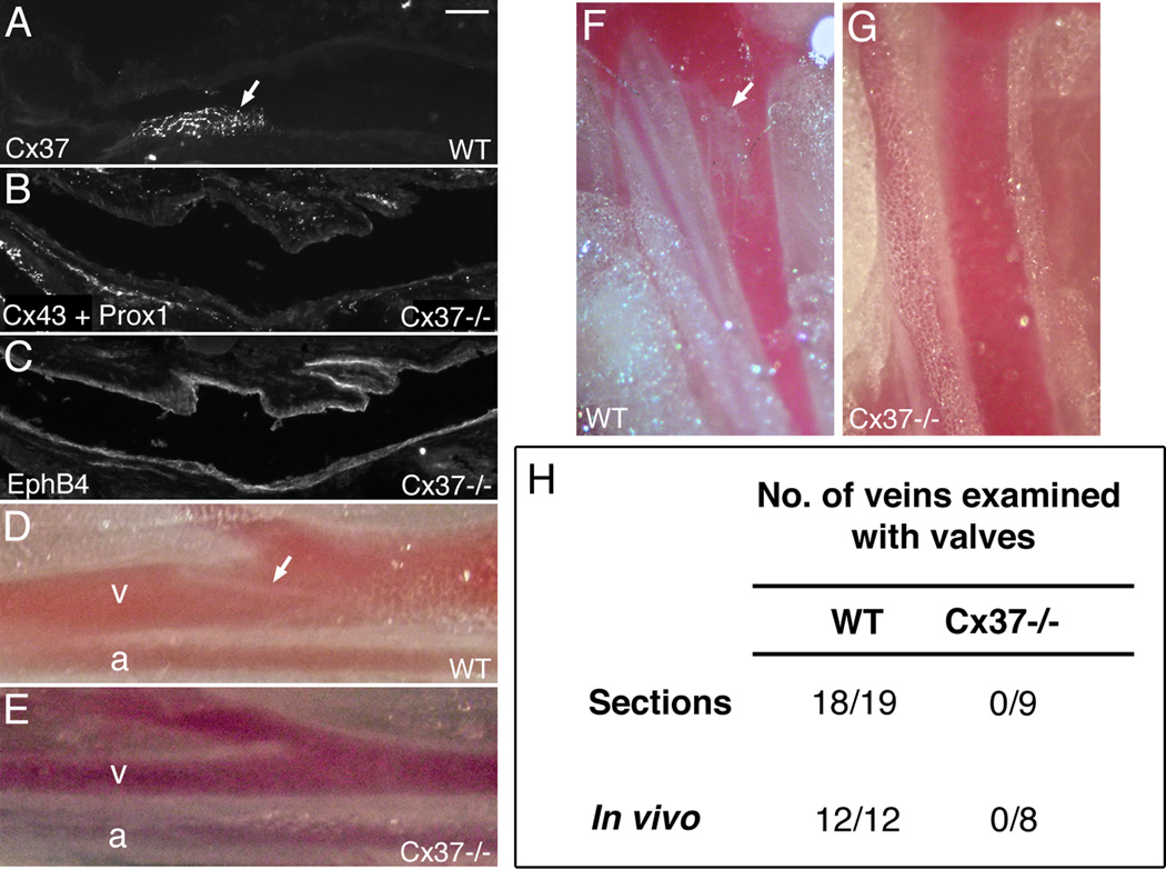
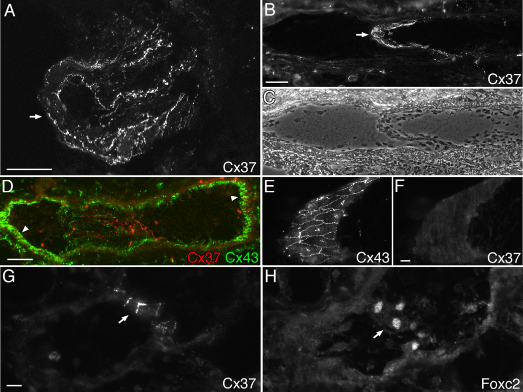
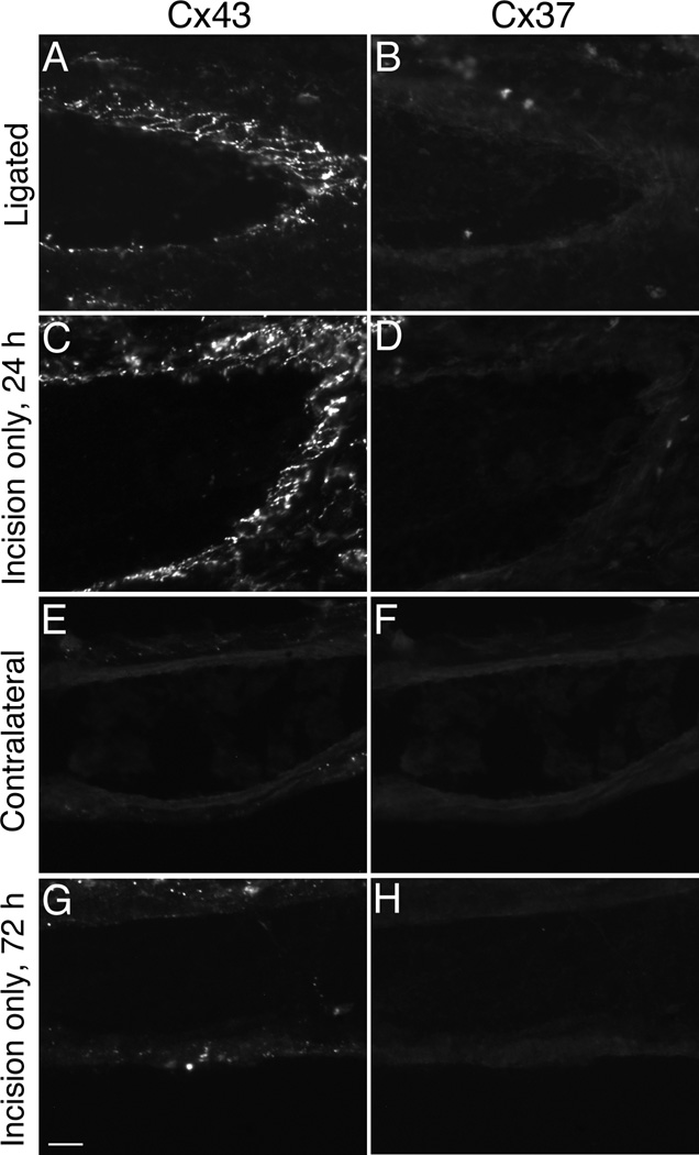
Venous valves play a crucial role in blood circulation, promoting the one-way movement of blood from superficial and deep veins towards the heart. By preventing retrograde flow, venous valves spare capillaries and venules from being subjected to damaging elevations in pressure, especially during skeletal muscle contraction. Pathologically, valvular incompetence or absence of valves are common features of venous disorders such as chronic venous insufficiency and varicose veins. The underlying causes of these conditions are not well understood, but congenital venous valve aplasia or agenesis may play a role in some cases. Despite progress in the study of cardiac and lymphatic valve morphogenesis, the molecular mechanisms controlling the development and maintenance of venous valves remain poorly understood. Here, we show that in valved veins of the mouse, three gap junction proteins (Connexins, Cxs), Cx37, Cx43, and Cx47, are expressed exclusively in the valves in a highly polarized fashion, with Cx43 on the upstream side of the valve leaflet and Cx37 on the downstream side. Surprisingly, Cx43 expression is strongly induced in the non-valve venous endothelium in superficial veins following wounding of the overlying skin. Moreover, we show that in Cx37-deficient mice, venous valves are entirely absent. Thus, Cx37, a protein involved in cell-cell communication, is one of only a few proteins identified so far as critical for the development or maintenance of venous valves. Because Cxs are necessary for the development of valves in lymphatic vessels as well, our results support the notion of common molecular pathways controlling valve development in veins and lymphatic vessels.


Figures

Figure 1
Figure 1. Cx37 is highly expressed at venous valves
(A) Cx37 immunostaining of a longitudinal section through the brachial vein shows that Cx37 is expressed exclusively at the venous valve (arrow). Note that Cx37 staining is not seen in the non-valve venous endothelium (arrowhead). (B) The same section as in (A) immunostained for Prox1 transcription factor, which is highly expressed in venous valves. (C) A merged image of panels A and B, showing Cx37 and Prox1 double labeling in the venous valve. (D) Phase contrast image of the brachial vein section shown in A–C. (E) A higher magnification view of the valve region shown in panel C. Prox1 exhibits a nuclear distribution whereas Cx37 is found at cell-cell appositions, as expected. (F) Cx37 and CD31 double labeling of a section of saphenous vein shows again that Cx37 is highly expressed at the valve (arrow). The CD31 immunostaining confirms that the vascular endothelium was intact throughout the vein section. (G,H) Additional examples of saphenous vein sections, showing high Cx37 expression at the venous valve (arrows). (I) Cx37 and Foxc2 double labeling at the valve (arrow) of the saphenous vein. Like Prox1, Foxc2 is a transcription factor that is enriched at venous valves and shows a nuclear localization. Note that the morphology of valves in this and other figures varies considerably depending on the exact orientation of the valve leaflets during sectioning. Blood flow is from left to right in the panels. Scale bars: 50 µm.
Figure 2
Figure 2. Cx37 and Cx43 are on opposite sides of the venous valve leaflets
(A) Cx37 immunostaining at a brachial vein valve (arrow) where the section presents an en face view of the relatively flat valve leaflet. (B) The same section as in panel A immunostained for Cx43, shows that Cx43, like Cx37, is expressed exclusively at the valve. (C) An overlay of Cx37 and Cx43 staining at the valve shows little or no overlap between the two immunosignals. (D) DIC image of the valve (arrow) shown in panels A–C and E. The box shows the area imaged by confocal microscopy in A–C. (E) Confocal z-stack imaging of the flattest part of the valve shown in A–D, which extends into the vessel lumen, colabeled for Cx37 and Cx43. In the center is the x–y projection through the z-stack. At the top is the x–z projection and on the right is the y–z projection, both showing strong separation of the Cx37 and Cx43 signals. The spatial separation of the signals in the x–z and y–z projections is not perfect perhaps because the valve is not exactly flat in the section, and there may also be a few Cx43-expressing leukocytes adhered to the valve leaflets on either side (isolated blotches of Cx43 signal). The orientation of this and other vein specimens was recorded, therefore we could determine that Cx43 is on the upstream side of the valve leaflet and Cx37 is on the downstream side. (F) A transverse section of a different brachial vein valve, where the valve is viewed in cross-section (end-on), shows in a different way that Cx37 and Cx43 are expressed on opposite sides of the valve leaflets. The Cx43 signal appears as short linear stretches because Cx43 is localized to endothelial cell-cell appositions. The Cx37 signal in this example was very strong and there may be some Cx37 present in the non-junctional membranes as well as at cell-cell appositions. (G) A lower magnification phase contrast image showing both leaflets (arrows) of the brachial vein valve immunostained in F. The boxed region shows the small area imaged in F. Blood flow is perpendicular to the plane of section in F and G. (H,I) Additional examples of the polarized expression of Cx37 and Cx43 on the two sides of venous valve leaflets (end-on views). Flow is from left to right. Taken together, these images show that Cx43 is present on the central, lumenal side of the valve leaflets (u, upstream side) and Cx37 is found on the sides of the leaflets facing the valve sinus or pocket (d, downstream side). A schematic illustration of the polarized expression of Cx43 and Cx37 on the upstream and downstream sides of venous valve leaflets is shown in J. Note that the illustration shows an idealized longitudinal section of a vein whereas the actual sections shown in the figure have differing orientations. Scale bars: 20 µm.
Figure 3
Figure 3. Cx47 expression is highly restricted to a small subset of cells in the venous valve, compared with Cx37 or Cx43
All of the panels in the figure show longitudinal sections through the same saphenous vein valve, where both valve leaflets (arrowheads) are visible in the lumen. Blood flow is from left to right. (A–E) A saphenous vein valve section double labeled for Cx37 and Cx43. (A) Phase contrast image of the valve. (B) Cx37 immunostaining of the valve. (C) Cx43 immunostaining of the same section as in B. (D) An overlay of Cx37 and Cx43 staining shown in B and C. The boxed region is shown at higher magnification in E. Both Cx37 and Cx43 are widely expressed throughout the valve leaflets, but on opposite sides. The orientation of the leaflets is known and Cx43 is again present on the central lumenal side of the valve leaflets (u, upstream side) and Cx37 is found on the sides of the leaflets facing the valve sinus or pocket (d, downstream side). (F–J) An adjacent section of the same saphenous vein valve shown in A–E, but double labeled for Cx37 and Cx47. (F) Phase contrast image of the valve. (G) Cx37 immunostaining. (H) Cx47 immunostaining of the same section as in G, showing that Cx47 expression is highly restricted to a small subset of cells in the valve. Arrows highlight two examples of areas where Cx47 is expressed. (I) An overlay of Cx37 and Cx47 staining shown in G and H. The boxed region is shown at higher magnification in J. Scale bars: 20 µm.
Figure 4
Figure 4. Colocalization studies reveal a complex spatial relationship between Cx47, Cx37, and Cx43 expression in the venous valve
(A) A saphenous vein valve (arrow) longitudinal section immunolabeled for Cx37. The section was a glancing one at the point where the valve leaflet starts to merge with the wall of the vein. (B) The same valve section as in A immunostained for Cx47. (C) An overlay of the Cx37 and Cx47 staining shown in A and B. A highly localized area of the valve showing intense, line-like Cx47 immunosignal is shown at higher magnification in D. Note that in this example, there was little or no overlap between the Cx37 and Cx47 signals. (E) A different saphenous vein valve (arrow) immunostained for Cx37. (F) The same valve section as in E immunolabeled for Cx47. (G) Overlay of the Cx37 and Cx47 staining shown in E and F. An area of the valve showing intense, line-like Cx47 staining is shown at higher magnification in H. In this case, there was significant overlap between the Cx37 and Cx47 signals. (I) A high magnification view of another saphenous vein valve (en face orientation) immunostained for Cx47. (J) The same valve section as in I immunostained for Cx43. (K) The overlay of I and J shows that the weaker punctate Cx47 signal colocalizes with Cx43. (L) A high magnification view of a different saphenous vein valve (en face orientation) immunolabeled for Cx47. (M) The same valve section as in L immunostained for Cx43. (N) Overlay of Cx47 and Cx43 staining shown in L and M. Note that the weaker, punctate Cx47 signal in the valve colocalizes with Cx43 but the intense, line-like Cx47 signal is distinct from the Cx43 signal. Blood flow is from left to right in A–H. Scale bars: (A–C) 20 µm; (D) 20 µm; (E–G) 20 µm; (H) 10 µm; (I–K) 10 µm; (L–N) 10 µm
Figure 5
Figure 5. Cx37 is required for the presence of venous valves in mice
(A) A longitudinal section of a wild-type (WT) saphenous vein immunostained for Cx37 showing the presence of a valve (arrow). (B) A representative section (one of a complete series of sections) of a Cx37−/− saphenous vein immunostained for Cx43 and Prox1 (combined staining) reveals the absence of a valve in the knockout specimen. (C) An adjacent section to the one in B stained for EphB4 confirms the vein identification and also shows the absence of a valve. (D) The saphenous vein valve (arrow) of a WT mouse is visible at a stereotypical location when the vein is exposed in vivo. (E) The exposed saphenous vein of a Cx37−/− mouse shows the absence of a valve at the same location. (F) A venous valve (arrow) is present in the iliac vein of a WT mouse but is absent in the iliac vein of a Cx37−/− mouse (G). Blood flow is from left to right in A–E and from bottom to top in F and G. (H) A summary of the data showing that the presence of venous valves in mice requires Cx37 expression. a, artery. v, vein. Scale bars: (A–C) 50 µm.
Figure 6
Figure 6. Cx37 expression at venous valves is maintained after a prolonged disruption of blood flow
(A) Cx37 immunostaining of a vein valve section from a WT saphenous vein ligated for 24 hrs shows that Cx37 expression persists at the valve (arrow) after a prolonged disruption of venous blood flow, although the Cx37 staining appears somewhat disorganized. (B) Cx37 expression at the valve (arrow) is still detected in a saphenous vein ligated for 72 hrs. (C) Phase contrast image of the vein shown in B. (D) At 24 hr post-ligation, Cx43 but not Cx37 expression is strongly induced in the non-valve endothelium (arrowheads) of the ligated saphenous vein, where there is normally no Cx43 expression. (E) At 8 hr post-ligation, Cx43 expression is already induced in the non-valve endothelium of the saphenous vein. The image shows a high magnification view of Cx43 staining in a glancing section of the non-valve endothelium. (F) The same section as in E immunostained for Cx37, shows that Cx37 is not induced by the 8 hr ligation. (G,H) Adjacent sections of a ligated saphenous vein immunostained for Cx37 (G) and Foxc2 (H), shows that Foxc2 expression is maintained at the valve (arrows) along with Cx37 when flow is disrupted. Blood flow would normally be from left to right in these specimens. Scale bars: (A) 50 µm; (B and C) 50 µm; (D) 50 µm; (E and F) 10 µm; (G and H) 10 µm.
Figure 7
Figure 7. Cx43 is strongly induced in the non-valve endothelium of the saphenous vein when the overlying skin is wounded by a surgical incision
(A,B) Double labeling shows that Cx43, but not Cx37, is strongly induced in the non-valve endothelium of the saphenous vein when the vein is ligated for 24 hrs. At higher magnification than is shown, a very small amount of Cx37 induction could sometimes be observed to colocalize with Cx43. (C,D) Surgical incision of the lower limb skin alone, without vessel ligation, is sufficient to induce Cx43, but not Cx37 expression in the non-valve endothelium of the saphenous vein, after 24 hrs. (E,F) Neither Cx43 nor Cx37 is induced in the saphenous vein of the contralateral, control limb opposite to the incised limb. (G,H) 72 hrs after a surgical incision of the lower limb skin, Cx43 expression in the non-valve endothelium of the saphenous vein is mostly gone, although a few scattered areas of the endothelium still showed some Cx43 (not shown). Cx37 was also not present in the non-valve endothelium at this time point. Scale bar: 20 µm.

Similar articles

Cited by

References

    1. Alonso F, Krattinger N, Mazzolai L, Simon A, Waeber G, Meda P, Haefliger JA. An angiotensin II- and NF-kappaB-dependent mechanism increases connexin 43 in murine arteries targeted by renin-dependent hypertension. Cardiovasc. Res. 2010;87:166–176. - PMC - PubMed
    1. Bazigou E, Lyons OT, Smith A, Venn GE, Cope C, Brown NA, Makinen T. Genes regulating lymphangiogenesis control venous valve formation and maintenance in mice. J. Clin. Invest. 2011;121:2984–2992. - PMC - PubMed
    1. Bazigou E, Xie S, Chen C, Weston A, Miura N, Sorokin L, Adams R, Muro AF, Sheppard D, Makinen T. Integrin-alpha9 is required for fibronectin matrix assembly during lymphatic valve morphogenesis. Dev. Cell. 2009;17:175–186. - PMC - PubMed
    1. Becker DL, Thrasivoulou C, Phillips AR. Connexins in wound healing; perspectives in diabetic patients. Biochim. Biophys. Acta. 2011;1818:2068–2075. - PubMed
    1. Bruzzone R, Haefliger JA, Gimlich RL, Paul DL. Connexin40, a component of gap junctions in vascular endothelium, is restricted in its ability to interact with other connexins. Mol. Biol. Cell. 1993;4:7–20. - PMC - PubMed

Publication types

LinkOut - more resources