. 2012 Dec;19(12):1266-72.
doi: 10.1038/nsmb.2435. Epub 2012 Nov 11.

Molecular basis for H3K36me3 recognition by the Tudor domain of PHF1

Affiliations

Molecular basis for H3K36me3 recognition by the Tudor domain of PHF1

Catherine A Musselman et al. Nat Struct Mol Biol. 2012 Dec.

Abstract

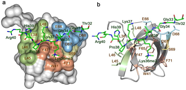
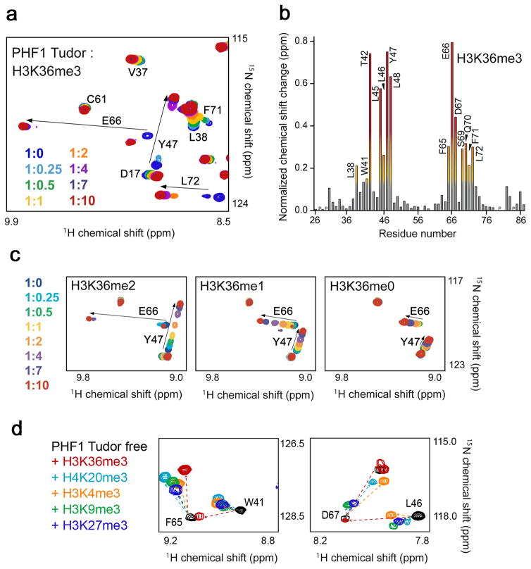
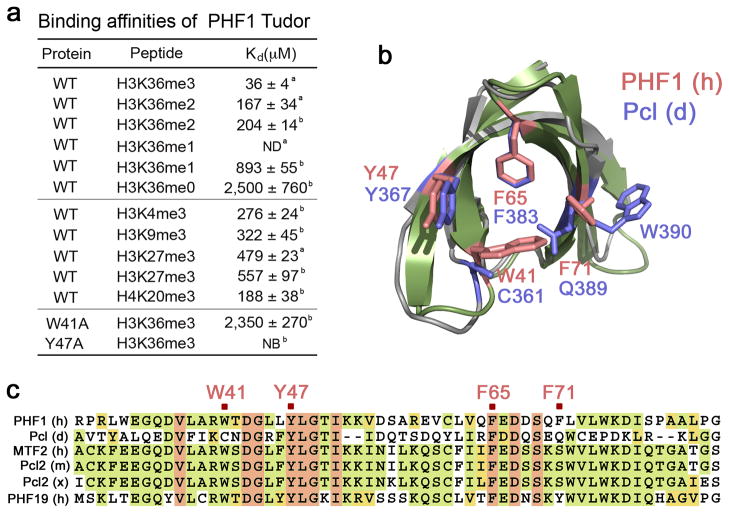
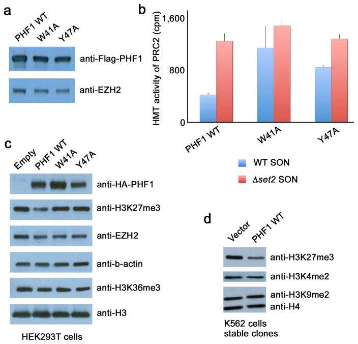
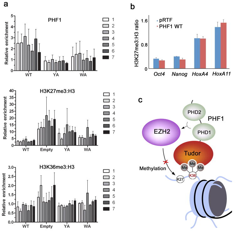
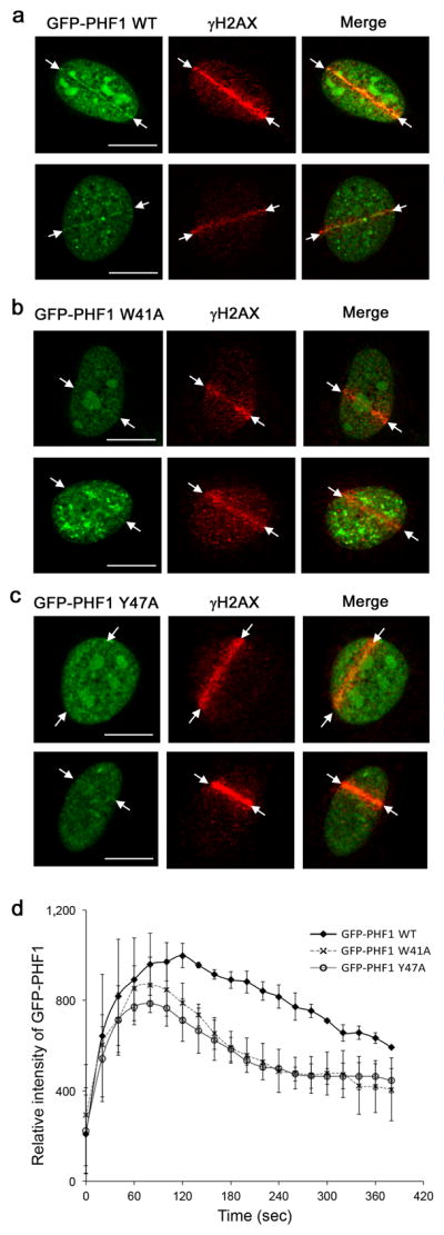
The PHD finger protein 1 (PHF1) is essential in epigenetic regulation and genome maintenance. Here we show that the Tudor domain of human PHF1 binds to histone H3 trimethylated at Lys36 (H3K36me3). We report a 1.9-Å resolution crystal structure of the Tudor domain in complex with H3K36me3 and describe the molecular mechanism of H3K36me3 recognition using NMR. Binding of PHF1 to H3K36me3 inhibits the ability of the Polycomb PRC2 complex to methylate Lys27 of histone H3 in vitro and in vivo. Laser microirradiation data show that PHF1 is transiently recruited to DNA double-strand breaks, and PHF1 mutants impaired in the H3K36me3 interaction exhibit reduced retention at double-strand break sites. Together, our findings suggest that PHF1 can mediate deposition of the repressive H3K27me3 mark and acts as a cofactor in early DNA-damage response.


Conflict of interest statement

COMPETING FINANCIAL INTERESTS

The authors declare no competing financial interests.

Figures

Figure 1
Figure 1
The crystal structure of the Tudor domain of PHF1 in complex with the H3K36me3 peptide. The Tudor domain is depicted as a solid surface (a) and a ribbon diagram (b) with the peptide shown as a stick model. The aromatic cage residues involved in the interaction with the methylated lysine residue are colored brown, those in the hydrophobic patch and the acidic groove are colored green and blue, respectively. H3 residues are labeled in green and Tudor residues are labeled in brown. Dashed lines represent intermolecular hydrogen bonds. For clarity, here and throughout the text, individual residues of the Tudor domain are denoted using a single-letter code, whereas individual residues of the histone peptide are denoted using a three-letter code.
Figure 2
Figure 2
The PHF1 Tudor domain recognizes H3K36me3. (a) Superimposed 1H,15N Heteronuclear single quantum coherence (HSQC) spectra of the Tudor domain collected upon titration of the H3K36me3 peptide. Spectra are color coded according to the protein:peptide molar ratio. (b) The normalized chemical shift changes observed in the corresponding (a) spectra of the Tudor domain as a function of residue. Residues showing large chemical shift differences are labeled. Differences greater than the average plus one standard deviation (SD), the average plus one-half SD, and the average are shown in red, orange and yellow, respectively. (c) 1H,15N HSQC overlays of the PHF1 Tudor domain in the presence of increasing concentrations of dimethylated, monomethylated or unmodified H3K36 peptides. Spectra are color coded by the protein:peptide molar ratio (legend at left). (d) 1H,15N HSQC overlays of the PHF1 Tudor domain in the free state (black) and in the presence of a 1:10 molar ratio of H3K36me3 (red), H3K4me3 (orange), H3K9me3 (green), H3K27me3 (dark blue) and H4K20me3 (light blue). The resonances of S69 and Q70 are omitted in the right panel for clarity.
Figure 3
Figure 3
Interaction of the PHF1 Tudor domain with H3K36me3 is specific. (a) Binding affinities of PHF1 Tudor for H3 and H4 peptides as measured by ITC a or NMR b. ND-not detected, NB-no binding. (b) Conservation of the aromatic cage residues across PHF1 homologues. Structural overlay of H3K36me3-bound PHF1 Tudor (histone peptide is not shown) with the NMR structure of the apo-state Drosophila homologue Pcl (PDB code 2XK0). The aromatic cage residues of PHF1 are colored salmon, and corresponding Pcl residues are colored blue. (c) Sequence alignment of PHF1 homologues shows that all four aromatic residues (marked in salmon), necessary for binding to methylated lysine, are conserved only in PHF19.
Figure 4
Figure 4
Recognition of H3K36me3 by PHF1 inhibits PRC2 methyltransferase activity. (a) Western analysis of wild type and mutated Flag-PHF1 and EZH2 in the PRC2 complexes. (b) HMT assays with PHF1-PRC2 complexes purified from HEK293T cells on native wild type chromatin (wt SON, blue) and chromatin lacking the H3K36me mark (Δset2 SON, red). Error bars represent SD based on three experiments. (c) Western analysis of whole cell extract from HEK293T cells 48 hours after transfection with wild type HA-PHF1 or W41A or Y47A mutants. Empty vector is control. (d) Western analysis of K562 cells stably expressing Flag-PHF1.
Figure 5
Figure 5
Binding of PHF1 to H3K36me3 decreases PRC2-mediated deposition of H3K27me3. (a) ChIP assays on chromatin from HEK293T cells fixed and harvested 48 hours after transfection with either empty vector, wild-type HA-PHF1 or W41A or Y47A mutants. The levels of HA-PHF1, H3K27me3, H3K36me3 and H3 were probed across the MYT1 promoter using 7 primer sets. The top panel shows occupancy of wild type and mutant HA-PHF1 and the bottom two panels show H3K27me3 and H3K36me3 levels normalized to H3 (H3K27me3:H3). All data are relative to the PCLB4 non-target control gene. Error bars represent SD based on three experiments. (b) ChIP assays on chromatin harvested from mouse ES cells transduced with empty vector (pRTF, blue) or vector containing Flag-PHF1 (PHF1, red). Using quantitative PCR (qPCR), the levels of H3K27me3 and H3 were probed at the Oct4, Nanog, HoxA4 and HoxA11 promoters. Data are presented as the ratio of H3K27me3 to total H3 signal to correct for possible differences in nucleosome density at the different loci examined. Error bars represent SD based on two experiments. (c) A model for inhibition of EZH2-PRC2 activity through recognition of H3K36me3 by the Tudor domain of PHF1.
Figure 6
Figure 6
Tudor-dependent accumulation and retention of PHF1 at laser-irradiated sites of DSBs. Co-localization of GFP-PHF1 wild type (a), GFP-PHF1 W41A (b) and GFP-PHF1 Y47A (c) at the DSB sites in U2OS cells 3 min after irradiation. Scale bars correspond to 10 μm. (d) Kinetic analysis of GFP-PHF1 intensity at laser irradiated sites. GFP-PHF1 wild type (closed diamond), GFP-PHF1 W41A (X) and GFP-PHF1 Y47A (open circle). Error bars represent SD based on three experiments.

Comment in

References

    1. Margueron R, Reinberg D. The Polycomb complex PRC2 and its mark in life. Nature. 2011;469:343–9. - PMC - PubMed
    1. Varambally S, et al. The polycomb group protein EZH2 is involved in progression of prostate cancer. Nature. 2002;419:624–9. - PubMed
    1. Morin RD, et al. Somatic mutations altering EZH2 (Tyr641) in follicular and diffuse large B-cell lymphomas of germinal-center origin. Nature genetics. 2010;42:181–5. - PMC - PubMed
    1. Cao R, et al. Role of histone H3 lysine 27 methylation in Polycomb-group silencing. Science. 2002;298:1039–43. - PubMed
    1. Kuzmichev A, Nishioka K, Erdjument-Bromage H, Tempst P, Reinberg D. Histone methyltransferase activity associated with a human multiprotein complex containing the Enhancer of Zeste protein. Genes Dev. 2002;16:2893–905. - PMC - PubMed

Publication types

Associated data