. 2012 Dec;122(12):4606-20.
doi: 10.1172/JCI63287. Epub 2012 Nov 12.

Intravaginal immunization with HPV vectors induces tissue-resident CD8+ T cell responses

Affiliations

Intravaginal immunization with HPV vectors induces tissue-resident CD8+ T cell responses

Nicolas Çuburu et al. J Clin Invest. 2012 Dec.

Abstract

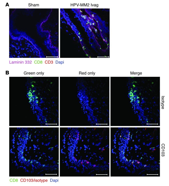
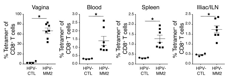
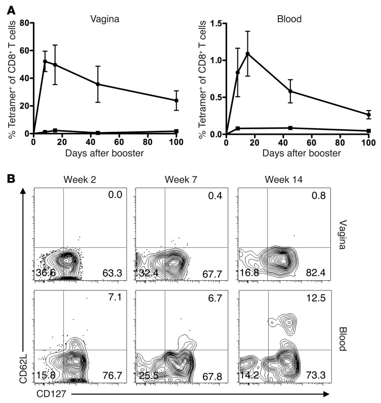
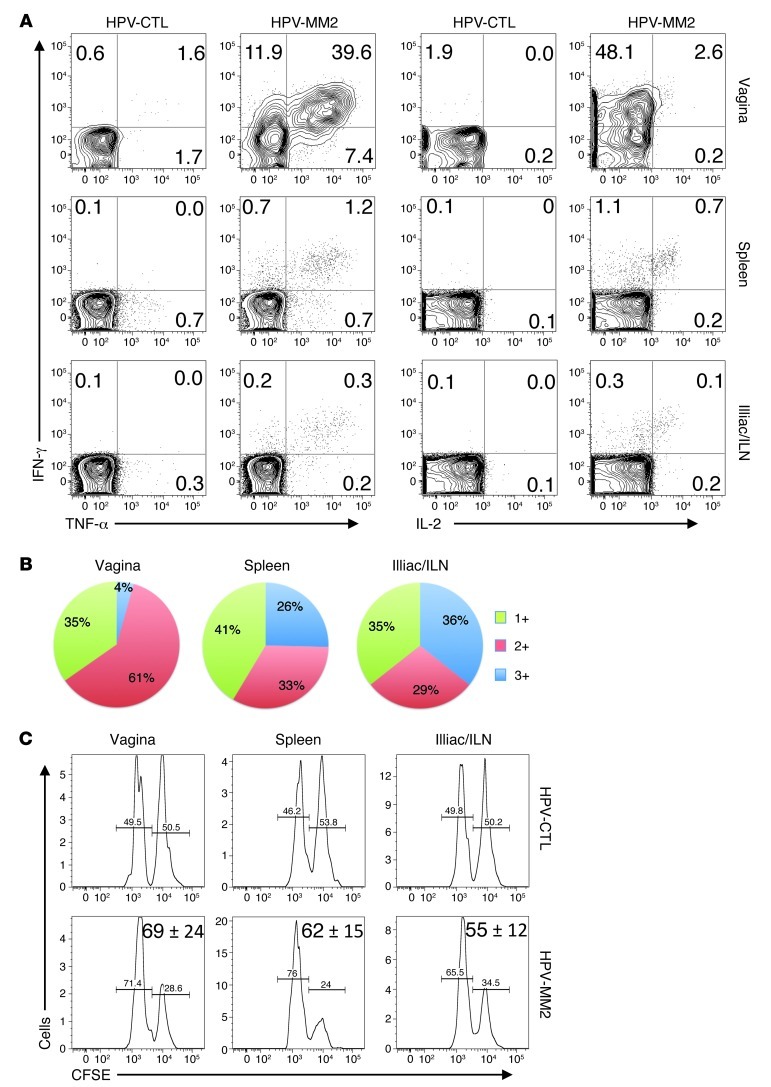
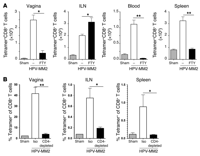
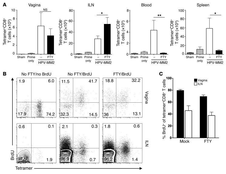
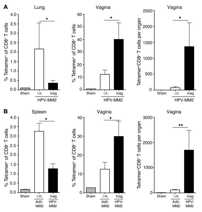
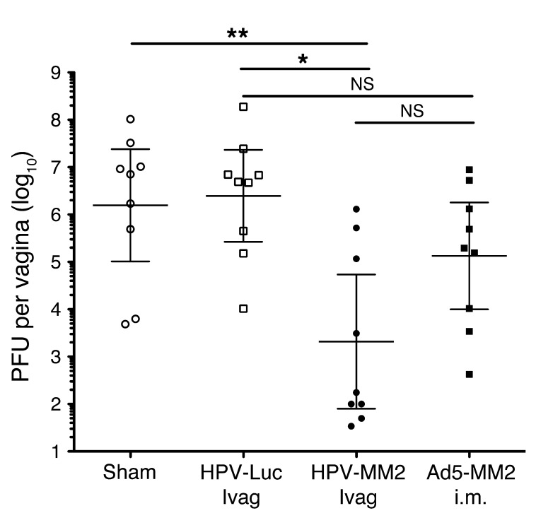
The induction of persistent intraepithelial CD8+ T cell responses may be key to the development of vaccines against mucosally transmitted pathogens, particularly for sexually transmitted diseases. Here we investigated CD8+ T cell responses in the female mouse cervicovaginal mucosa after intravaginal immunization with human papillomavirus vectors (HPV pseudoviruses) that transiently expressed a model antigen, respiratory syncytial virus (RSV) M/M2, in cervicovaginal keratinocytes. An HPV intravaginal prime/boost with different HPV serotypes induced 10-fold more cervicovaginal antigen-specific CD8+ T cells than priming alone. Antigen-specific T cell numbers decreased only 2-fold after 6 months. Most genital antigen-specific CD8+ T cells were intra- or subepithelial, expressed αE-integrin CD103, produced IFN-γ and TNF-α, and displayed in vivo cytotoxicity. Using a sphingosine-1-phosphate analog (FTY720), we found that the primed CD8+ T cells proliferated in the cervicovaginal mucosa upon HPV intravaginal boost. Intravaginal HPV prime/boost reduced cervicovaginal viral titers 1,000-fold after intravaginal challenge with vaccinia virus expressing the CD8 epitope M2. In contrast, intramuscular prime/boost with an adenovirus type 5 vector induced a higher level of systemic CD8+ T cells but failed to induce intraepithelial CD103+CD8+ T cells or protect against recombinant vaccinia vaginal challenge. Thus, HPV vectors are attractive gene-delivery platforms for inducing durable intraepithelial cervicovaginal CD8+ T cell responses by promoting local proliferation and retention of primed antigen-specific CD8+ T cells.


Figures

Figure 1
Figure 1. Ivag heterologous prime/boost immunization regimen overcomes type-specific antibody neutralization and enhances CD8+ T cell response in the genital tract.
(A) Depo-Provera–treated BALB/c mice (n = 5/group) were inoculated Ivag with 5 × 10⁷ IU HPV16 PsV expressing luciferase (16Luc) or sham treated. One month after initial 16Luc PsV inoculation, mice were challenged Ivag with 5 × 10⁷ IU 16Luc or HPV45 PsV expressing luciferase (45Luc). In vivo luciferase expression was measured 2 days after Ivag challenge and is expressed as mean luminescence in photons per second + SD (**P < 0.01, Mann Whitney U test). (B) Depo-Provera–treated mice were immunized Ivag with 5 × 10⁷ IU HPV PsV expressing the RSV fusion protein M/M2. For the single immunization regimen, mice were immunized with 5 × 10⁷ IU HPV16MM2 PsV, and for the prime/boost regimen, they were immunized with 5 × 10⁷ IU HPV16MM2; 1 month later the primed animals were immunized with 5 × 10⁷ IU HPV45MM2. Two weeks after the final immunization, vaginal cell suspensions were analyzed by flow cytometry for the presence of KdM282-tetramer+CD8+ T lymphocytes. Data are expressed as the mean of the total number of cells per organ + SD (*P < 0.05, **P < 0.01, Kruskal-Wallis/Dunn’s test).
Figure 2
Figure 2. HPV Ivag prime/boost immunization induces high numbers of cervicovaginal CD8+ T cells.
Depo-Provera–treated mice (n = 5/group) were immunized with 5 × 10⁷ IU HPV16MM2 or HPV16 control (HPV-CTL) Ivag, and 1 month later, primed mice were immunized with 5 × 10⁷ IU HPV45MM2 or HPV45 control, respectively. Two weeks after the second immunization, cervicovaginal cell suspensions were analyzed by flow cytometry for the presence of KdM282-tetramer+CD8+ T lymphocytes in HPVMM2-immunized and sham-treated animals. (A) CD8 and CD4 expression on gated vaginal cells with a lymphocyte morphology (contour plot) and KdM282-tetramer binding on gated CD8+ T cells (histogram plot); the percentage of each gate and marker of the parent population is indicated in each plot. (B) Total CD8+ T lymphocytes and KdM282-tetramer+CD8+ T lymphocytes per cervicovaginal mucosa. Data are representative of more than 3 experiments (*P < 0.05, **P < 0.01, Mann-Whitney U test).
Figure 3
Figure 3. HPV Ivag prime/boost immunization induces CD103+ cervicovaginal intraepithelial CD8+ T cells.
Depo-Provera–treated mice (n = 5/group) were immunized with 5 × 10⁷ IU HPV16MM2 or HPV16 control Ivag, and 1 month later, primed mice were immunized with 5 × 10⁷ IU HPV45MM2 or HPV45 control, respectively. Two weeks after the second immunization, the distribution of CD8+ T cells was analyzed in cervicovaginal tissue section from HPVMM2-immunized and sham-treated animals. (A) Cervicovaginal tissue section stained with CD8 (green), CD3 (red), laminin 332 (magenta), and DAPI (blue). (B) Cervicovaginal tissue sections from HPVMM2-immunized mice were stained with CD8 (green), isotype control (upper images), or CD103 (lower images; red) antibodies and DAPI (blue). Single channel for CD8 staining (green only) and CD103/isotype control (red only) and merge. Scale bars: 50 μm. Images are representative of 5 animals tested for each condition examined.
Figure 4
Figure 4. HPV Ivag prime/boost immunization induces local and systemic CD8+ T cell responses.
Depo-Provera–treated mice were immunized with 5 × 10⁷ IU HPV16MM2 or HPV16 control Ivag, and 1 month later, primed mice were immunized with 5 × 10⁷ IU HPV45MM2 or HPV45 control, respectively. Two weeks after the final immunization, cervicovaginal, blood, spleen, and ILN cell suspensions were analyzed by flow cytometry for the presence of KdM282-tetramer+CD8+ T cells. Data are representative of more than 3 experiments (*P < 0.05, Mann-Whitney U test).
Figure 5
Figure 5. HPV PsV Ivag prime/boost immunization induces durable CD8+ T cell responses with an effector memory phenotype after HPV PsV Ivag immunization.
Depo-Provera–treated mice were immunized with 5 × 10⁷ IU HPV16MM2 or HPV16 control Ivag, and 1 month later mice were immunized with 5 × 10⁷ IU HPV45MM2 or HPV45 control, respectively. (A) Cervicovaginal and blood cell suspensions were obtained from week 1 to week 14 and analyzed by flow cytometry for the presence of KdM282-tetramer+CD8+ T lymphocytes in HPVMM2 (squares) and HPV control (circles). Data are expressed as the mean percentage of KdM282-tetramer+CD8+ T lymphocytes in total CD8+ T lymphocytes + SD. (B) Representative plots of CD62L and CD127 expression of KdM282-tetramer+CD8+ T lymphocytes in cervicovaginal and blood cells suspensions collected at weeks 2, 7, and 14. The percentage of CD8+ T cells in each quadrant is indicated in each plot. Data are representative of 3 experiments.
Figure 6
Figure 6. HPV Ivag prime/boost immunization induces polyfunctional CD8+ T cells with cytotoxic activity.
Depo-Provera–treated mice were immunized with 5 × 10⁷ IU HPV16MM2 or HPV16 control Ivag, and 1 month later, mice were immunized with 5 × 10⁷ IU HPV45MM2 or HPV45 control, respectively. (A) Two weeks after the last immunization, the production of IFN-γ, TNF-α, and IL-2 by CD8+ T cells was measured after in vitro stimulation with M282–90 peptide in cervicovaginal, spleen, and ILN cell suspensions. The percentage of CD8+ T cells in each quadrant is indicated in each plot. (B) Relative proportion of single-, double-, and triple-cytokine-producing CD8+ T cells restimulated with M282–90 peptide after subtraction of background cytokine production of unstimulated CD8+ T cells. (C) In vivo cytotoxic activity was assessed 2 weeks after the last immunization. Target cells consisting of an equal mixture of two splenocyte populations labeled with a high and a low concentration of CFSE were pulsed with M282–90 peptide or remained unpulsed, respectively, were injected Ivag and i.v. Twenty-four hours later, cervicovaginal, spleen, and ILN cell suspensions were analyzed by flow cytometry. Representative histograms gated on CFSE-positive cells. Mean percentage ± SD of M282–90 specific lysis is indicated.
Figure 7
Figure 7. Primary CD8+ T cell responses are induced in the ILN and are abrogated in the absence of CD4+ T cell help.
Mice (n = 5/group) were treated with FTY720 in drinking water from day 1 to day 14 after single or booster immunization (A). On day 14, vagina, ILN, spleen, and blood cell suspensions were analyzed by flow cytometry for the presence KdM282-tetramer+CD8+ T lymphocytes. Data are expressed as mean + SD of the total number of KdM282-tetramer+CD8+ T lymphocytes per organ or per milliliter of blood. (B) To evaluate the role of CD4+ T cell help in the induction of CD8+ T cell responses, mice were injected i.p. every second day with rat anti-mouse CD4 or isotype control (iso) antibody from day –3 to day 9 relative to the day of immunization. Two weeks after immunization, vagina, ILN, and spleen were analyzed by flow cytometry for the presence KdM282-tetramer+CD8+ T cells. Data are expressed as the mean + SD percentage of KdM282-tetramer+CD8+ T cells in CD8+ T cells. Data are representative of 3 experiments (*P < 0.05, **P < 0.01, Kruskal-Wallis/Dunn’s test).
Figure 8
Figure 8. Memory CD8+ T lymphocytes can expand locally in the vagina upon booster Ivag immunization with HPV.
Depo-Provera–treated mice (n = 5/group) were immunized Ivag with 5 × 10⁷ IU HPV16MM2, followed 1 month later by a second immunization with 5 × 10⁷ IU. On day 14, vagina, ILN, spleen, and blood cell suspensions were analyzed by flow cytometry for the presence KdM282-tetramer+CD8+ T lymphocytes (A). Data are expressed as mean + SD of the total number of KdM282-tetramer+CD8+ T lymphocytes per organ or per ml of blood. FTY, FTY720. (B and C) Mice received BrdU per os after the boost to measure in vivo proliferation of KdM282-tetramer+CD8+ T cells in FTY720- or mock-treated mice upon secondary immunization. Two weeks after boost, incorporation of BrdU by KdM282-tetramer+CD8+ T cells was measured by intracellular staining of cervicovaginal and ILN cell suspensions and further analyzed by flow cytometry. (B) Representative plot of BrdU and KdM282-tetramer staining of CD8+ T cells (percentage of CD8+ T cells in each quadrant is indicated). (C) Mean + SD percentage of BrdU+ relative to KdM282-tetramer+CD8+ T cells. Data are representative of 2 (A) and 3 (B) experiments (*P < 0.05, **P < 0.01, Kruskal-Wallis/Dunn’s test).
Figure 9
Figure 9. HPV Ivag prime/boost immunization preferentially induces genital CD8+ T lymphocyte responses compared with HPV IN and Ad5 IM immunization.
Depo-Provera–treated mice were immunized Ivag or i.n. with 5 × 10⁷ IU HPV16MM2 and 1 month later received a second immunization of 5 × 10⁷ IU HPV45MM2, or mice were immunized i.m. twice, 1 month apart, with 5 × 10⁶ PFU Ad5-MM2. Two weeks after the last immunization, lung, spleen, and cervicovaginal cell suspensions were analyzed by flow cytometry for the presence of KdM282-tetramer+CD8+ T lymphocytes. Data are expressed as the mean ± SD percentage or total number per organ of KdM282-tetramer+ of CD8+ T lymphocytes. Data are representative of 2 (A) and 3 (B) experiments (*P < 0.05, **P <0.01, Kruskal-Wallis/Dunn’s test).
Figure 10
Figure 10. HPV Ivag, but not Ad5 i.m., prime/boost immunization induces intraepithelial CD8+ T cells in the cervicovaginal epithelium.
Depo-Provera–treated mice were immunized with 5 × 10⁷ IU HPV16MM2 Ivag or with 5 × 10⁷ PFU Ad5-MM2 i.m., and 1 month later mice were immunized with 5 × 10⁷ IU HPV45MM2 or Ad5-MM2 i.m., respectively. (A) Two weeks after the last immunization, cervicovaginal tissue sections were co-stained for CD8 (green), CD3 (red), laminin 332 (magenta), and nuclei (blue). Scale bars: 50 μm. Images are representative of 5 animals tested for each condition examined. (B) In separate experiments, CD103 expression by KdM282-tetramer+CD8+ T lymphocytes was analyzed by flow cytometry in cervicovaginal, ILN, and blood cell suspensions from HPV Ivag and Ad5 i.m. immunized animals. Control isotype (red lines) and CD103 antibody (blue lines) staining is shown in B, and the percentage of CD103+ cells is indicated in the histogram plots. Data are representative of 3 experiments.
Figure 11
Figure 11. HPV Ivag, but not Ad5 i.m., prime/boost immunization reduces viral titers in the cervicovaginal mucosa after vaginal challenge with recombinant M282–90–VV.
Depo-Provera–treated mice were immunized with 5 × 10⁷ IU HPV16MM2 Ivag or with 5 × 10⁷ PFU Ad5-MM2 i.m., and 1 month later mice were immunized with 5 × 10⁷ IU HPV45MM2 or Ad5-MM2 i.m., respectively. Two months after the last immunization, mice were challenged Ivag with 1 × 10⁷ PFU of recombinant M282–90–VV. Three days after challenge, vaginal tissue was collected, and recombinant VV titers were determined by plaque assays. Results are expressed as PFU per vagina for individual mice (symbols) and geometric mean (horizontal bars). Data are representative of 3 experiments (*P < 0.05, **P < 0.01, Kruskal-Wallis/Dunn’s test).

References

    1. Zinkernagel RM, Doherty PC. Restriction of in vitro T cell-mediated cytotoxicity in lymphocytic choriomeningitis within a syngeneic or semiallogeneic system. Nature. 1974;248(450):701–702. doi: 10.1038/248701a0. - DOI - PubMed
    1. Bjorkman PJ, Saper MA, Samraoui B, Bennett WS, Strominger JL, Wiley DC. The foreign antigen binding site and T cell recognition regions of class I histocompatibility antigens. Nature. 1987;329(6139):512–518. doi: 10.1038/329512a0. - DOI - PubMed
    1. Black CA, et al. Vaginal mucosa serves as an inductive site for tolerance. J Immunol. 2000;165(9):5077–5083. - PubMed
    1. Dudek T, Knipe DM. Replication-defective viruses as vaccines and vaccine vectors. Virology. 2006;344(1):230–239. doi: 10.1016/j.virol.2005.09.020. - DOI - PubMed
    1. Rollier CS, Reyes-Sandoval A, Cottingham MG, Ewer K, Hill AV. Viral vectors as vaccine platforms: deployment in sight. Curr Opin Immunol. 2011;23(3):377–382. doi: 10.1016/j.coi.2011.03.006. - DOI - PubMed

Publication types

MeSH terms