

Differentiated parietal connectivity of frontal regions for "what" and "where" memory
- PMID: 23143344
- PMCID: PMC3825581
- DOI: 10.1007/s00429-012-0476-4
Differentiated parietal connectivity of frontal regions for "what" and "where" memory
Abstract
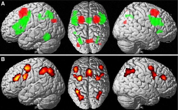
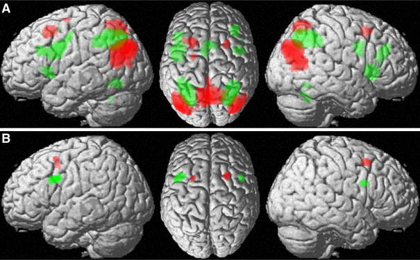
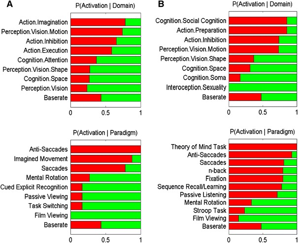
In a previous meta-analysis across almost 200 neuroimaging experiments, working memory for object location showed significantly stronger convergence on the posterior superior frontal gyrus, whereas working memory for identity showed stronger convergence on the posterior inferior frontal gyrus (dorsal to, but overlapping with Brodmann's area BA 44). As similar locations have been discussed as part of a dorsal frontal-superior parietal reach system and an inferior frontal grasp system, the aim of the present study was to test whether the regions of working-memory related "what" and "where" processing show a similar distinction in parietal connectivity. The regions that were found in the previous meta-analysis were used as seeds for functional connectivity analyses using task-based meta-analytic connectivity modelling and task-independent resting state correlations. While the ventral seed showed significantly stronger connectivity with the bilateral intraparietal sulcus (IPS), the dorsal seed showed stronger connectivity with the bilateral posterior inferior parietal and the medial superior parietal lobule. The observed connections of regions involved in memory for object location and identity thus clearly demonstrate a distinction into separate pathways that resemble the parietal connectivity patterns of the dorsal and ventral premotor cortex in non-human primates and humans. It may hence be speculated that memory for a particular location and reaching towards it as well as object memory and finger positioning for manipulation may rely on shared neural systems. Moreover, the ensuing regions, in turn, featured differential connectivity with the bilateral ventral and dorsal extrastriate cortex, suggesting largely segregated bilateral connectivity pathways from the dorsal visual cortex via the superior and inferior parietal lobules to the dorsal posterior frontal cortex and from the ventral visual cortex via the IPS to the ventral posterior frontal cortex that may underlie action and cognition.
Figures

Similar articles
-
Visual Short-Term Memory Activity in Parietal Lobe Reflects Cognitive Processes beyond Attentional Selection.J Neurosci. 2018 Feb 7;38(6):1511-1519. doi: 10.1523/JNEUROSCI.1716-17.2017. Epub 2018 Jan 8. J Neurosci. 2018. PMID: 29311140 Free PMC article.
-
Dissociation of mnemonic and perceptual processes during spatial and nonspatial working memory using fMRI.Hum Brain Mapp. 1998;6(1):14-32. doi: 10.1002/(SICI)1097-0193(1998)6:1<14::AID-HBM2>3.0.CO;2-O. Hum Brain Mapp. 1998. PMID: 9673660 Free PMC article. Clinical Trial.
-
A coordinate-based meta-analysis of the n-back working memory paradigm using activation likelihood estimation.Brain Cogn. 2019 Jun;132:1-12. doi: 10.1016/j.bandc.2019.01.002. Epub 2019 Jan 30. Brain Cogn. 2019. PMID: 30708115
-
A brief comparative review of primate posterior parietal cortex: A novel hypothesis on the human toolmaker.Neuropsychologia. 2017 Oct;105:123-134. doi: 10.1016/j.neuropsychologia.2017.01.034. Epub 2017 Jan 31. Neuropsychologia. 2017. PMID: 28159617 Free PMC article. Review.
-
Development of a superior frontal-intraparietal network for visuo-spatial working memory.Neuropsychologia. 2006;44(11):2171-7. doi: 10.1016/j.neuropsychologia.2005.11.019. Epub 2006 Jan 6. Neuropsychologia. 2006. PMID: 16405923 Review.
Cited by
-
Comprehensive investigation of predictive processing: A cross- and within-cognitive domains fMRI meta-analytic approach.Hum Brain Mapp. 2024 Aug 15;45(12):e26817. doi: 10.1002/hbm.26817. Hum Brain Mapp. 2024. PMID: 39169641 Free PMC article.
-
The neural correlates of spatial and object working memory in elderly and Parkinson's disease subjects.Behav Neurol. 2015;2015:123636. doi: 10.1155/2015/123636. Epub 2015 Mar 16. Behav Neurol. 2015. PMID: 25861157 Free PMC article.
-
The ability of patients with Parkinson's disease to recognize masked faces during the COVID-19 pandemic.Dement Neuropsychol. 2022 Jul-Sep;16(3):309-315. doi: 10.1590/1980-5764-DN-2021-0117. Epub 2022 Aug 15. Dement Neuropsychol. 2022. PMID: 36619841 Free PMC article.
-
Aging and response conflict solution: behavioural and functional connectivity changes.Brain Struct Funct. 2015;220(3):1739-57. doi: 10.1007/s00429-014-0758-0. Epub 2014 Apr 10. Brain Struct Funct. 2015. PMID: 24718622 Free PMC article.
-
Functional Hemispheric (A)symmetries in the Aged Brain-Relevance for Working Memory.Front Aging Neurosci. 2018 Mar 12;10:58. doi: 10.3389/fnagi.2018.00058. eCollection 2018. Front Aging Neurosci. 2018. PMID: 29593523 Free PMC article.
References
-
- Abe M, Hanakawa T. Functional coupling underlying motor and cognitive functions of the dorsal premotor cortex. Behav Brain Res. 2009;198:13–23. - PubMed
-
- Amunts K, Schleicher A, Burgel U, Mohlberg H, Uylings HB, Zilles K. Broca’s region revisited: cytoarchitecture and intersubject variability. J Comp Neurol. 1999;412:319–341. - PubMed
-
- Arnott SR, Binns MA, Grady CL, Alain C. Assessing the auditory dual-pathway model in humans. Neuroimage. 2004;22:401–408. - PubMed
-
- Ashburner J, Friston KJ. Unified segmentation. Neuroimage. 2005;26:839–851. - PubMed
Publication types
MeSH terms
Grants and funding
LinkOut - more resources
Full Text Sources
Research Materials
