

Granular cell tumor of the cervical esophagus: case report and literature review of an unusual cause of Dysphagia
- PMID: 23143390
- PMCID: PMC3738753
- DOI: 10.1007/s12105-012-0408-x
Granular cell tumor of the cervical esophagus: case report and literature review of an unusual cause of Dysphagia
Abstract
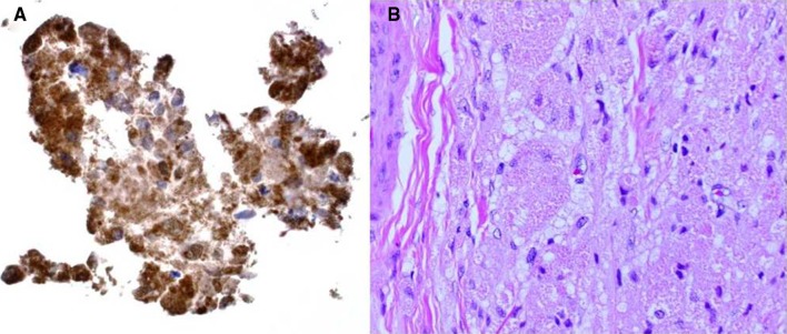
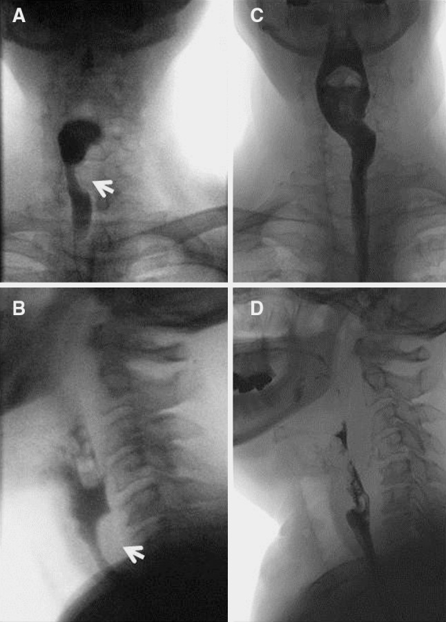
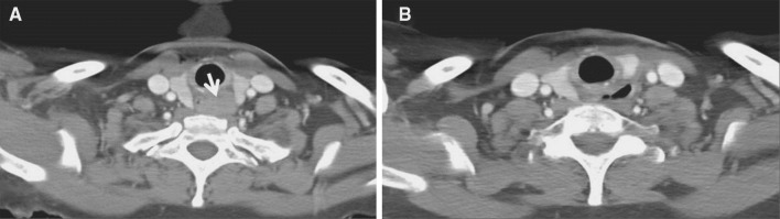
Granular cell tumors (GCT) of the head and neck are not uncommon; however, involvement of the cervical esophagus is rare. Characterized by an infiltrative growth pattern, these benign tumors are historically difficult to surgically excise and are radioresistant. We present here a case of dysphagia caused by a GCT of the cervical esophagus. Work up with ultrasound-guided fine needle aspiration was suggestive of a GCT due to the presence of cohesive cells with granular cytoplasm that were S-100 and CD68 positive with immunostaining, and PAS positive with histochemistry. Resection required removal of a portion of the muscular wall of the esophagus sparing the overlying mucosa. The patient is currently asymptomatic and without recurrence after 10 month follow-up. Review of the literature revealed 19 reports of cervical esophageal GCTs. There is a female preponderance (75%), with an average age of 41 years. Dysphagia and weight loss are the most common presenting symptoms. The average tumor size on presentation was 2.7 cm, with symptomatic tumors being significantly larger than asymptomatic lesions; the latter was present in 25% of patients. Concurrent GCTs in the upper aerodigestive tract were identified in 35% of cases. Approximately 30% of tumors required segmental cervical esophageal resection. The purpose of this report is to describe the epidemiology and treatment of GCTs of the cervical esophagus. Lesions should be addressed early with complete surgical excision to prevent growth necessitating more morbid surgery. Due to the high rate of concurrent GCTs, upper endoscopy is advised in the workup of these patients.
Figures
Similar articles
-
Granular cell tumor of the gastrointestinal tract: histologic and immunohistochemical analysis of 98 cases.Hum Pathol. 2015 Jun;46(6):813-9. doi: 10.1016/j.humpath.2015.02.005. Epub 2015 Feb 26. Hum Pathol. 2015. PMID: 25882927
-
Coexistence of esophageal granular cell tumor and squamous cell carcinoma: a case report.Dis Esophagus. 2002;15(1):88-92. doi: 10.1046/j.1442-2050.2002.00232.x. Dis Esophagus. 2002. PMID: 12060050 Review.
-
Granular cell tumor of the esophagus: a clinicopathological study of 31 cases.Int J Clin Exp Pathol. 2014 Jun 15;7(7):4000-7. eCollection 2014. Int J Clin Exp Pathol. 2014. PMID: 25120777 Free PMC article.
-
Granular cell tumors of the esophagus: a clinical and pathologic study of 13 cases.Ann Thorac Surg. 1996 Sep;62(3):860-5. doi: 10.1016/s0003-4975(96)00443-2. Ann Thorac Surg. 1996. PMID: 8784020
-
Esophageal Granular Cell Tumor in Children: A Clinicopathologic Study of 11 Cases and Review of the Literature.Am J Clin Pathol. 2023 Jul 5;160(1):106-112. doi: 10.1093/ajcp/aqad025. Am J Clin Pathol. 2023. PMID: 37026754 Review.
Cited by
-
Large Mid-Esophageal Granular Cell Tumor: Benign Versus Malignant.Rare Tumors. 2015 Jun 26;7(2):5772. doi: 10.4081/rt.2015.5772. eCollection 2015 May 5. Rare Tumors. 2015. PMID: 26266012 Free PMC article.
-
Gastrointestinal and biliary granular cell tumor: diagnosis and management.Ann Gastroenterol. 2018 Jul-Aug;31(4):439-447. doi: 10.20524/aog.2018.0275. Epub 2018 May 9. Ann Gastroenterol. 2018. PMID: 29991888 Free PMC article. Review.
-
Atypical esophageal granular cell tumor: Case report.Radiol Case Rep. 2021 Oct 22;16(12):3995-3999. doi: 10.1016/j.radcr.2021.09.040. eCollection 2021 Dec. Radiol Case Rep. 2021. PMID: 34745406 Free PMC article.
-
Solitary, multiple, benign, atypical, or malignant: the "Granular Cell Tumor" puzzle.Virchows Arch. 2016 May;468(5):527-38. doi: 10.1007/s00428-015-1877-6. Epub 2015 Dec 5. Virchows Arch. 2016. PMID: 26637199 Review.
References
-
- Barnes L. Surgical Pathology of the Head and Neck: Third Edition. Informa Healthcare, 2009, p. 698–701.
Publication types
MeSH terms
Substances
LinkOut - more resources
Full Text Sources
Medical
