. 2012 Dec;15(12):1645-54.
doi: 10.1038/nn.3249. Epub 2012 Nov 11.

A chemical genetic approach reveals distinct EphB signaling mechanisms during brain development

Affiliations

A chemical genetic approach reveals distinct EphB signaling mechanisms during brain development

Michael J Soskis et al. Nat Neurosci. 2012 Dec.

Abstract

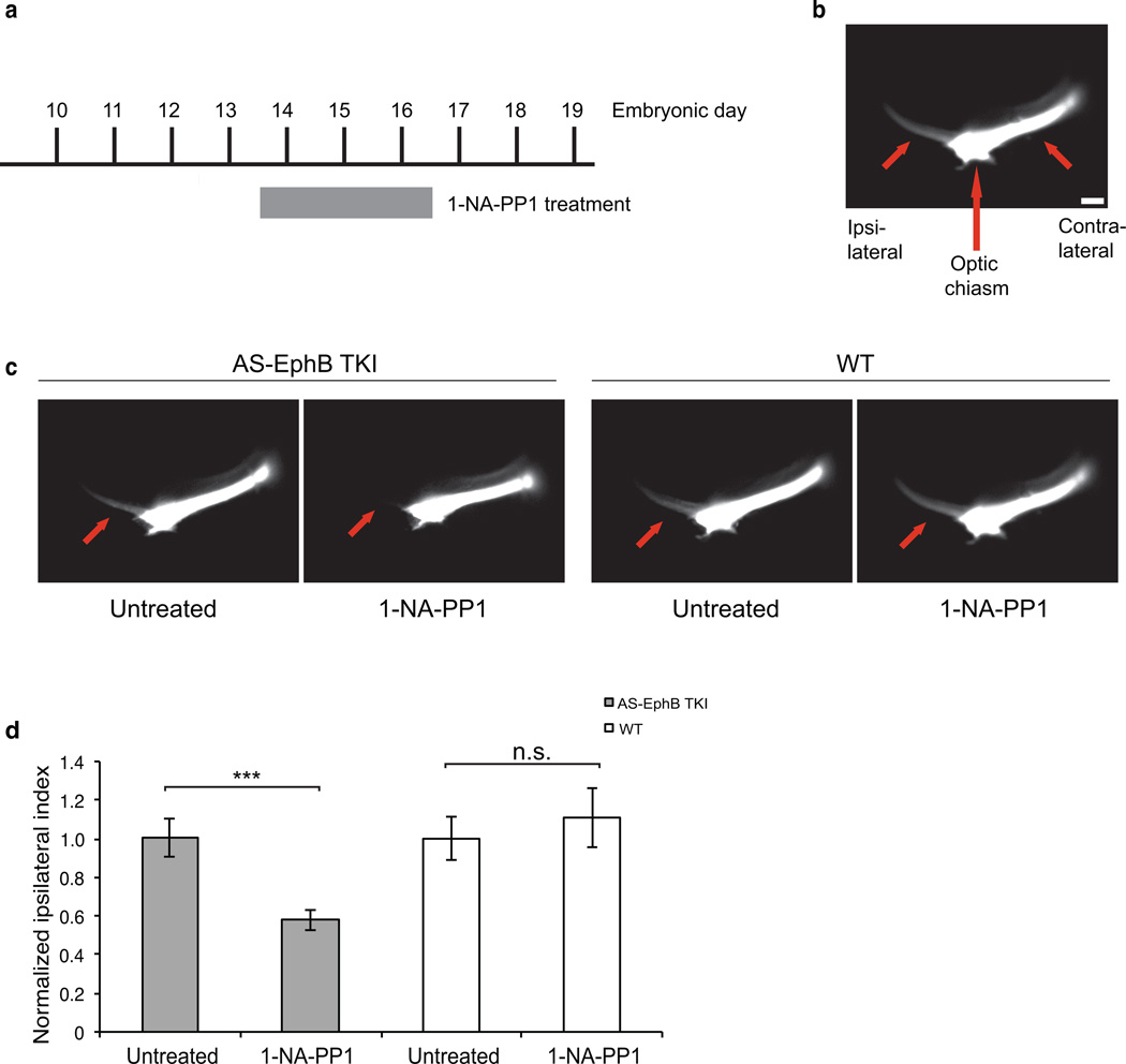
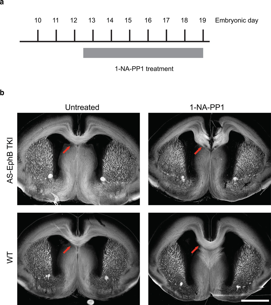
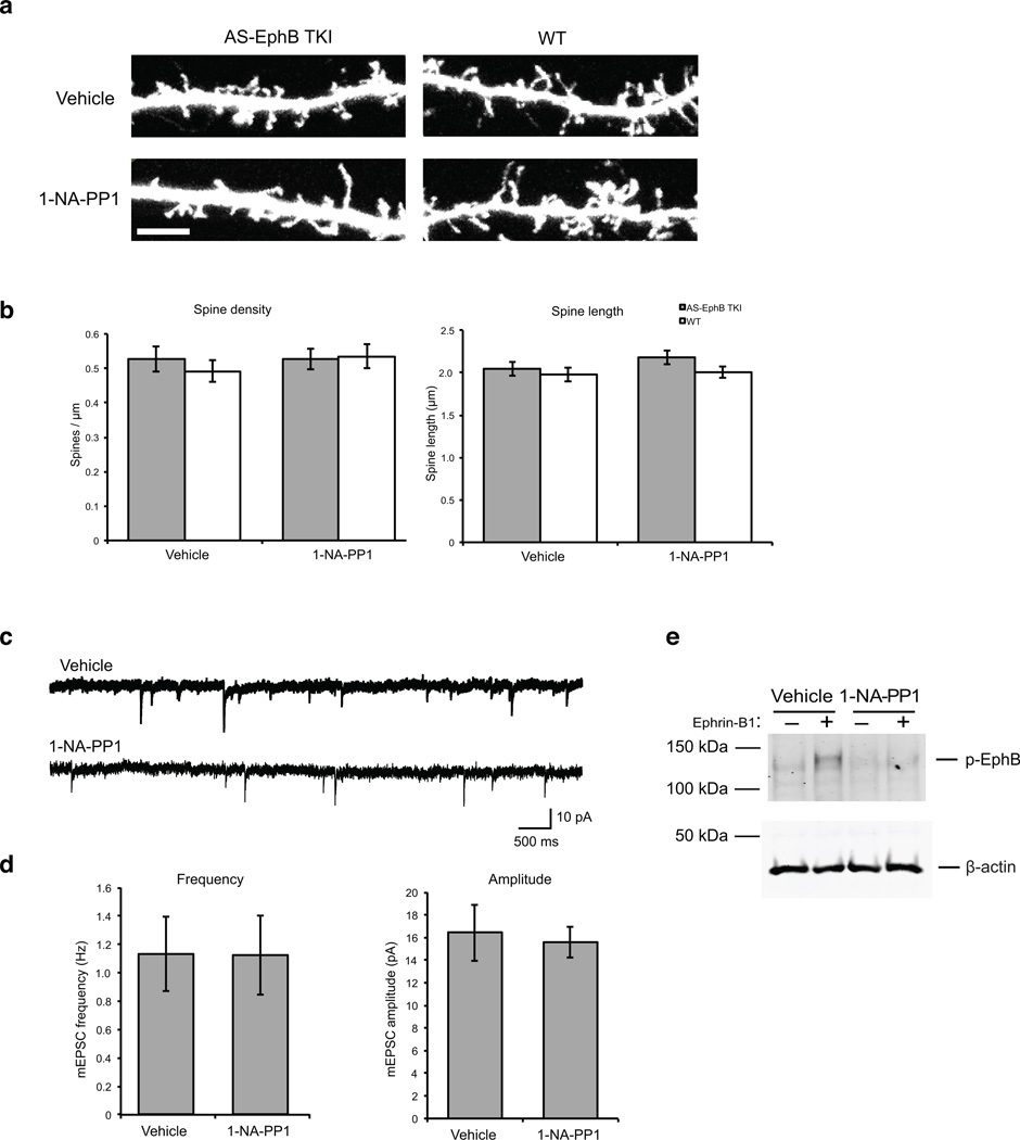
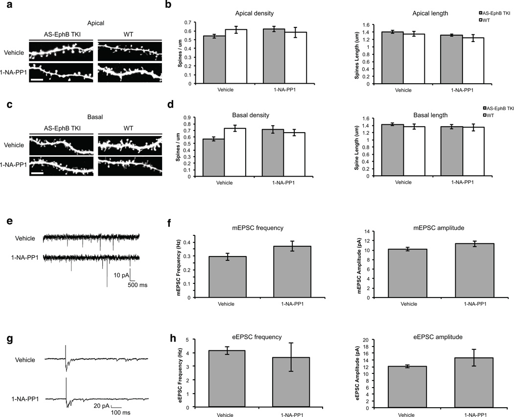
EphB receptor tyrosine kinases control multiple steps in nervous system development. However, it remains unclear whether EphBs regulate these different developmental processes directly or indirectly. In addition, given that EphBs signal through multiple mechanisms, it has been challenging to define which signaling functions of EphBs regulate particular developmental events. To address these issues, we engineered triple knock-in mice in which the kinase activity of three neuronally expressed EphBs can be rapidly, reversibly and specifically blocked. We found that the tyrosine kinase activity of EphBs was required for axon guidance in vivo. In contrast, EphB-mediated synaptogenesis occurred normally when the kinase activity of EphBs was inhibited, suggesting that EphBs mediate synapse development by an EphB tyrosine kinase-independent mechanism. Taken together, our data indicate that EphBs control axon guidance and synaptogenesis by distinct mechanisms and provide a new mouse model for dissecting EphB function in development and disease.


Figures

Figure 1
Figure 1. A chemical genetic approach to studying EphB signaling
(a) Chemical structures of the Src inhibitor PP1 and its analogs 1-NA-PP1 and 3-MB-PP1. (b) Amino acid alignment of kinase domains of mouse EphBs with those of avian vSrc and mouse TrkB. The gatekeeper residue is highlighted in red and the PP1 analog-sensitive (AS) mutation made in the EphBs is shown on the right. (c) Inhibition of the kinase function of AS-EphB proteins. HEK 293 cells expressing the AS or wild-type versions of EphB1, EphB2 and EphB3 were incubated with 1-NA-PP1 (250 nM) or 3-MB-PP1 (1 µM) for 1 hour. Cell lysates were analyzed by Western blotting for total EphBs or tyrosine phosphorylated EphBs. (d) Time course of AS-EphB1 inhibition after 1-NA-PP1 (250 nM) addition to AS-EphB1-expressing HEK 293 cells. (e) Time course of recovery of AS-EphB1 auto-phosphorylation after 1-NA-PP1 (250 nM) washout from AS-EphB1-expressing HEK 293 cells. The zero time point reflects the moment of 1-NA-PP1 washout after an initial 1-hour incubation. (f) Effect of 1-NA-PP1 on the ability of AS-EphB1 to bind Grb2 or Pick1. HEK 293 cells expressing AS-EphB1 were incubated with either vehicle or 1-NA-PP1. Cell lysates were incubated with GST, GST-Grb2 or GST-Pick1 proteins immobilized on glutathione beads. Proteins bound to the beads were analyzed by Western blotting for EphB1 (top of gel). The same gel (bottom) was stained with Coomassie blue to verify that similar amounts of GST fusion proteins were present in the binding reactions. Uncropped blots are shown in Supplementary Figure 7.
Figure 2
Figure 2. Generation of AS-EphB TKI mice
(a) Sequencing reads from AS-EphB knockin mouse embryonic stem cells showing the gatekeeper (AS) mutation in the Ephb1, Ephb2, and Ephb3 genes. The mutated amino acids are shown in red. The arrows indicate the DNA base substitutions. (b) Relative mRNA expression of EphB1, EphB2, and EphB3 normalized to β-actin in AS-EphB TKI vs. wild-type cultured cortical neurons. Data are mean of two biological replicates ± SEM.
Figure 3
Figure 3. Selective inhibition of the kinase function of EphBs in AS-EphB TKI mice
(a) Inhibition of the EphB kinase activity in AS-EphB TKI neurons. 15 day in vitro (DIV) dissociated AS-EphB TKI embryonic cortical neurons were pre-incubated with vehicle, 250 nM 1-NA-PP1, or 1 µM 3-MB-PP1 for 1 hour before a 30-minute ephrin-B1 stimulation. Cell lysates were then analyzed by Western blotting for phospho-EphB or β-actin. (b) Effect of PP1 analogs on the kinase activity of EphA4. 4 DIV cortical neurons were pre-incubated with vehicle, 250 nM 1-NA-PP1, or 1 µM 3-MB-PP1 for 1 hour before a 30-minute ephrin-A1 stimulation. Lysates were immunoprecipitated with an anti-EphA4 antibody and blotted for phospho-EphA4 and EphA4. (c) Effect of PP1 analogs on ephrin-B1-induced Vav2 phosphorylation. 4 DIV AS-Eph TKI or wild-type cortical neurons were stimulated with ephrin-B1, immunoprecipitated with an anti-Vav2 antibody, and blotted with a pan-phospho-tyrosine (pY99) or Vav2 antibody. Cells were pre-incubated with vehicle, 250 nM 1-NA-PP1, or 1 µM 3-MB-PP1 for 1 hour before a 30-min ephrin-B1 stimulation. (d) Quantification of phospho-Vav2/Vav2 band intensity from quantitative western blots in (c). Data are mean ± SEM (n=3 biological replicates). Values were normalized to the unstimulated AS-EphB TKI vehicle-treated condition. Uncropped blots are shown in Supplementary Figure 7.
Figure 4
Figure 4. The kinase function of EphBs is required for growth cone collapse in ventrotemporal (VT) retinal ganglion cells
All data are mean ± SEM. N=4–8 explants (biological replicates) per condition for each experiment. (a) Effect of PP1 analog on RGC growth cone collapse. Embryonic day 14 (E14) VT retinal explants were treated with vehicle, 250 nM 1-NA-PP1, or 1 µM 3-MB-PP1 before ephrin-B2 stimulation. Explants were stained for neurofilament (red), phospho-EphB (white) and labeled with phalloidin to visualize F-actin (green). White arrows denote clusters of phospho-EphB staining. Scale bar represents 10 µm. (b) Quantification of the percentage of collapsed growth cones from (a). A 2-way ANOVA revealed a significant interaction between genotype and inhibitor treatment in each condition, indicating that the effects of inhibitors were significantly greater in AS-EphB TKI neurons than in wild-type neurons: vehicle vs. 1-NA-PP1: F(1,20)=6.66, p=.018; vehicle vs. 3-MB-PP1: F(1,22)=7.41, p=.012. (c) Quantification of the average growth cone width from (a). A 2-way ANOVA revealed a significant interaction between genotype and inhibitor treatment in each of the conditions: vehicle vs. 1-NA-PP1: F(1,20)=11.75, p=.0027; vehicle vs. 3-MB-PP1: F(1,22)=11.49, p=.0026. (d) Quantification of the percentage of collapsed growth cones after 1-NA-PP1 washout. Retinal explants were treated with 250 nM1-NA-PP1 as in (a), but 15 minutes into a 30-minute ephrin-B2 stimulation, media were removed and replaced with fresh media containing vehicle. Percentage of collapsed growth cones was then quantified as in (b).
Figure 5
Figure 5. The kinase function of EphBs is required for the formation of the ipsilateral retinal projection in vivo
(a) Schedule of in vivo 1-NA-PP1 administration. Twice-daily subcutaneous injections of 80 mg/kg 1-NA-PP1 were administered to pregnant females from E13.5–16.5. (b) Representative image demonstrating the orientation of the ipsilateral and contralateral retinal projections (short red arrows) of the optic tract with respect to the optic chiasm (long red arrow) as visualized by DiI labeling (white). Scale bar represents 100 µm. (c) Representative images of DiI-filled retinal projections at E16.5. Pregnant AS-EphB and C57BL/6 wild-type mice were treated as described in (a). Red arrows denote the ipsilateral projection. (d) Quantification of the ipsilateral phenotype shown in (c). The Ipsilateral Index is defined as: ipsilateral / (ipsilateral + contralateral) fluorescence intensity and normalized for each genotype. The number of embryos examined was as follows: Untreated AS-EphB TKI (n=30), 1-NA-PP1 treated AS-EphB TKI (n=30), Untreated wild-type (n=15), 1-NA-PP1-treated wild-type (n=17). ***, p<.001; n.s., not significant by Student’s t-test. A 2-way ANOVA revealed a significant interaction between genotype and inhibitor treatment, indicating that 1-NA-PP1 treatment effects were significantly greater in AS-EphB TKI embryos than in wild-type embryos (F(1,88)=6.5, p=.0125). Data are mean ± SEM. Samples are biological replicates.
Figure 6
Figure 6. The kinase activity of EphBs is required for the formation of the corpus callosum in vivo
(a) Schedule of in vivo 1-NA-PP1 administration. Twice-daily subcutaneous injections of 80 mg/kg 1-NA-PP1 were administered to pregnant mice from E12.5–E19. (b) Representative images of brain sections from E19 embryos stained with L1-CAM antibody (white) to visualize axon tracts. Red arrows denote the corpus callosum. Scale bar represents 1 mm.
Figure 7
Figure 7. The kinase activity of EphBs is dispensable for the formation of dendritic spines and functional excitatory synapses in culture
(a) Representative images of dendritic spines from cultured AS-EphB TKI and wild-type cortical neurons treated with vehicle or 1 µM 1-NA-PP1 from 10–21 DIV. Neurons were transfected at 10 DIV with GFP and stained with an anti-GFP antibody. Scale bar represents 5 µm (b) Quantification of spine density and spine length from (a). Data are mean ± SEM. N = 25–33 neurons from independent biological replicates/condition. (c) Representative traces of recordings of miniature postsynaptic excitatory currents (mEPSCs) from dissociated cortical neurons from AS-EphB TKI embryos at 10–12 DIV. Cultures were treated with vehicle or 1-NA-PP1 (1 µM) from 3 DIV until the time of recording. (d) Quantification of mEPSC frequency and amplitude from vehicle and 1-NA-PP1 treated neurons. Data are mean ± SEM. N=13 cells from independent biological replicates/condition. (e) The tyrosine kinase activity of AS-EphBs is inhibited by 1-NA-PP1. Cultures concurrent with those used in (d) were treated with 1-NA-PP1 under identical culture conditions then stimulated for 30 minutes with ephrin-B1 at 10 DIV. Cell lysates were analyzed by western blotting for phospho-EphB and β-actin. Uncropped blots are shown in Supplementary Figure 7.
Figure 8
Figure 8. The kinase activity of EphBs is dispensable for the formation of dendritic spines and functional excitatory synapses
All experiments were done in organotypic hippocampal slices. The concentration of 1-NA-PP1 is 1µM. All data are mean ± SEM. Samples are independent biological replicates. (a) Representative images of apical spines from AS-EphB TKI slices. Slices from P5–7 mice were treated with vehicle or 1-NA-PP1 from 0–8 DIV. Scale bar represents 5 µm (b) Quantification of apical spine density and spine length from (a). N (neurons) = wild-type Vehicle (21), wild-type 1-NA-PP1 (11), AS-EphB TKI Vehicle (30), AS-EphB TKI1-NA-PP1 (16). (c) Representative images of basal spines from slices as described in (a). (d) Quantification of basal spine density and spine length from (c). (e) Representative traces from recordings of mEPSCs from P5–7 AS-EphB TKI slices at 12–14 DIV. (f) Quantification of mEPSC frequency and amplitude from (e). N (neurons) = AS-EphB TKI Vehicle (16), AS-EphB TKI 1-NA-PP1 (19).1-NA-PP1 did not produce a significant change in mEPSC frequency or amplitude by Student's t-test. (g) Representative traces from recordings of eEPSCs from P5–7 AS-EphB TKI slices at 12–14 DIV. (h) Quantification of eEPSC frequency and amplitude in hippocampal slices. N (neurons) = AS-EphB TKI Vehicle (7), AS-EphB TKI 1-NA-PP1 (7). 1-NA-PP1 did not produce a significant change in eEPSC frequency or amplitude by Student's t-test.

Comment in

References

    1. Klein R. Eph/ephrin signaling in morphogenesis, neural development and plasticity. Curr Opin Cell Biol. 2004;16:580–589. - PubMed
    1. Lai KO, Ip NY. Synapse development and plasticity: roles of ephrin/Eph receptor signaling. Curr Opin Neurobiol. 2009;19:275–283. - PubMed
    1. Genander M, Frisen J. Ephrins and Eph receptors in stem cells and cancer. Curr Opin Cell Biol. 2010;22:611–616. - PubMed
    1. Sanders SJ, et al. De novo mutations revealed by whole-exome sequencing are strongly associated with autism. Nature. 2012;485:237–241. - PMC - PubMed
    1. Merlos-Suarez A, Batlle E. Eph-ephrin signalling in adult tissues and cancer. Curr Opin Cell Biol. 2008;20:194–200. - PubMed

Publication types

MeSH terms

Substances