

Interhemispheric temporal lobe connectivity predicts language impairment in adolescents born preterm
- PMID: 23144265
- PMCID: PMC4031625
- DOI: 10.1093/brain/aws276
Interhemispheric temporal lobe connectivity predicts language impairment in adolescents born preterm
Abstract
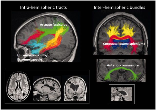
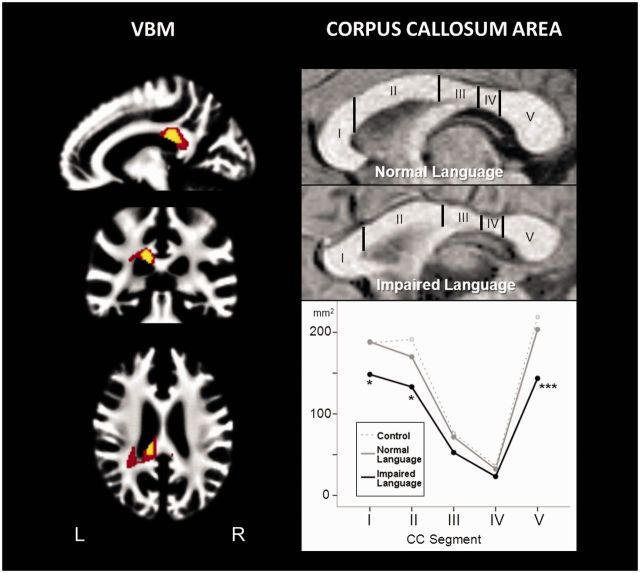
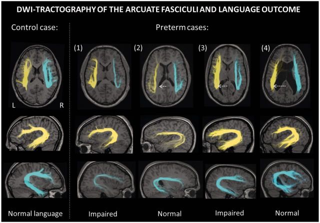
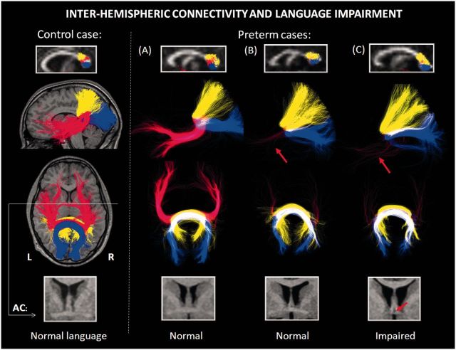
Although language difficulties are common in children born prematurely, robust neuroanatomical correlates of these impairments remain to be established. This study investigated whether the greater prevalence of language problems in preterm (versus term-born) children might reflect injury to major intra- or interhemispheric white matter pathways connecting frontal and temporal language regions. To investigate this, we performed a comprehensive assessment of language and academic abilities in a group of adolescents born prematurely, some of whom had evidence of brain injury at birth (n = 50, mean age: 16 years, mean gestational age: 27 weeks) and compared them to a term-born control group (n = 30). Detailed structural magnetic resonance imaging and diffusion-tractography analyses of intrahemispheric and interhemispheric white matter bundles were performed. Analysis of intrahemispheric pathways included the arcuate fasciculus (dorsal language pathway) and uncinate fasciculus/extreme capsule (ventral language pathway). Analysis of interhemispheric pathways (in particular, connections between the temporal lobes) included the two major commissural bundles: the corpus callosum and anterior commissure. We found language impairment in 38% of adolescents born preterm. Language impairment was not related to abnormalities of the arcuate fasciculus (or its subsegments), but was associated with bilateral volume reductions in the ventral language pathway. However, the most significant volume reduction was detected in the posterior corpus callosum (splenium), which contains interhemispheric connections between the occipital, parietal and temporal lobes. Diffusion tractography showed that of the three groups of interhemispheric fibres within the splenium, only those connecting the temporal lobes were reduced. Crucially, we found that language impairment was only detectable if the anterior commissure (a second temporal lobe commissural pathway) was also small. Regression analyses showed that a combination of anatomical measures of temporal interhemispheric connectivity (through the splenium of the corpus callosum and anterior commissure) explained 57% of the variance in language abilities. This supports recent theories emphasizing the importance of interhemispheric connections for language, particularly in the developing brain.
Figures
Similar articles
-
Interhemispheric microstructural connectivity in bitemporal lobe epilepsy with hippocampal sclerosis.Cortex. 2015 Jun;67:106-21. doi: 10.1016/j.cortex.2015.03.018. Epub 2015 Apr 1. Cortex. 2015. PMID: 25955498
-
Diffusion tensor imaging in autism spectrum disorders: preliminary evidence of abnormal neural connectivity.Aust N Z J Psychiatry. 2011 Feb;45(2):153-62. doi: 10.3109/00048674.2010.534069. Epub 2010 Dec 6. Aust N Z J Psychiatry. 2011. PMID: 21128874 Free PMC article.
-
Rewiring the extremely preterm brain: Altered structural connectivity relates to language function.Neuroimage Clin. 2020;25:102194. doi: 10.1016/j.nicl.2020.102194. Epub 2020 Jan 22. Neuroimage Clin. 2020. PMID: 32032818 Free PMC article.
-
Visual interhemispheric communication and callosal connections of the occipital lobes.Cortex. 2014 Jul;56:1-13. doi: 10.1016/j.cortex.2013.02.001. Epub 2013 Feb 13. Cortex. 2014. PMID: 23489777 Review.
-
The connectional anatomy of the temporal lobe.Handb Clin Neurol. 2022;187:3-16. doi: 10.1016/B978-0-12-823493-8.00001-8. Handb Clin Neurol. 2022. PMID: 35964979 Review.
Cited by
-
Asymmetry of planum temporale constrains interhemispheric language plasticity in children with focal epilepsy.Brain. 2013 Oct;136(Pt 10):3163-75. doi: 10.1093/brain/awt225. Epub 2013 Sep 10. Brain. 2013. PMID: 24022474 Free PMC article.
-
Cognition and connectomes in nondementia idiopathic Parkinson's disease.Netw Neurosci. 2018 Mar 1;2(1):106-124. doi: 10.1162/NETN_a_00027. eCollection 2018 Spring. Netw Neurosci. 2018. PMID: 29911667 Free PMC article.
-
White matter abnormalities and impaired attention abilities in children born very preterm.Neuroimage. 2016 Jan 1;124(Pt A):75-84. doi: 10.1016/j.neuroimage.2015.08.044. Epub 2015 Aug 28. Neuroimage. 2016. PMID: 26318524 Free PMC article.
-
Early development of structural networks and the impact of prematurity on brain connectivity.Neuroimage. 2017 Apr 1;149:379-392. doi: 10.1016/j.neuroimage.2017.01.065. Epub 2017 Jan 30. Neuroimage. 2017. PMID: 28153637 Free PMC article.
-
Adaptive mechanisms of developing brain: cerebral lateralization in the prematurely-born.Neuroimage. 2015 Mar;108:144-50. doi: 10.1016/j.neuroimage.2014.12.032. Epub 2014 Dec 17. Neuroimage. 2015. PMID: 25528658 Free PMC article.
References
-
- Allin M, Nosarti C, Narberhaus A, Walshe M, Frearson S, Kalpakidou A, et al. Growth of the corpus callosum in adolescents born preterm. Arch Pediatr Adolesc Med. 2007;161:1183–9. - PubMed
-
- Anwander A, Tittgemeyer M, von Cramon DY, Friederici AD, Knosche TR. Connectivity-based parcellation of Broca’s area. Cereb Cortex. 2007;17:816–25. - PubMed
-
- Baldeweg T, Liegeois F. Functional MRI in pediatric epilepsy surgery. In: Cataltepe O, Jallo G, editors. Pediatric epilepsy surgery: preoperative assessment and surgical treatment. New York: Thieme Medical Publishers; 2010. pp. 74–81.
-
- Barre N, Morgan A, Doyle LW, Anderson PJ. Language abilities in children who were very preterm and/or very low birth weight: a meta-analysis. J Pediatr. 2011;158:766–74. - PubMed
Publication types
MeSH terms
LinkOut - more resources
Full Text Sources
Other Literature Sources
